Journal of Shanghai Jiao Tong University (Medical Science) ›› 2023, Vol. 43 ›› Issue (7): 848-859.doi: 10.3969/j.issn.1674-8115.2023.07.007
• Basic research • Previous Articles
WU Qiqian( ), HU Yanqin, CHEN Jiale, LI Muchen, ZHAO Yougan, WU Jingwen()
Received:2023-01-11
Accepted:2023-06-20
Online:2023-07-28
Published:2023-07-28
Contact:
WU Jingwen
E-mail:garrywuqiqian@163.com;zpwujw@shsmu.edu.cn
Supported by:CLC Number:
WU Qiqian, HU Yanqin, CHEN Jiale, LI Muchen, ZHAO Yougan, WU Jingwen. Establishment and phenotype verification of mouse oviductal epithelial organoids[J]. Journal of Shanghai Jiao Tong University (Medical Science), 2023, 43(7): 848-859.
Add to citation manager EndNote|Ris|BibTeX
URL: https://xuebao.shsmu.edu.cn/EN/10.3969/j.issn.1674-8115.2023.07.007
| Primer | Sequence (5′→3′) |
|---|---|
| Foxj1-F | AAGGAGGCAGAAATCCGGTG |
| Foxj1-R | TTGTAGCCTCCCTTGTGCAG |
| Pax8-F | CCCTTCGCCATAAAGCAGGA |
| Pax8-R | AGCATGGGGAAAGGCATTGA |
| Tubb4a-F | AACCCGGCACCATGGACTCTGT |
| Tubb4a-R | TGCCTGCTCCGGATTGACCAAATA |
| Ovgp1-F | TGGACCCCTTTCTTTGTACG |
| Ovgp1-R | TGGACAGCAGTGTTTTCAGC |
| Gapdh-F | TGGAAAGCTGTGGCGTGAT |
| Gapdh-R | GGGTAGGAACACGGAAGGC |
Tab 1 Primer sequences in RT-qPCR
| Primer | Sequence (5′→3′) |
|---|---|
| Foxj1-F | AAGGAGGCAGAAATCCGGTG |
| Foxj1-R | TTGTAGCCTCCCTTGTGCAG |
| Pax8-F | CCCTTCGCCATAAAGCAGGA |
| Pax8-R | AGCATGGGGAAAGGCATTGA |
| Tubb4a-F | AACCCGGCACCATGGACTCTGT |
| Tubb4a-R | TGCCTGCTCCGGATTGACCAAATA |
| Ovgp1-F | TGGACCCCTTTCTTTGTACG |
| Ovgp1-R | TGGACAGCAGTGTTTTCAGC |
| Gapdh-F | TGGAAAGCTGTGGCGTGAT |
| Gapdh-R | GGGTAGGAACACGGAAGGC |

Fig 1 Morphological comparison of oviductal epithelial organoids from WT mice and dKO mice
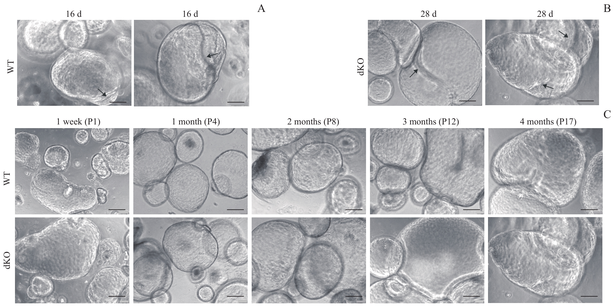
Fig 2 Long-term culture and passage of oviductal epithelial organoids
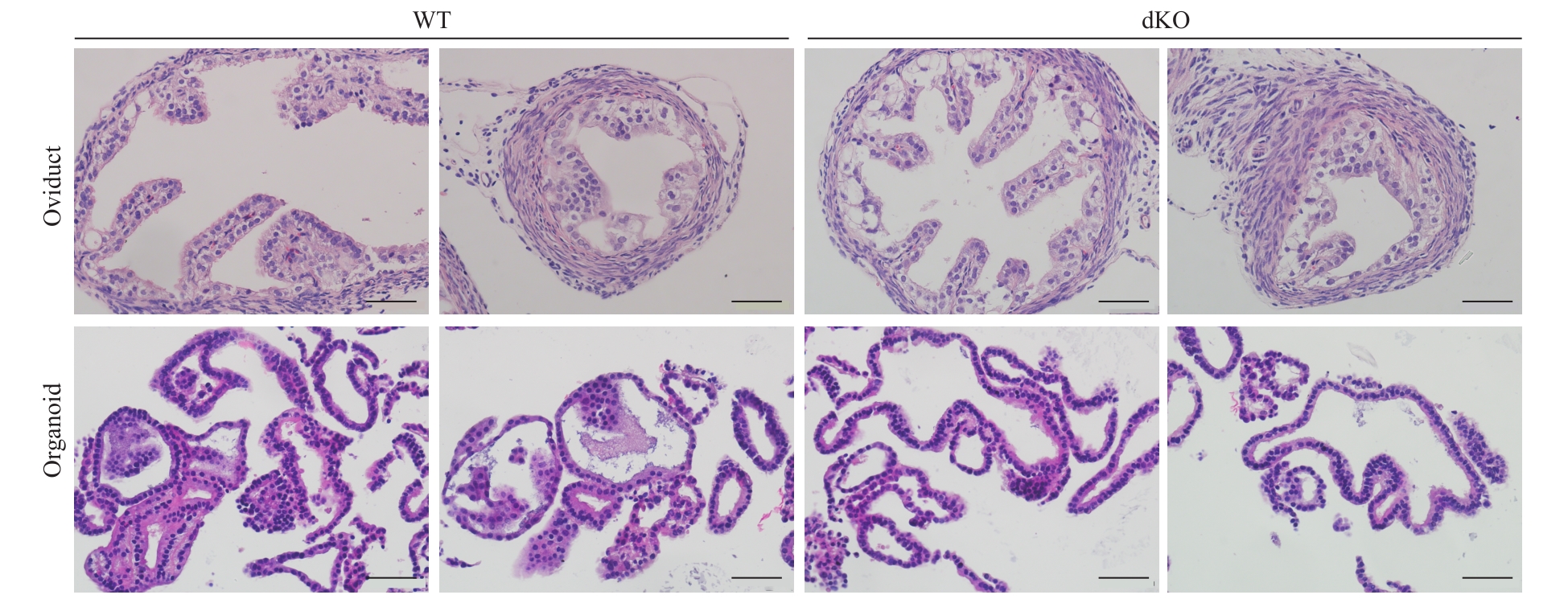
Fig 3 Structure of the oviductal epithelial organoids under light microscopy (H-E staining, ×400)

Fig 4 Microstructures of ciliated cells and secretory cells in the oviductal epithelial organoids
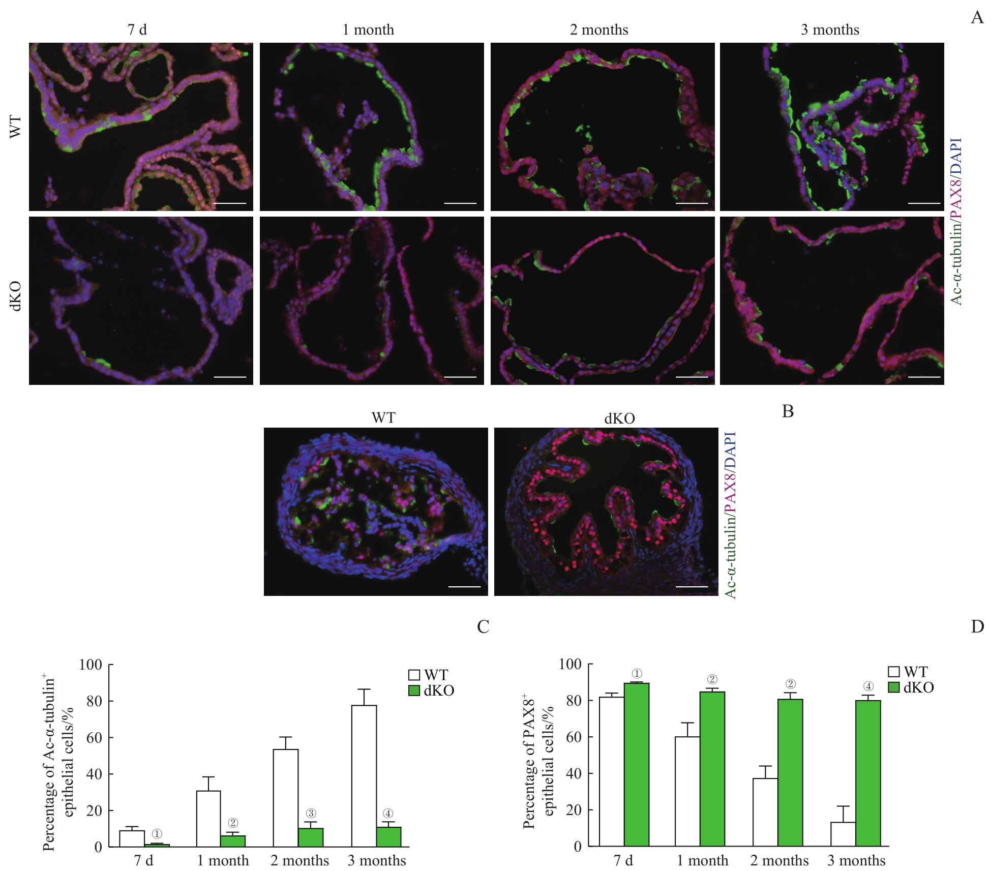
Fig 5 Cell composition of the oviductal epithelial organoids

Fig 6 Expression levels of ciliated cell- and secretory cell-related genes in oviductal epithelial organoids
| 1 | LI S, WINUTHAYANON W. Oviduct: roles in fertilization and early embryo development[J]. J Endocrinol, 2017, 232(1): R1-R26. |
| 2 | TALBOT P, SHUR B D, MYLES D G. Cell adhesion and fertilization: steps in oocyte transport, sperm-zona pellucida interactions, and sperm-egg fusion[J]. Biol Reprod, 2003, 68(1): 1-9. |
| 3 | KÖLLE S, DUBIELZIG S, REESE S, et al. Ciliary transport, gamete interaction, and effects of the early embryo in the oviduct: ex vivo analyses using a new digital videomicroscopic system in the cow[J]. Biol Reprod, 2009, 81(2): 267-274. |
| 4 | CHEN S, SCHOEN J. Air-liquid interface cell culture: from airway epithelium to the female reproductive tract[J]. Reprod Domest Anim, 2019, 54(Suppl 3): 38-45. |
| 5 | CUI Y T, ZHAO H Q, WU S W, et al. Human female reproductive system organoids: applications in developmental biology, disease modelling, and drug discovery[J]. Stem Cell Rev Rep, 2020, 16(6): 1173-1184. |
| 6 | KLASVOGT S, ZUSCHRATTER W, SCHMIDT A, et al. Air-liquid interface enhances oxidative phosphorylation in intestinal epithelial cell line IPEC-J2[J]. Cell Death Discov, 2017, 3: 17001. |
| 7 | ANDREI G, DURAFFOUR S, VAN DEN OORD J, et al. Epithelial raft cultures for investigations of virus growth, pathogenesis and efficacy of antiviral agents[J]. Antiviral Res, 2010, 85(3): 431-449. |
| 8 | LI D D, LI H, WANG Y, et al. Development and characterization of a polarized human endometrial cell epithelia in an air-liquid interface state[J]. Stem Cell Res Ther, 2018, 9(1): 209. |
| 9 | LEVANON K, NG V, PIAO H Y, et al. Primary ex vivo cultures of human fallopian tube epithelium as a model for serous ovarian carcinogenesis[J]. Oncogene, 2010, 29(8): 1103-1113. |
| 10 | CHEN S, PALMA-VERA S E, KEMPISTY B, et al. In vitro mimicking of estrous cycle stages: dissecting the impact of estradiol and progesterone on oviduct epithelium[J]. Endocrinology, 2018, 159(9): 3421-3432. |
| 11 | ZHU M B, IWANO T, TAKEDA S. Fallopian tube basal stem cells reproducing the epithelial sheets in vitro-stem cell of fallopian epithelium[J]. Biomolecules, 2020, 10(9): 1270. |
| 12 | RAJAGOPAL M, TOLLNER T L, FINKBEINER W E, et al. Differentiated structure and function of primary cultures of monkey oviductal epithelium[J]. In Vitro Cell Dev Biol Anim, 2006, 42(8/9): 248-254. |
| 13 | DVORAK A, TILLEY A E, SHAYKHIEV R, et al. Do airway epithelium air-liquid cultures represent the in vivo airway epithelium transcriptome?[J]. Am J Respir Cell Mol Biol, 2011, 44(4): 465-473. |
| 14 | ALZAMIL L, NIKOLAKOPOULOU K, TURCO M Y. Organoid systems to study the human female reproductive tract and pregnancy[J]. Cell Death Differ, 2021, 28(1): 35-51. |
| 15 | ROSSI G, MANFRIN A, LUTOLF M P. Progress and potential in organoid research[J]. Nat Rev Genet, 2018, 19(11): 671-687. |
| 16 | CLEVERS H. Modeling development and disease with organoids[J]. Cell, 2016, 165(7): 1586-1597. |
| 17 | HUCH M, KNOBLICH J A, LUTOLF M P, et al. The hope and the hype of organoid research[J]. Development, 2017, 144(6): 938-941. |
| 18 | MEBARKI M, BENNACEUR A, BONHOMME-FAIVRE L. Human-cell-derived organoids as a new ex vivo model for drug assays in oncology[J]. Drug Discov Today, 2018, 23(4): 857-863. |
| 19 | KESSLER M, HOFFMANN K, BRINKMANN V, et al. The Notch and Wnt pathways regulate stemness and differentiation in human fallopian tube organoids[J]. Nat Commun, 2015, 6: 8989. |
| 20 | XIE Y, PARK E S, XIANG D X, et al. Long-term organoid culture reveals enrichment of organoid-forming epithelial cells in the fimbrial portion of mouse fallopian tube[J]. Stem Cell Res, 2018, 32: 51-60. |
| 21 | WU J W, BAO J Q, KIM M, et al. Two miRNA clusters, miR-34b/c and miR-449, are essential for normal brain development, motile ciliogenesis, and spermatogenesis[J]. Proc Natl Acad Sci U S A, 2014, 111(28): E2851-E2857. |
| 22 | YUAN S Q, WANG Z Q, PENG H Y, et al. Oviductal motile cilia are essential for oocyte pickup but dispensable for sperm and embryo transport[J]. Proc Natl Acad Sci U S A, 2021, 118(22): e2102940118. |
| 23 | LOUKAS I, SKAMNELOU M, TSARIDOU S, et al. Fine-tuning multiciliated cell differentiation at the post-transcriptional level: contribution of miR-34/449 family members[J]. Biol Rev Camb Philos Soc, 2021, 96(5): 2321-2332. |
| 24 | MERCEY O, POPA A, CAVARD A, et al. Characterizing isomiR variants within the microRNA-34/449 family[J]. FEBS Lett, 2017, 591(5): 693-705. |
| 25 | LABIDI-GALY S I, PAPP E, HALLBERG D, et al. High grade serous ovarian carcinomas originate in the fallopian tube[J]. Nat Commun, 2017, 8(1): 1093. |
| 26 | PERETS R, WYANT G A, MUTO K W, et al. Transformation of the fallopian tube secretory epithelium leads to high-grade serous ovarian cancer in Brca;Tp53;Pten models[J]. Cancer Cell, 2013, 24(6): 751-765. |
| 27 | ISHIGURO T, OHATA H, SATO A, et al. Tumor-derived spheroids: relevance to cancer stem cells and clinical applications[J]. Cancer Sci, 2017, 108(3): 283-289. |
| 28 | ZHANG S, DOLGALEV I, ZHANG T, et al. Both fallopian tube and ovarian surface epithelium are cells-of-origin for high-grade serous ovarian carcinoma[J]. Nat Commun, 2019, 10(1): 5367. |
| [1] | ZHENG Xiaoyan, WANG Xingyun, ZHANG Yongjun. Improvement of alveolarization arrest in newborn rats with bronchopulmonary dysplasia via inhibiting alveolar epithelial cell pyroptosis [J]. Journal of Shanghai Jiao Tong University (Medical Science), 2023, 43(2): 171-179. |
| [2] | XIE Lin, CHENG Ye, ZHENG Qimin, ZHANG Xi, FU Lili, CHEN Min, WANG Yi, MEI Changlin, XIE Jingyuan, GU Xiangchen. Preventive effect of icariin on transition from acute kidney injury to chronic kidney disease in mouse model [J]. Journal of Shanghai Jiao Tong University (Medical Science), 2023, 43(1): 8-19. |
| [3] | XIE Xinyi, YANG Yumeng, WANG Shaowei, SUN Na, SHI Chuandao, LIU Qiling, ZHANG Rongqiang, LI Junjie. Genomic regulatory network of human olfactory neuroepithelial cells infected with novel coronavirus (SARS-COV-2) [J]. Journal of Shanghai Jiao Tong University (Medical Science), 2022, 42(11): 1524-1533. |
| [4] | Tian-hao ZHOU, Zhao-chen XIN, Shao-qian DU, Yuan CAO, Jing-xuan XU, Zeng-hong LAO, Hong-xia WANG. Establishment and optimization of co-culture technology for breast cancer organoids [J]. JOURNAL OF SHANGHAI JIAOTONG UNIVERSITY (MEDICAL SCIENCE), 2021, 41(8): 1017-1024. |
| [5] | WANG Yu-ting, LIU Jin-yan, SHI Ce, ZHAO Jun-tao, XIANG Ming-jie. Knocking out ERG3 gene of Candida albicans and its effect on drug resistance [J]. , 2020, 40(2): 163-. |
| [6] | WANG Ling-xiao, LIU Ting-ting, YANG Xiao-hui, YAO Zhi-qing, CAI Hui-zhen. Effect of Lycium barbarum polysaccharides on inflammatory cytokines in type 2 diabetes mellitus model mice without myeloid differentiation factor 88 gene [J]. , 2019, 39(2): 136-. |
| [7] | WANG Pei, XU Ting-ting, ZHAO Qing, WANG Zhen . Application and development of genetic knockout animal models in researches of obsessive-compulsive disorder#br# [J]. , 2017, 37(9): 1292-. |
| [8] | WANG Jian-ru, LIU Ping . Advances in differences between ApoE -/- and LDLR -/- atherosclerotic model mice#br# [J]. , 2017, 37(7): 1033-. |
| [9] | LI Wen-jing, LIU Jin-yan, SHI Ce, et al. Knock out FLO8 gene in Candida albicans by fusion PCR combined with homologous recombination [J]. , 2016, 36(3): 334-. |
| [10] | YING Ji, LIU Jian, ZHANG Li-wen, ZHONG Fang, ZHOU Fang-fang, WANG Wei-ming. Generation and identification of renal tubular epithelial cell C/EBPα conditional knockout mice [J]. , 2016, 36(10): 1403-. |
| [11] | WANG Yu-xuan, GAN Hua. Effects of angiotensin Ⅱ on necroptosis of rat renal tubular epithelial cells [J]. , 2015, 35(6): 813-. |
| [12] | JIANG Shi-zhong, YAN Ya-bin, XIE Fei, et al. Model establishment of transgenic mouse mammary epithelial cells for research of gene expression [J]. , 2012, 32(7): 955-. |
| [13] | ZHENG Tao, YANG Zu-jing, QIAN Lin-xi. Mechanism of lactogenic effect of Medulla Tetrapanacis in lactation [J]. , 2012, 32(6): 689-. |
| [14] | WANG Luo-wen, DENG Lian-fu, ZHU Ya-ping, et al. Regulation of interaction of osteoblasts and osteoclasts by hypoxia/hypoxia-inducible factor-1&alpha|pathway [J]. , 2012, 32(3): 274-. |
| [15] | LI Hong-li, DU Lian-fang, LI Feng-hua, et al. Effects of ultrasound exposure and sodium butyrate on transfection of adeno-associated virus to rat retinal pigment epithelial cells [J]. , 2012, 32(10): 1288-. |
| Viewed | ||||||
|
Full text |
|
|||||
|
Abstract |
|
|||||