Journal of Shanghai Jiao Tong University (Medical Science) ›› 2024, Vol. 44 ›› Issue (5): 543-551.doi: 10.3969/j.issn.1674-8115.2024.05.001
• High-risk?pregnancy column •
MA Ruilin1( ), LIU Yu1(), XU Guixiang1, SHI Haoran1, CUI Jianjian1, YANG Zejun1, MAO Yan1, ZHAO Yin1,2()
Received:2023-10-16
Accepted:2024-03-04
Online:2024-05-28
Published:2024-05-28
Contact:
ZHAO Yin
E-mail:maruilin202076100@163.com;lyu_9618@126. com;Zhaoyin@hust.edu.cn
Supported by:CLC Number:
MA Ruilin, LIU Yu, XU Guixiang, SHI Haoran, CUI Jianjian, YANG Zejun, MAO Yan, ZHAO Yin. Relationship between Doppler ultrasound parameters of uterine artery, umbilical artery, middle cerebral artery and placental vasculopathology and pregnancy outcome in preeclampsia rat model[J]. Journal of Shanghai Jiao Tong University (Medical Science), 2024, 44(5): 543-551.
Add to citation manager EndNote|Ris|BibTeX
URL: https://xuebao.shsmu.edu.cn/EN/10.3969/j.issn.1674-8115.2024.05.001
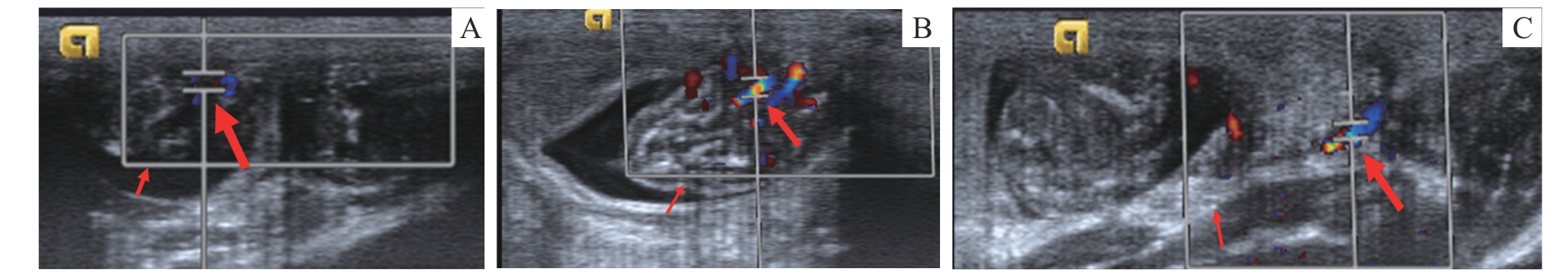
Fig 1 Ultrasound imaging of the umbilical artery, middle cerebral artery and uterine artery in SD rats
| Item | |||
|---|---|---|---|
Tab 1 Comparison of tail artery blood pressure and urinary protein between the two groups
| Item | |||
|---|---|---|---|
| Item | NP | PE | P value |
|---|---|---|---|
| 7.05±1.51 | 10.51±2.74 | 0.008 | |
| 2.08±0.78 | 1.59±0.71 | 0.203 | |
| 4.26±0.94 | 5.25±1.16 | 0.083 | |
| 3.61±0.97 | 13.95±18.00 | 0.127 | |
| 1.18±0.16 | 1.72±0.13 | 0.000 | |
| 0.71±0.07 | 0.88±0.06 | 0.000 |
Tab 2 Comparison of fetal umbilical artery hemodynamics using ultrasound evaluation between the two groups
| Item | NP | PE | P value |
|---|---|---|---|
| 7.05±1.51 | 10.51±2.74 | 0.008 | |
| 2.08±0.78 | 1.59±0.71 | 0.203 | |
| 4.26±0.94 | 5.25±1.16 | 0.083 | |
| 3.61±0.97 | 13.95±18.00 | 0.127 | |
| 1.18±0.16 | 1.72±0.13 | 0.000 | |
| 0.71±0.07 | 0.88±0.06 | 0.000 |
| Item | NP group | PE group | P value |
|---|---|---|---|
| 16.27±3.93 | 0.203 | ||
| 7.98±2.50 | 0.067 | ||
| 11.05±3.26 | 0.751 | ||
| 2.05±0.16 | 0.000 | ||
| 0.77±0.10 | 0.000 | ||
| 0.51±0.44 | 0.000 |
Tab 3 Comparison of uterine artery hemodynamics using ultrasound evaluation between the two groups
| Item | NP group | PE group | P value |
|---|---|---|---|
| 16.27±3.93 | 0.203 | ||
| 7.98±2.50 | 0.067 | ||
| 11.05±3.26 | 0.751 | ||
| 2.05±0.16 | 0.000 | ||
| 0.77±0.10 | 0.000 | ||
| 0.51±0.44 | 0.000 |
| Item | NP | PE | P value |
|---|---|---|---|
| 0.264 | |||
| 0.000 | |||
| 0.073 | |||
| 0.000 | |||
| 0.000 | |||
| 0.000 |
Tab 4 Comparison of fetal middle cerebral artery hemodynamics using ultrasound evaluation between the two groups
| Item | NP | PE | P value |
|---|---|---|---|
| 0.264 | |||
| 0.000 | |||
| 0.073 | |||
| 0.000 | |||
| 0.000 | |||
| 0.000 |
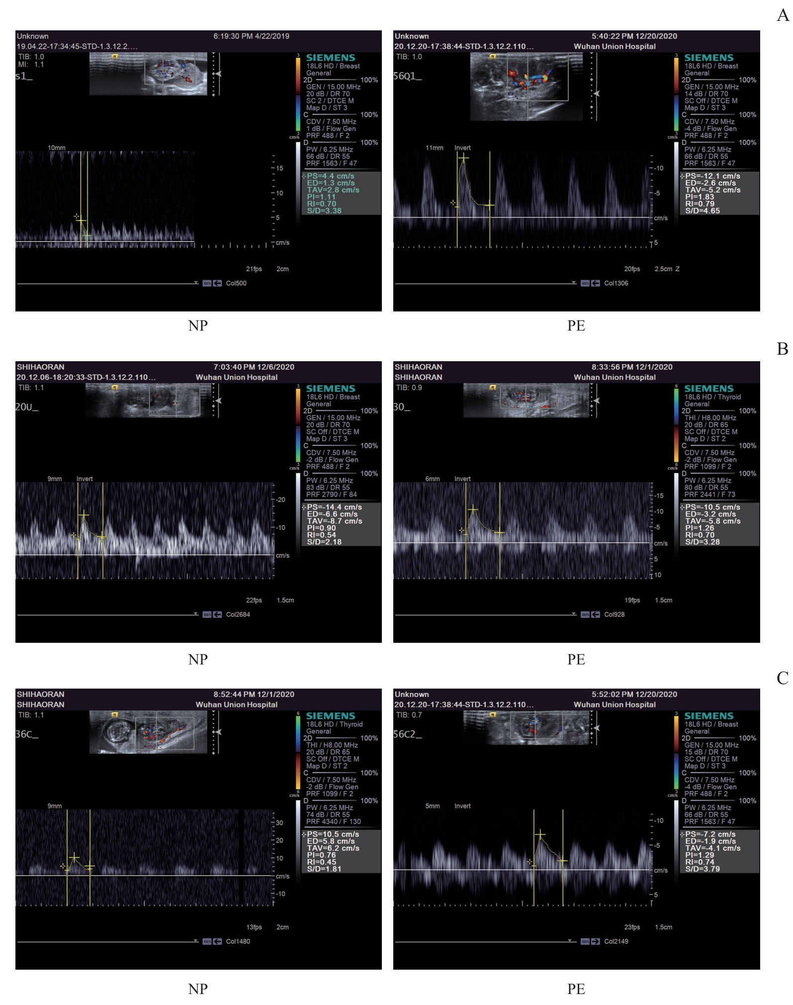
Fig 2 Ultrasound Doppler spectrogram between the two groups
| Item | NP group | PE group | P value |
|---|---|---|---|
| Placental quality/ | 0.006 | ||
| Fetal rat quality/ | 4.20 | 0.000 | |
| Fetal number/n | 0.526 |
Tab 5 Comparison of placental quality, fetal rat number and fetal rat quality between the two groups
| Item | NP group | PE group | P value |
|---|---|---|---|
| Placental quality/ | 0.006 | ||
| Fetal rat quality/ | 4.20 | 0.000 | |
| Fetal number/n | 0.526 |
| P value | |||
|---|---|---|---|
Tab 6 Comparison of MVD between the two groups
| P value | |||
|---|---|---|---|
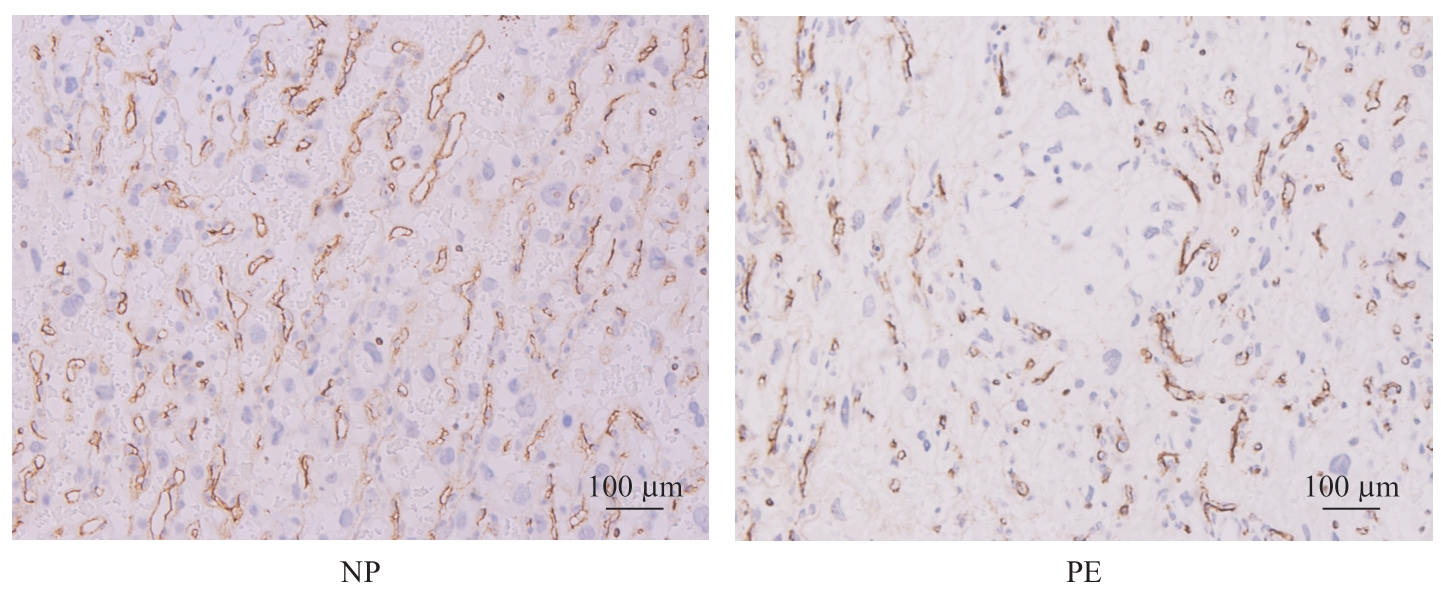
Fig 3 Comparison of MVD between two groups
| Item | MVD | Placental quality | Fetal rat quality |
|---|---|---|---|
| PS | -0.657 (0.006) | -0.160 (0.554) | -0.638 (0.008) |
| ED | 0.304 (0.253) | 0.522 (0.038) | 0.354 (0.178) |
| TAV | -0.466 (0.069) | -0.182 (0.500) | -0.424 (0.102) |
| S/D | -0.284 (0.287) | -0.698 (0.003) | -0.353 (0.179) |
| PI | -0.850 (0.000) | -0.450 (0.081) | -0.901 (0.000) |
| RI | -0.758 (0.001) | -0.740 (0.001) | -0.832 (0.000) |
Tab 7 Correlation analysis between umbilical artery parameters and placental quality, fetal rat quality and placental MVD [r (P)]
| Item | MVD | Placental quality | Fetal rat quality |
|---|---|---|---|
| PS | -0.657 (0.006) | -0.160 (0.554) | -0.638 (0.008) |
| ED | 0.304 (0.253) | 0.522 (0.038) | 0.354 (0.178) |
| TAV | -0.466 (0.069) | -0.182 (0.500) | -0.424 (0.102) |
| S/D | -0.284 (0.287) | -0.698 (0.003) | -0.353 (0.179) |
| PI | -0.850 (0.000) | -0.450 (0.081) | -0.901 (0.000) |
| RI | -0.758 (0.001) | -0.740 (0.001) | -0.832 (0.000) |
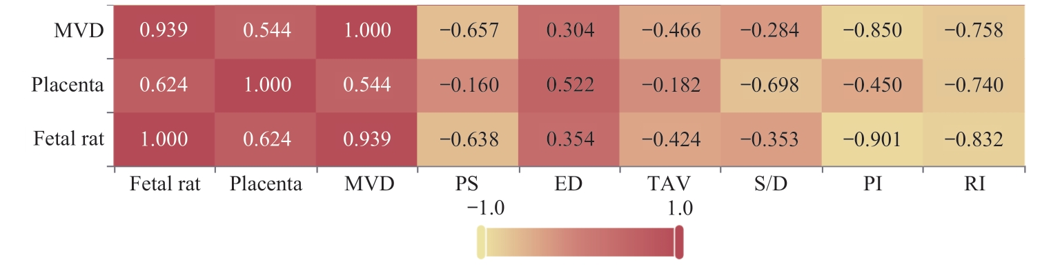
Fig 4 Heatmap of the correlation analysis between umbilical artery parameter indices and placental quality, fetal rat quality and placental MVD
| Item | MVD | Placental quality | Fetal rat quality |
|---|---|---|---|
| PS | -0.332 (0.209) | -0.381 (0.145) | -0.302 (0.256) |
| ED | 0.447 (0.083) | 0.291 (0.274) | 0.487 (0.056) |
| TAV | 0.078 (0.775) | -0.061 (0.824) | 0.123 (0.650) |
| S/D | -0.871 (0.000) | -0.638 (0.008) | -0.870 (0.000) |
| PI | -0.892 (0.000) | -0.615 (0.011) | -0.922 (0.000) |
| RI | -0.874 (0.000) | -0.621 (0.010) | -0.902 (0.000) |
Tab 8 Correlation analysis between uterine artery parameters and placental quality, fetal rat quality and placental MVD [r (P)]
| Item | MVD | Placental quality | Fetal rat quality |
|---|---|---|---|
| PS | -0.332 (0.209) | -0.381 (0.145) | -0.302 (0.256) |
| ED | 0.447 (0.083) | 0.291 (0.274) | 0.487 (0.056) |
| TAV | 0.078 (0.775) | -0.061 (0.824) | 0.123 (0.650) |
| S/D | -0.871 (0.000) | -0.638 (0.008) | -0.870 (0.000) |
| PI | -0.892 (0.000) | -0.615 (0.011) | -0.922 (0.000) |
| RI | -0.874 (0.000) | -0.621 (0.010) | -0.902 (0.000) |
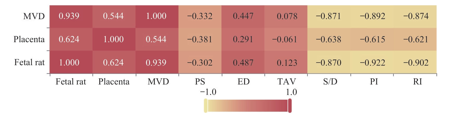
Fig 5 Heatmap of the correlation analysis between uterine artery parameter indices and placental quality, fetal rat quality and placental MVD
| Item | MVD | Placental quality | Fetal rat quality |
|---|---|---|---|
| PS | -0.148 (0.586) | -0.196 (0.467) | -0.300 (0.260) |
| ED | -0.749 (0.001) | -0.575 (0.020) | -0.802 (0.000) |
| TAV | -0.333 (0.207) | -0.342 (0.195) | -0.451 (0.079) |
| S/D | 0.923 (0.000) | 0.582 (0.018) | 0.915 (0.000) |
| PI | 0.895 (0.000) | 0.571 (0.021) | 0.850 (0.000) |
| RI | 0.914 (0.000) | 0.569 (0.022) | 0.888 (0.000) |
Tab 9 Correlation coefficients between middle cerebral artery parameters and placental quality, fetal rat quality, and placental MVD
| Item | MVD | Placental quality | Fetal rat quality |
|---|---|---|---|
| PS | -0.148 (0.586) | -0.196 (0.467) | -0.300 (0.260) |
| ED | -0.749 (0.001) | -0.575 (0.020) | -0.802 (0.000) |
| TAV | -0.333 (0.207) | -0.342 (0.195) | -0.451 (0.079) |
| S/D | 0.923 (0.000) | 0.582 (0.018) | 0.915 (0.000) |
| PI | 0.895 (0.000) | 0.571 (0.021) | 0.850 (0.000) |
| RI | 0.914 (0.000) | 0.569 (0.022) | 0.888 (0.000) |
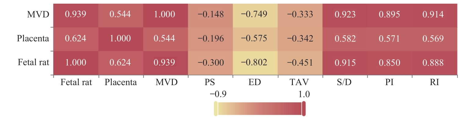
Fig 6 Heatmap of the correlation analysis between middle cerebral artery parameter indices and placental quality, fetal rat quality and placental MVD
| 1 | DIMITRIADIS E, ROLNIK D L, ZHOU W, et al. Pre-eclampsia[J]. Nat Rev Dis Primers, 2023, 9(1): 8. |
| 2 | 朱玥霖, 李倩倩, 王雁玲, 等. 胎盘发育和子痫前期[J]. 生物医学转化, 2022, 3(4): 22-29. |
| ZHU Y L, LI Q Q, WANG Y L, et al.Placenta development and preeclampsia[J]. Biomedical Transformation, 2022, 3(4): 22-29. | |
| 3 | VATISH M, POWYS V R, CERDEIRA A S. Novel therapeutic and diagnostic approaches for preeclampsia[J]. Curr Opin Nephrol Hypertens, 2023, 32(2): 124-133. |
| 4 | CARTER A M. Comparative studies of placentation and immunology in non-human primates suggest a scenario for the evolution of deep trophoblast invasion and an explanation for human pregnancy disorders[J]. Reproduction, 2011, 141(4): 391-396. |
| 5 | CARTER A M, PIJNENBORG R. Evolution of invasive placentation with special reference to non-human primates[J]. Best Pract Res Clin Obstet Gynaecol, 2011, 25(3): 249-257. |
| 6 | BAKRANIA B A, GEORGE E M, GRANGER J P. Animal models of preeclampsia: investigating pathophysiology and therapeutic targets[J]. Am J Obstet Gynecol, 2022, 226(2S): S973-S987. |
| 7 | PIJNENBORG R, ROBERTSON W B, BROSENS I, et al. Review article: trophoblast invasion and the establishment of haemochorial placentation in man and laboratory animals[J]. Placenta, 1981, 2(1): 71-91. |
| 8 | VERKESTE C M, SLANGEN B F, DAEMEN M, et al. The extent of trophoblast invasion in the preplacental vasculature of the guinea-pig[J]. Placenta, 1998, 19(1): 49-54. |
| 9 | SOARES M J, CHAKRABORTY D, KARIM RUMI M A, et al. Rat placentation: an experimental model for investigating the hemochorial maternal-fetal interface[J]. Placenta, 2012, 33(4): 233-243. |
| 10 | SHU W, LI H Y, GONG H, et al. Evaluation of blood vessel injury, oxidative stress and circulating inflammatory factors in an L-NAME-induced preeclampsia-like rat model[J]. Exp Ther Med, 2018, 16(2): 585-594. |
| 11 | GINOSAR Y, GIELCHINSKY Y, NACHMANSSON N, et al. BOLD-MRI demonstrates acute placental and fetal organ hypoperfusion with fetal brain sparing during hypercapnia[J]. Placenta, 2018, 63: 53-60. |
| 12 | MARKOVIC S, FAGES A, ROUSSEL T, et al. Placental physiology monitored by hyperpolarized dynamic 13C magnetic resonance[J]. Proc Natl Acad Sci USA, 2018, 115(10): E2429-E2436. |
| 13 | ARTHUIS C J, MENDES V, MÊME S, et al. Comparative determination of placental perfusion by magnetic resonance imaging and contrast-enhanced ultrasound in a murine model of intrauterine growth restriction[J]. Placenta, 2018, 69: 74-81. |
| 14 | DINES V, SUVAKOV S, KATTAH A, et al. Preeclampsia and the kidney: pathophysiology and clinical implications[J]. Compr Physiol, 2023, 13(1): 4231-4267. |
| 15 | FEBRES-CORDERO D A, YOUNG B C. Hypertensive disorders of pregnancy[J]. NeoReviews, 2021, 22(11): e760-e766. |
| 16 | POON L C, SHENNAN A, HYETT J A, et al. The International Federation of Gynecology and Obstetrics (FIGO) initiative on pre-eclampsia: a pragmatic guide for first-trimester screening and prevention[J]. Int J Gynaecol Obstet, 2019, 145(Suppl 1): 1-33. |
| 17 | 谢幸, 孔北华, 段涛. 妇产科学[M]. 9版. 北京: 人民卫生出版社, 2018. |
| XIE X, KONG B H, DUAN T. Obstetrics and Gynecology[M]. The 9th edition. Beijing: People′s Medical Publishing House, 2018. | |
| 18 | USTA A, TURAN G, SANCAKLI USTA C, et al. Placental fractalkine immunoreactivity in preeclampsia and its correlation with histopathological changes in the placenta and adverse pregnancy outcomes[J]. J Matern Fetal Neonatal Med, 2020, 33(5): 806-815. |
| 19 | TICA O S, TICA A A, COJOCARU D, et al. Maternal steroids on fetal doppler indices, in growth-restricted fetuses with abnormal umbilical flow from pregnancies complicated with early-onset severe preeclampsia[J]. Diagnostics, 2023, 13(3): 428. |
| 20 | SHI M T, YANG X F, SUN L, et al. Comparison of different modified operations in the reduced uteroplacental perfusion pressure rat model of preeclampsia[J]. J Reprod Immunol, 2023, 156: 103815. |
| 21 | SAKOWICZ A, BRALEWSKA M, KAMOLA P, et al. Reliability of rodent and rabbit models in preeclampsia research[J]. Int J Mol Sci, 2022, 23(22): 14344. |
| 22 | Zeisler H, Llurba E, Chantraine F, et al. Predictive value of the sFlt-1:PlGF ratio in women with suspected preeclampsia[J]. N Engl J Med, 2016, 374(1): 13-22. |
| 23 | ARTHUIS C J, NOVELL A, ESCOFFRE J M, et al. New insights into uteroplacental perfusion: quantitative analysis using doppler and contrast-enhanced ultrasound imaging[J]. Placenta, 2013, 34(5): 424-431. |
| 24 | SPRADLEY F T, GE Y, GRANGER J P, et al. Utero-placental vascular remodeling during late gestation in Sprague-Dawley rats[J]. Pregnancy Hypertens, 2020, 20: 36-43. |
| 25 | TRAVIS O K, TARDO G A, GIACHELLI C, et al. Interferon γ neutralization reduces blood pressure, uterine artery resistance index, and placental oxidative stress in placental ischemic rats[J]. Am J Physiol Regul Integr Comp Physiol, 2021, 321(2): R112-R124. |
| 26 | BIBEAU K, SICOTTE B, BÉLAND M, et al. Placental underperfusion in a rat model of intrauterine growth restriction induced by a reduced plasma volume expansion[J]. PLoS One, 2016, 11(1): e0145982. |
| 27 | PAPAGEORGHIOU A T, YU C K H, ERASMUS I E, et al. Assessment of risk for the development of pre-eclampsia by maternal characteristics and uterine artery doppler[J]. BJOG, 2005, 112(6): 703-709. |
| 28 | ZHOU P, SUN Y, TAN Y P, et al. Fetal and neonatal middle cerebral artery hemodynamic changes and significance under ultrasound detection in hypertensive disorder complicating pregnancy patients with different severities[J]. Comput Math Methods Med, 2022, 2022: 6110228. |
| Viewed | ||||||
|
Full text |
|
|||||
|
Abstract |
|
|||||