
Journal of Shanghai Jiao Tong University (Medical Science) ›› 2026, Vol. 46 ›› Issue (10): 1375-1382.doi: 10.3969/j.issn.1674-8115.2026.10.007
• Clinical research • Previous Articles
Xiao Huoyuan, Quyang Danzeng, Zhang Qingchen, Feng Rui, Liu Yuting, Kang Sang, Pan Jingwei( )
Received:2025-11-28
Accepted:2026-04-22
Online:2026-10-28
Published:2026-10-28
Contact:
Pan Jingwei
E-mail:jwpan@sjtu.edu.cn
Supported by:CLC Number:
Xiao Huoyuan, Quyang Danzeng, Zhang Qingchen, Feng Rui, Liu Yuting, Kang Sang, Pan Jingwei. A radiomic model based on cardiac magnetic resonance for the classification of COVID-19-associated myocardial injury[J]. Journal of Shanghai Jiao Tong University (Medical Science), 2026,(10): 1375-1382.
Add to citation manager EndNote|Ris|BibTeX
URL: https://xuebao.shsmu.edu.cn/EN/10.3969/j.issn.1674-8115.2026.10.007
| Item | Elevated troponin group (n=24) | Normal troponin group (n=46) | Healthy control group (n=70) | P value① | P value② | P value③ |
|---|---|---|---|---|---|---|
| Age/year | 38.0 (29.0, 52.0) | 35.5 (26.8, 41.0) | 38.0 (28.0, 56.8) | 0.294 | 0.766 | 0.159 |
| Hematocrit/% | 41.3±5.4 | 42.0±4.9 | 43.1±5.2 | 0.599 | 0.203 | 0.323 |
| hs-cTnI/(ng·L-1) | 1 366.0 (114.0, 4 707.7) | 2.0 (2.0, 3.0) | 2.0 (1.9, 2.0) | <0.001 | <0.001 | 0.001 |
| NT-proBNP/(pg·mL-1) | 639.5 (112.7, 2 194.1) | 54.8 (36.8, 98.2) | 22.9 (22.9, 39.1) | <0.001 | <0.001 | <0.001 |
| LVEF/% | 61.7 (55.0, 63.7) | 57.6 (55.0, 63.2) | 61.0 (56.1, 65.0) | 0.444 | 0.402 | 0.121 |
| T1/ms | 1 279 (1 225, 1 319) | 1 246 (1 219, 1 268) | 1 198 (1 159, 1 234) | 0.017 | <0.001 | <0.001 |
| T2/ms | 43 (41, 45) | 44 (41, 47) | 41 (40, 43) | 0.774 | 0.034 | 0.007 |
| ECV/% | 35±4 | 32±4 | 30±4 | 0.035 | <0.001 | 0.011 |
Tab 1 Comparison of baseline clinical characteristics and CMR imaging data among the three groups
| Item | Elevated troponin group (n=24) | Normal troponin group (n=46) | Healthy control group (n=70) | P value① | P value② | P value③ |
|---|---|---|---|---|---|---|
| Age/year | 38.0 (29.0, 52.0) | 35.5 (26.8, 41.0) | 38.0 (28.0, 56.8) | 0.294 | 0.766 | 0.159 |
| Hematocrit/% | 41.3±5.4 | 42.0±4.9 | 43.1±5.2 | 0.599 | 0.203 | 0.323 |
| hs-cTnI/(ng·L-1) | 1 366.0 (114.0, 4 707.7) | 2.0 (2.0, 3.0) | 2.0 (1.9, 2.0) | <0.001 | <0.001 | 0.001 |
| NT-proBNP/(pg·mL-1) | 639.5 (112.7, 2 194.1) | 54.8 (36.8, 98.2) | 22.9 (22.9, 39.1) | <0.001 | <0.001 | <0.001 |
| LVEF/% | 61.7 (55.0, 63.7) | 57.6 (55.0, 63.2) | 61.0 (56.1, 65.0) | 0.444 | 0.402 | 0.121 |
| T1/ms | 1 279 (1 225, 1 319) | 1 246 (1 219, 1 268) | 1 198 (1 159, 1 234) | 0.017 | <0.001 | <0.001 |
| T2/ms | 43 (41, 45) | 44 (41, 47) | 41 (40, 43) | 0.774 | 0.034 | 0.007 |
| ECV/% | 35±4 | 32±4 | 30±4 | 0.035 | <0.001 | 0.011 |
| Item | Training set (n=98) | Test set (n=42) | P value |
|---|---|---|---|
| Age/year | 38.0 (28.0, 51.0) | 36.5 (26.3, 49.3) | 0.896 |
| Hematocrit/% | 41.9±5.3 | 43.2±4.7 | 0.202 |
| hs-cTnI/(ng·L-1) | 2.0 (2.0, 3.7) | 2.0 (1.9, 3.0) | 0.567 |
| NT-proBNP/(pg·mL-1) | 39.9 (22.9, 70.5) | 39.4 (22.9, 83.4) | 0.891 |
| LVEF/% | 60.3 (56.0, 64.0) | 61.0 (54.0, 64.1) | 0.842 |
| T1/ms | 1 224 (1 173, 1 268) | 1 228 (1 209, 1 275) | 0.260 |
| T2/ms | 42 (40, 45) | 41 (39, 45) | 0.180 |
| ECV/% | 32±5 | 32±4 | 0.894 |
Tab 2 Comparison of baseline clinical characteristics and CMR imaging data between the training and test sets
| Item | Training set (n=98) | Test set (n=42) | P value |
|---|---|---|---|
| Age/year | 38.0 (28.0, 51.0) | 36.5 (26.3, 49.3) | 0.896 |
| Hematocrit/% | 41.9±5.3 | 43.2±4.7 | 0.202 |
| hs-cTnI/(ng·L-1) | 2.0 (2.0, 3.7) | 2.0 (1.9, 3.0) | 0.567 |
| NT-proBNP/(pg·mL-1) | 39.9 (22.9, 70.5) | 39.4 (22.9, 83.4) | 0.891 |
| LVEF/% | 60.3 (56.0, 64.0) | 61.0 (54.0, 64.1) | 0.842 |
| T1/ms | 1 224 (1 173, 1 268) | 1 228 (1 209, 1 275) | 0.260 |
| T2/ms | 42 (40, 45) | 41 (39, 45) | 0.180 |
| ECV/% | 32±5 | 32±4 | 0.894 |
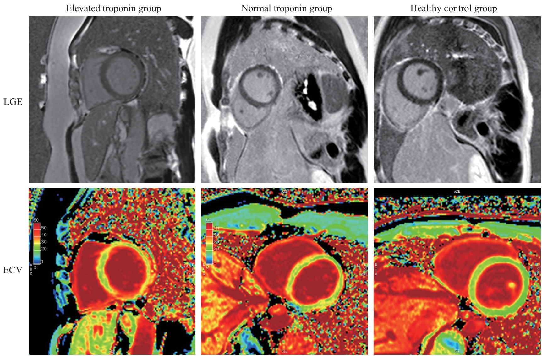
Fig 1 Representative LGE and ECV images of participants in the three groups
| Feature | Feature class | Filter | Definition |
|---|---|---|---|
| Autocorrelation | GLCM | Wavelet-HHL | Measures the correlation between the intensities of neighboring voxels |
| JointAverage | GLCM | Wavelet-HHL | Measures the mean of the joint gray-level distribution of neighboring voxel pairs |
| Strength | NGTDM | Log.sigma.2.0mm | Measures of the strength/distinctiveness of texture primitives |
| SumAverage | GLCM | Wavelet-HHL | Measures the mean of the sum of gray levels for neighboring intensity pairs |
| LargeDependenceLowGrayLevelEmphasis | GLDM | Log.sigma.1.0mm | Measures the extent of large, contiguous low-gray-level regions |
Tab 3 Explications of the features selected for CMR radiomic model construction
| Feature | Feature class | Filter | Definition |
|---|---|---|---|
| Autocorrelation | GLCM | Wavelet-HHL | Measures the correlation between the intensities of neighboring voxels |
| JointAverage | GLCM | Wavelet-HHL | Measures the mean of the joint gray-level distribution of neighboring voxel pairs |
| Strength | NGTDM | Log.sigma.2.0mm | Measures of the strength/distinctiveness of texture primitives |
| SumAverage | GLCM | Wavelet-HHL | Measures the mean of the sum of gray levels for neighboring intensity pairs |
| LargeDependenceLowGrayLevelEmphasis | GLDM | Log.sigma.1.0mm | Measures the extent of large, contiguous low-gray-level regions |
| Item | Accuracy/% | Precision/% | Sensitivity/% | Specificity/% | F1 | AUC |
|---|---|---|---|---|---|---|
| Training cohort | ||||||
| Elevated troponin group | ||||||
| CMR radiomic model | 91.84 | 73.68 | 82.35 | 93.83 | 0.77 | 0.944 |
| ECV control model | 70.41 | 32.35 | 64.71 | 71.60 | 0.43 | 0.741 |
| Normal troponin group | ||||||
| CMR radiomic model | 83.67 | 78.57 | 68.75 | 90.91 | 0.73 | 0.903 |
| ECV control model | 66.33 | 46.67 | 21.88 | 87.88 | 0.29 | 0.552 |
| Healthy control group | ||||||
| CMR radiomic model | 87.76 | 86.27 | 89.80 | 85.71 | 0.88 | 0.929 |
| ECV control model | 63.27 | 63.27 | 63.27 | 63.27 | 0.63 | 0.684 |
| Test cohort | ||||||
| Elevated troponin group | ||||||
| CMR radiomic model | 83.33 | 50.00 | 71.43 | 85.71 | 0.58 | 0.865 |
| ECV control model | 71.43 | 30.77 | 57.14 | 74.29 | 0.40 | 0.708 |
| Normal troponin group | ||||||
| CMR radiomic model | 73.81 | 61.54 | 57.14 | 82.14 | 0.59 | 0.691 |
| ECV control model | 66.67 | 50.00 | 21.43 | 89.29 | 0.30 | 0.557 |
| Healthy control group | ||||||
| CMR radiomic model | 85.71 | 89.47 | 80.95 | 90.48 | 0.85 | 0.873 |
| ECV control model | 66.67 | 65.22 | 71.43 | 61.90 | 0.68 | 0.667 |
Tab 4 Comparison of the performance of CMR radiomic model and ECV control model in discriminating among the elevated troponin, normal troponin, and healthy control groups in the training and test sets
| Item | Accuracy/% | Precision/% | Sensitivity/% | Specificity/% | F1 | AUC |
|---|---|---|---|---|---|---|
| Training cohort | ||||||
| Elevated troponin group | ||||||
| CMR radiomic model | 91.84 | 73.68 | 82.35 | 93.83 | 0.77 | 0.944 |
| ECV control model | 70.41 | 32.35 | 64.71 | 71.60 | 0.43 | 0.741 |
| Normal troponin group | ||||||
| CMR radiomic model | 83.67 | 78.57 | 68.75 | 90.91 | 0.73 | 0.903 |
| ECV control model | 66.33 | 46.67 | 21.88 | 87.88 | 0.29 | 0.552 |
| Healthy control group | ||||||
| CMR radiomic model | 87.76 | 86.27 | 89.80 | 85.71 | 0.88 | 0.929 |
| ECV control model | 63.27 | 63.27 | 63.27 | 63.27 | 0.63 | 0.684 |
| Test cohort | ||||||
| Elevated troponin group | ||||||
| CMR radiomic model | 83.33 | 50.00 | 71.43 | 85.71 | 0.58 | 0.865 |
| ECV control model | 71.43 | 30.77 | 57.14 | 74.29 | 0.40 | 0.708 |
| Normal troponin group | ||||||
| CMR radiomic model | 73.81 | 61.54 | 57.14 | 82.14 | 0.59 | 0.691 |
| ECV control model | 66.67 | 50.00 | 21.43 | 89.29 | 0.30 | 0.557 |
| Healthy control group | ||||||
| CMR radiomic model | 85.71 | 89.47 | 80.95 | 90.48 | 0.85 | 0.873 |
| ECV control model | 66.67 | 65.22 | 71.43 | 61.90 | 0.68 | 0.667 |
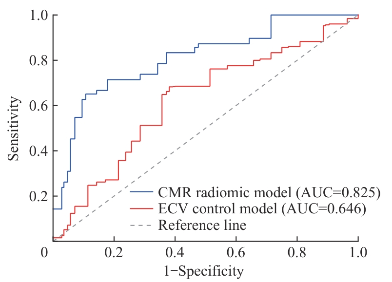
Fig 2 Comparison of the overall diagnostic performance between the CMR radiomic model and the ECV control model in the test set
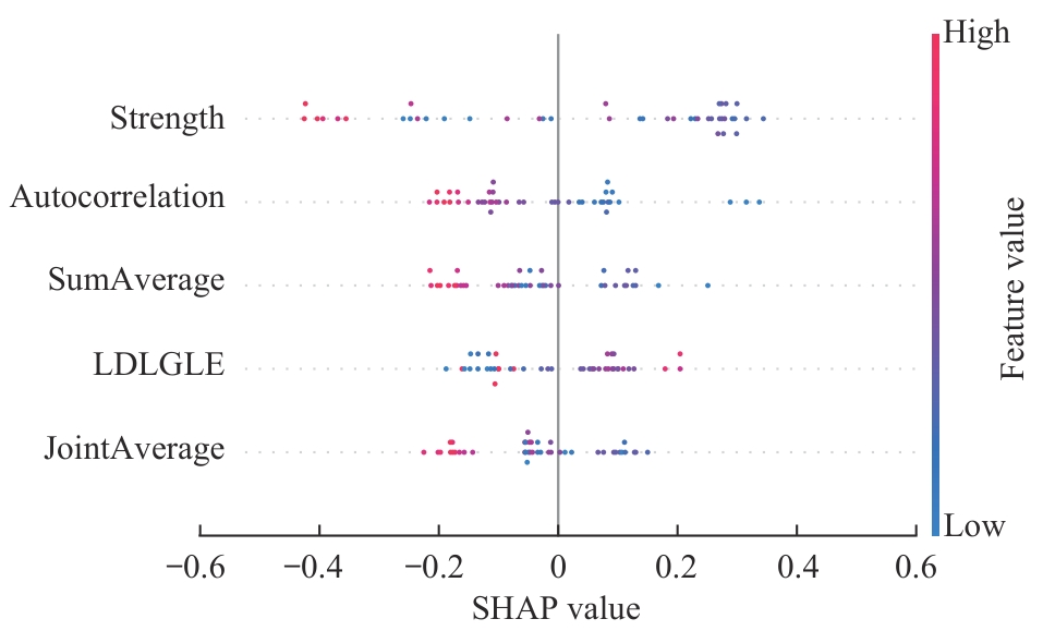
Fig 3 SHAP beeswarm plot for interpreting the CMR radiomic model in the test set
| [1] | Harrison S L, Buckley B J R, Rivera-Caravaca J M, et al. Cardiovascular risk factors, cardiovascular disease, and COVID-19: an umbrella review of systematic reviews[J]. Eur Heart J Qual Care Clin Outcomes, 2021, 7(4): 330-339. |
| [2] | Bavishi C, Bonow R O, Trivedi V, et al. Special Article-Acute myocardial injury in patients hospitalized with COVID-19 infection: a review[J]. Prog Cardiovasc Dis, 2020, 63(5): 682-689. |
| [3] | He J, Zhang B C, Zhou Q, et al. The prognostic value of myocardial injury in COVID-19 patients and associated characteristics[J]. Immun Inflamm Dis, 2021, 9(4): 1358-1369. |
| [4] | Lockie T, Nagel E, Redwood S, et al. Use of cardiovascular magnetic resonance imaging in acute coronary syndromes[J]. Circulation, 2009, 119(12): 1671-1681. |
| [5] | Rudski L, Januzzi J L, Rigolin V H, et al. Multimodality imaging in evaluation of cardiovascular complications in patients with COVID-19: JACC scientific expert panel[J]. J Am Coll Cardiol, 2020, 76(11): 1345-1357. |
| [6] | Phelan D, Kim J H, Elliott M D, et al. Screening of potential cardiac involvement in competitive athletes recovering from COVID-19: an expert consensus statement[J]. JACC Cardiovasc Imaging, 2020, 13(12): 2635-2652. |
| [7] | Task Force for the management of COVID-19 of the European Society of Cardiology. ESC guidance for the diagnosis and management of cardiovascular disease during the COVID-19 pandemic: part 2-care pathways, treatment, and follow-up[J]. Eur Heart J, 2022, 43(11): 1059-1103. |
| [8] | Artico J, Shiwani H, Moon J C, et al. Myocardial involvement after hospitalization for COVID-19 complicated by troponin elevation: a prospective, multicenter, observational study[J]. Circulation, 2023, 147(5): 364-374. |
| [9] | Chen B H, Shi N N, Wu C W, et al. Early cardiac involvement in patients with acute COVID-19 infection identified by multiparametric cardiovascular magnetic resonance imaging[J]. Eur Heart J Cardiovasc Imaging, 2021, 22(8): 844-851. |
| [10] | Shafiabadi Hassani N, Talakoob H, Karim H, et al. Cardiac magnetic resonance imaging findings in 2954 COVID-19 adult survivors: a comprehensive systematic review[J]. J Magn Reson Imaging, 2022, 55(3): 866-880. |
| [11] | Tijmes F S, Marschner C, Thavendiranathan P, et al. Magnetic resonance imaging of cardiovascular manifestations following COVID-19[J]. J Magn Reson Imaging, 2023, 58(1): 26-43. |
| [12] | Neisius U, El-Rewaidy H, Nakamori S, et al. Radiomic analysis of myocardial native T1 imaging discriminates between hypertensive heart disease and hypertrophic cardiomyopathy[J]. JACC Cardiovasc Imaging, 2019, 12(10): 1946-1954. |
| [13] | Pujadas E R, Raisi-Estabragh Z, Szabo L, et al. Prediction of incident cardiovascular events using machine learning and CMR radiomics[J]. Eur Radiol, 2023, 33(5): 3488-3500. |
| [14] | Durmaz E S, Karabacak M, Ozkara B B, et al. Radiomics-based machine learning models in STEMI: a promising tool for the prediction of major adverse cardiac events[J]. Eur Radiol, 2023, 33(7): 4611-4620. |
| [15] | Baessler B, Luecke C, Lurz J, et al. Cardiac MRI and texture analysis of myocardial T1 and T2 maps in myocarditis with acute versus chronic symptoms of heart failure[J]. Radiology, 2019, 292(3): 608-617. |
| [16] | Baessler B, Luecke C, Lurz J, et al. Cardiac MRI texture analysis of T1 and T2 maps in patients with infarctlike acute myocarditis[J]. Radiology, 2018, 289(2): 357-365. |
| [17] | van Griethuysen J J M, Fedorov A, Parmar C, et al. Computational radiomics system to decode the radiographic phenotype[J]. Cancer Res, 2017, 77(21): e104-e107. |
| [1] | Quyang Danzeng, Xiao Huoyuan, Zhang Qingchen, Liu Yuting, Kang Sang, Feng Rui, Pan Jingwei. Diagnostic value of cardiac magnetic resonance for myocardial injury in patients with mild COVID-19 infection [J]. Journal of Shanghai Jiao Tong University (Medical Science), 2026, 46(3): 322-331. |
| [2] | Zhang Yeting, Zheng Yuxuan, Tao Pengjie, Zhou Wenqin, Lü Dongchao. Overview of new advances in the treatment of myocardial injury with circular RNA [J]. Journal of Shanghai Jiao Tong University (Medical Science), 2026, 46(3): 391-399. |
| [3] | LI Yaomin, XU Jianguo, YU Xia. Brugada phenocopy induced by heatstroke: a case report [J]. Journal of Shanghai Jiao Tong University (Medical Science), 2025, 45(4): 523-528. |
| [4] | LI Wenli, JIN Lixing, ZHAO Yichao, ZHONG Fangyuan, SHI Yao, LEI Jie, PU Jun, GE Heng. Impact of left ventricular myocardial strain injury on secondary tricuspid regurgitation in acute STEMI assessed by cardiac magnetic resonance [J]. Journal of Shanghai Jiao Tong University (Medical Science), 2025, 45(12): 1578-1588. |
| [5] | LIU Qiming, LU Qifan, CHAI Yezi, JIANG Meng, PU Jun. Short-axis cine cardiac magnetic resonance images-derived radiomics for hypertrophic cardiomyopathy and healthy control classification [J]. Journal of Shanghai Jiao Tong University (Medical Science), 2024, 44(1): 79-86. |
| [6] | LIU Qiming, LU Qifan, CHAI Yezi, JIANG Meng, PU Jun. Radiomics-based left ventricular ejection fraction prediction: a feasibility study [J]. Journal of Shanghai Jiao Tong University (Medical Science), 2023, 43(9): 1162-1168. |
| [7] | MA Ben, ZHAO Cheng, SHU Yijun, DONG Ping. Application progress of CT radiomics in gastrointestinal stromal tumor [J]. Journal of Shanghai Jiao Tong University (Medical Science), 2023, 43(7): 923-930. |
| [8] | CHENG Ran, HU Jiajia, LI Biao. Advances in the application of 18F-FDG PET/CT radiomics for diagnosis, treatment and prognosis prediction of lymphoma [J]. Journal of Shanghai Jiao Tong University (Medical Science), 2023, 43(6): 781-787. |
| [9] | ZHANG Yutang, JIN Yijie, ZHANG Fengchun, XU Yingchun. Exploration on rationalization of diagnosis and treatment of breast cancer patients combined with COVID-19 [J]. Journal of Shanghai Jiao Tong University (Medical Science), 2022, 42(12): 1745-1750. |
| [10] | Jun-lin HE, Qing LU, Xin XU, Shu-dong HU. Value of CT radiomic features in preoperative prediction of cervical lymph node metastasis in patients with papillary thyroid carcinoma [J]. JOURNAL OF SHANGHAI JIAOTONG UNIVERSITY (MEDICAL SCIENCE), 2021, 41(9): 1233-1239. |
| [11] | Jiang YUE, Yong ZHOU, Hua XU, Wen LIU, Xiao-feng HAN, Qing MAO, Ji-dong ZHANG, Jing MA, Han-dong JIANG, Wei LIU. Characteristic analysis and comparison of glycolipid metabolism in patients with coronavirus disease 2019 in common condition and severe cases [J]. JOURNAL OF SHANGHAI JIAOTONG UNIVERSITY (MEDICAL SCIENCE), 2021, 41(3): 355-359. |
| [12] | Ze-hao FENG, Ye-zi CHAI, Xuan SU, Bao-hang-xing SUN, Qi-ming LIU, Meng JIANG, Jun PU. Association between body mass index and myocardial involvements in patients with systemic lupus erythematosus [J]. JOURNAL OF SHANGHAI JIAOTONG UNIVERSITY (MEDICAL SCIENCE), 2021, 41(2): 180-186. |
| [13] | Pei-kun HU, Jie HE, Lian-ming WU, Heng GE, Jian-rong XU, Jun PU. Effect of microvascular obstruction on left ventricle function and prognosis in patients with ST-segment elevation myocardial infarction [J]. JOURNAL OF SHANGHAI JIAOTONG UNIVERSITY (MEDICAL SCIENCE), 2021, 41(2): 173-179. |
| [14] | Huan LI, Pei-qiang YI, Jun SU, Pei-zhan CHEN, Cheng XU, Lu CAO, Jia-yi CHEN, Min LI. Protective effect of pituitary adenylate cyclase-activating polypeptide 38 on acute radiation-induced myocardial injury [J]. JOURNAL OF SHANGHAI JIAOTONG UNIVERSITY (MEDICAL SCIENCE), 2021, 41(2): 129-133. |
| [15] | Jian-xun DONG, Lai WEI, Jie HE, Ling-cong KONG, Heng GE, Jun PU. Progress of cardiac magnetic resonance in assessment of left ventricular mechanical dyssynchrony [J]. JOURNAL OF SHANGHAI JIAOTONG UNIVERSITY (MEDICAL SCIENCE), 2021, 41(12): 1698-1702. |
| Viewed | ||||||
|
Full text |
|
|||||
|
Abstract |
|
|||||
