
JOURNAL OF SHANGHAI JIAOTONG UNIVERSITY (MEDICAL SCIENCE) ›› 2021, Vol. 41 ›› Issue (11): 1478-1484.doi: 10.3969/j.issn.1674-8115.2021.11.011
• Clinical research • Previous Articles Next Articles
Ya-jie GAO1( ), Wen-kun MA1, Cheng-jie GAO2, Yi ZHOU1, Jing-wei PAN1()
Online:2021-11-28
Published:2021-12-03
Contact:
Jing-wei PAN
E-mail:13333110348@163.com;jwpan@sjtu.edu.cn
Supported by:CLC Number:
Ya-jie GAO, Wen-kun MA, Cheng-jie GAO, Yi ZHOU, Jing-wei PAN. Exploration of the predictive value of myocardial strain on ventricular remodeling after acute ST-segment elevation myocardial infarction[J]. JOURNAL OF SHANGHAI JIAOTONG UNIVERSITY (MEDICAL SCIENCE), 2021,(11): 1478-1484.
Add to citation manager EndNote|Ris|BibTeX
URL: https://xuebao.shsmu.edu.cn/EN/10.3969/j.issn.1674-8115.2021.11.011
| Item | Total (n=47) | LVR group (n=16) | non-LVR group (n=31) | P value |
|---|---|---|---|---|
| Age/year | 58.28±11.59 | 59.06±10.00 | 57.87±12.47 | 0.742 |
| Male/n(%) | 41 (87.2) | 13 (81.3) | 28 (90.3) | 0.637 |
| BMI/(kg·m-2) | 25.65±4.00 | 25.18±2.34 | 25.89±4.65 | 0.568 |
| BSA/m2 | 1.84±0.16 | 1.83±0.18 | 1.84±0.16 | 0.839 |
| HR/min-1 | 70.23±11.56 | 76.94±12.98 | 66.77±9.18 | 0.003 |
| Follow-up time/month | 12.54±7.00 | 12.29±7.24 | 12.66±6.99 | 0.867 |
| Systolic blood pressure/mmHg | 128.02±23.70 | 131.44±25.84 | 126.26±22.75 | 0.484 |
| Diastolic blood pressure/mmHg | 77.40±14.12 | 77.06±17.90 | 77.58±12.06 | 0.907 |
| Hypertension/n(%) | 29 (61.7) | 10 (62.5) | 19 (61.3) | 0.936 |
| Diabetes/n(%) | 16 (34.0) | 7 (43.8) | 9 (29.0) | 0.313 |
| Hyperlipidemia/n(%) | 11 (23.4) | 4 (25.0) | 7 (22.6) | 1.000 |
| Smoking/n(%) | 23 (48.9) | 7 (43.8) | 16 (51.6) | 0.609 |
| Peak hs-cTnI/(μg·L-1) | 61.96±50.75 | 84.40±63.42 | 50.38±39.13 | 0.121 |
| Peak proBNP/(ng·L-1) | 986.00 (620.00, 1 762.00) | 1 267.50 (641.25, 1 958.50) | 944.00 (588.00, 1 701.00) | 0.686 |
| Culprit vessel | ||||
| LAD/n(%) | 43 (91.5) | 15 (93.8) | 28 (90.3) | 1.000 |
| LCX/n(%) | 29 (61.7) | 10 (62.5) | 19 (61.3) | 0.936 |
| RCA/n(%) | 21 (44.7) | 7 (43.8) | 14 (45.2) | 0.927 |
| Medication | ||||
| Antiplatelet/n(%) | 34 (72.3) | 11 (68.8) | 23 (74.2) | 0.693 |
| Stations/n(%) | 45 (95.7) | 15 (93.8) | 30 (96.8) | 1.000 |
| β-blocker/n(%) | 45 (95.7) | 16 (100.0) | 29 (93.6) | 0.541 |
| ACEI/ARB/ARNI/n(%) | 37 (78.7) | 12 (75.0) | 25 (80.7) | 0.943 |
| Diuretics/n(%) | 22 (46.8) | 8 (50.0) | 14 (45.2) | 0.753 |
| Nitrates/n(%) | 15 (31.9) | 5 (31.3) | 10 (32.3) | 0.944 |
| CMR parameter | ||||
| LVESV/mL | 74.65±25.04 | 66.72±20.09 | 78.75±26.62 | 0.119 |
| LVSV/mL | 68.74±20.28 | 60.08±11.27 | 73.21±22.50 | 0.034 |
| LVEF/% | 49.22±10.60 | 48.14±8.41 | 49.78±11.67 | 0.621 |
| LVMI/(g·m-2) | 70.40±13.41 | 76.30±13.35 | 67.36±12.58 | 0.029 |
| GLS | -10.15±2.80 | -8.50±2.18 | -11.00±2.73 | 0.003 |
| GCS | -12.12±2.70 | -10.82±2.30 | -12.79±2.68 | 0.016 |
| GRS | 18.11±4.91 | 15.81±4.30 | 19.30±4.84 | 0.019 |
Tab 1 Comparison of baseline data between the LVR group and the non-LVR group in the model where LVEDV increased by ≥20%
| Item | Total (n=47) | LVR group (n=16) | non-LVR group (n=31) | P value |
|---|---|---|---|---|
| Age/year | 58.28±11.59 | 59.06±10.00 | 57.87±12.47 | 0.742 |
| Male/n(%) | 41 (87.2) | 13 (81.3) | 28 (90.3) | 0.637 |
| BMI/(kg·m-2) | 25.65±4.00 | 25.18±2.34 | 25.89±4.65 | 0.568 |
| BSA/m2 | 1.84±0.16 | 1.83±0.18 | 1.84±0.16 | 0.839 |
| HR/min-1 | 70.23±11.56 | 76.94±12.98 | 66.77±9.18 | 0.003 |
| Follow-up time/month | 12.54±7.00 | 12.29±7.24 | 12.66±6.99 | 0.867 |
| Systolic blood pressure/mmHg | 128.02±23.70 | 131.44±25.84 | 126.26±22.75 | 0.484 |
| Diastolic blood pressure/mmHg | 77.40±14.12 | 77.06±17.90 | 77.58±12.06 | 0.907 |
| Hypertension/n(%) | 29 (61.7) | 10 (62.5) | 19 (61.3) | 0.936 |
| Diabetes/n(%) | 16 (34.0) | 7 (43.8) | 9 (29.0) | 0.313 |
| Hyperlipidemia/n(%) | 11 (23.4) | 4 (25.0) | 7 (22.6) | 1.000 |
| Smoking/n(%) | 23 (48.9) | 7 (43.8) | 16 (51.6) | 0.609 |
| Peak hs-cTnI/(μg·L-1) | 61.96±50.75 | 84.40±63.42 | 50.38±39.13 | 0.121 |
| Peak proBNP/(ng·L-1) | 986.00 (620.00, 1 762.00) | 1 267.50 (641.25, 1 958.50) | 944.00 (588.00, 1 701.00) | 0.686 |
| Culprit vessel | ||||
| LAD/n(%) | 43 (91.5) | 15 (93.8) | 28 (90.3) | 1.000 |
| LCX/n(%) | 29 (61.7) | 10 (62.5) | 19 (61.3) | 0.936 |
| RCA/n(%) | 21 (44.7) | 7 (43.8) | 14 (45.2) | 0.927 |
| Medication | ||||
| Antiplatelet/n(%) | 34 (72.3) | 11 (68.8) | 23 (74.2) | 0.693 |
| Stations/n(%) | 45 (95.7) | 15 (93.8) | 30 (96.8) | 1.000 |
| β-blocker/n(%) | 45 (95.7) | 16 (100.0) | 29 (93.6) | 0.541 |
| ACEI/ARB/ARNI/n(%) | 37 (78.7) | 12 (75.0) | 25 (80.7) | 0.943 |
| Diuretics/n(%) | 22 (46.8) | 8 (50.0) | 14 (45.2) | 0.753 |
| Nitrates/n(%) | 15 (31.9) | 5 (31.3) | 10 (32.3) | 0.944 |
| CMR parameter | ||||
| LVESV/mL | 74.65±25.04 | 66.72±20.09 | 78.75±26.62 | 0.119 |
| LVSV/mL | 68.74±20.28 | 60.08±11.27 | 73.21±22.50 | 0.034 |
| LVEF/% | 49.22±10.60 | 48.14±8.41 | 49.78±11.67 | 0.621 |
| LVMI/(g·m-2) | 70.40±13.41 | 76.30±13.35 | 67.36±12.58 | 0.029 |
| GLS | -10.15±2.80 | -8.50±2.18 | -11.00±2.73 | 0.003 |
| GCS | -12.12±2.70 | -10.82±2.30 | -12.79±2.68 | 0.016 |
| GRS | 18.11±4.91 | 15.81±4.30 | 19.30±4.84 | 0.019 |
| Variable | β | SE | OR | 95%CI | P value |
|---|---|---|---|---|---|
| HR | -0.070 | 0.035 | 0.932 | 0.871‒0.998 | 0.043 |
| GLS | -0.334 | 0.155 | 0.716 | 0.528‒0.971 | 0.031 |
Tab 2 Logistic regression analysis of risk factors for LVR in the model where LVEDV increased by ≥20%
| Variable | β | SE | OR | 95%CI | P value |
|---|---|---|---|---|---|
| HR | -0.070 | 0.035 | 0.932 | 0.871‒0.998 | 0.043 |
| GLS | -0.334 | 0.155 | 0.716 | 0.528‒0.971 | 0.031 |
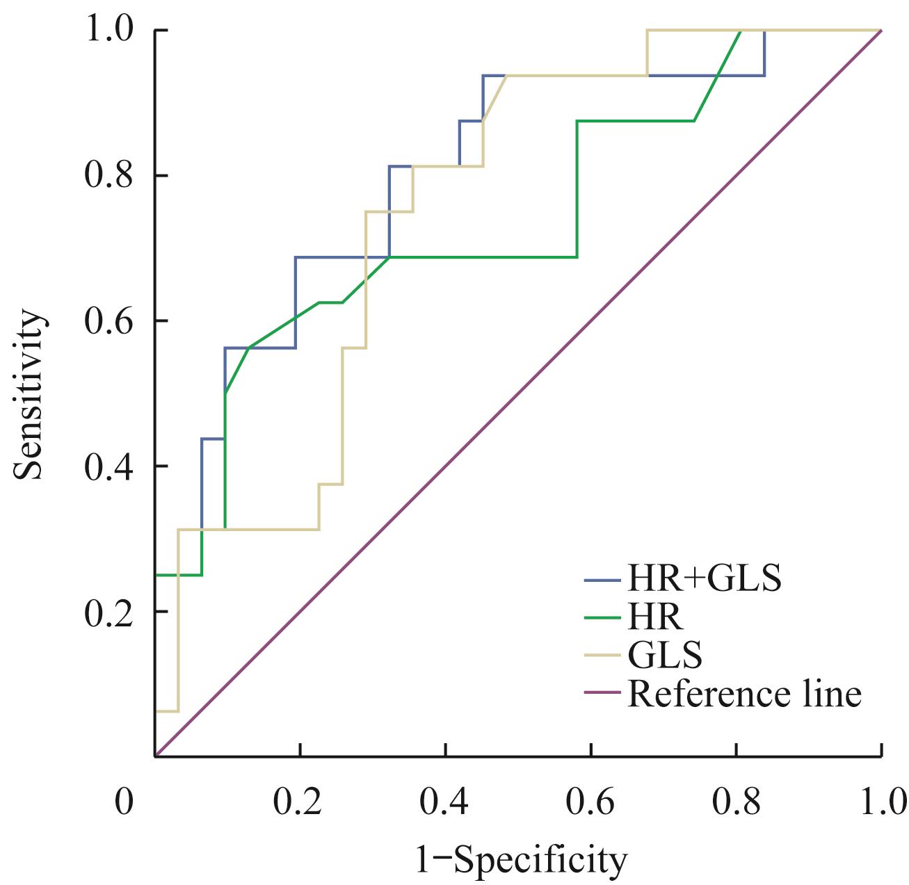
Fig 1 ROC curve analysis of HR and GLS diagnostic performance in the model where LVEDV increased by ≥20%
| Item | Total (n=47) | LVR group (n=17) | non-LVR group (n=30) | P value |
|---|---|---|---|---|
| Age/year | 58.28±11.59 | 60.29±10.43 | 57.13±12.21 | 0.375 |
| Male/n(%) | 41 (87.2) | 14 (82.4) | 27 (90.0) | 0.764 |
| BMI/(kg·m-2) | 25.65±4.00 | 26.09±3.46 | 25.40±4.32 | 0.574 |
| BSA/m2 | 1.84±0.16 | 1.85±0.21 | 1.83±0.13 | 0.599 |
| HR/min-1 | 70.23±11.56 | 72.47±12.28 | 68.97±11.14 | 0.323 |
| Follow-up time/month | 12.54±7.00 | 11.74±3.42 | 12.99±8.41 | 0.413 |
| Systolic blood pressure/mmHg | 128.02±23.70 | 127.53±25.11 | 128.30±23.29 | 0.916 |
| Diastolic blood pressure/mmHg | 77.40±14.12 | 77.47±16.29 | 77.37±13.03 | 0.981 |
| Hypertension/n(%) | 29 (61.7) | 12 (70.6) | 17 (56.7) | 0.345 |
| Diabetes/n(%) | 16 (34.0) | 7 (41.2) | 9 (30.0) | 0.437 |
| Hyperlipidemia/n(%) | 11 (23.4) | 3 (17.7) | 8 (26.7) | 0.731 |
| Smoking/n(%) | 23 (48.9) | 7 (41.2) | 16 (53.3) | 0.423 |
| Peak hs-cTnI/(μg·L-1) | 61.96±50.75 | 86.68±62.28 | 47.96±37.21 | 0.035 |
| Peak proBNP/(ng·L-1) | 986.00 (620.00, 1 762.00) | 1 571.00 (891.50, 2 643.50) | 930.00 (536.50, 1 589.25) | 0.044 |
| Culprit vessel | ||||
| LAD/n(%) | 43 (91.5) | 16 (94.1) | 27 (90.0) | 1.000 |
| LCX/n(%) | 29 (61.7) | 12 (70.6) | 17 (56.7) | 0.345 |
| RCA/n(%) | 21 (44.7) | 7 (41.2) | 14 (46.7) | 0.716 |
| Medication | ||||
| Antiplatelet/n(%) | 34 (72.3) | 15 (88.2) | 19 (63.3) | 0.135 |
| Stations/n(%) | 45 (95.7) | 17 (100.0) | 28 (93.3) | 0.528 |
| β-blocker/n(%) | 45 (95.7) | 17 (100.0) | 28 (93.3) | 0.528 |
| ACEI/ARB/ARNI/n(%) | 37 (78.7) | 14 (82.4) | 23 (76.7) | 0.931 |
| Diuretics/n(%) | 22 (46.8) | 8 (47.1) | 14 (46.7) | 0.979 |
| Nitrates/n(%) | 15 (31.9) | 4 (23.5) | 11 (36.7) | 0.547 |
| CMR parameter | ||||
| LVEDV/mL | 145.52±30.54 | 138.72±28.87 | 149.38±31.25 | 0.255 |
| LVSV/mL | 68.74±20.28 | 61.41±23.08 | 72.90±17.58 | 0.061 |
| LVEF/% | 49.22±10.60 | 48.54±12.15 | 49.61±9.83 | 0.745 |
| LVMI/(g·m-2) | 70.40±13.41 | 75.87±14.70 | 67.30±11.76 | 0.034 |
| GLS | -10.15±2.80 | -8.78±2.11 | -10.93±2.87 | 0.010 |
| GCS | -12.12±2.70 | -11.25±2.39 | -12.62±2.77 | 0.094 |
| GRS | 18.11±4.91 | 16.55±4.45 | 18.99±5.00 | 0.102 |
Tab 3 Comparison of baseline data between the LVR group and the non-LVR group in the model where LVESV increased by ≥15%
| Item | Total (n=47) | LVR group (n=17) | non-LVR group (n=30) | P value |
|---|---|---|---|---|
| Age/year | 58.28±11.59 | 60.29±10.43 | 57.13±12.21 | 0.375 |
| Male/n(%) | 41 (87.2) | 14 (82.4) | 27 (90.0) | 0.764 |
| BMI/(kg·m-2) | 25.65±4.00 | 26.09±3.46 | 25.40±4.32 | 0.574 |
| BSA/m2 | 1.84±0.16 | 1.85±0.21 | 1.83±0.13 | 0.599 |
| HR/min-1 | 70.23±11.56 | 72.47±12.28 | 68.97±11.14 | 0.323 |
| Follow-up time/month | 12.54±7.00 | 11.74±3.42 | 12.99±8.41 | 0.413 |
| Systolic blood pressure/mmHg | 128.02±23.70 | 127.53±25.11 | 128.30±23.29 | 0.916 |
| Diastolic blood pressure/mmHg | 77.40±14.12 | 77.47±16.29 | 77.37±13.03 | 0.981 |
| Hypertension/n(%) | 29 (61.7) | 12 (70.6) | 17 (56.7) | 0.345 |
| Diabetes/n(%) | 16 (34.0) | 7 (41.2) | 9 (30.0) | 0.437 |
| Hyperlipidemia/n(%) | 11 (23.4) | 3 (17.7) | 8 (26.7) | 0.731 |
| Smoking/n(%) | 23 (48.9) | 7 (41.2) | 16 (53.3) | 0.423 |
| Peak hs-cTnI/(μg·L-1) | 61.96±50.75 | 86.68±62.28 | 47.96±37.21 | 0.035 |
| Peak proBNP/(ng·L-1) | 986.00 (620.00, 1 762.00) | 1 571.00 (891.50, 2 643.50) | 930.00 (536.50, 1 589.25) | 0.044 |
| Culprit vessel | ||||
| LAD/n(%) | 43 (91.5) | 16 (94.1) | 27 (90.0) | 1.000 |
| LCX/n(%) | 29 (61.7) | 12 (70.6) | 17 (56.7) | 0.345 |
| RCA/n(%) | 21 (44.7) | 7 (41.2) | 14 (46.7) | 0.716 |
| Medication | ||||
| Antiplatelet/n(%) | 34 (72.3) | 15 (88.2) | 19 (63.3) | 0.135 |
| Stations/n(%) | 45 (95.7) | 17 (100.0) | 28 (93.3) | 0.528 |
| β-blocker/n(%) | 45 (95.7) | 17 (100.0) | 28 (93.3) | 0.528 |
| ACEI/ARB/ARNI/n(%) | 37 (78.7) | 14 (82.4) | 23 (76.7) | 0.931 |
| Diuretics/n(%) | 22 (46.8) | 8 (47.1) | 14 (46.7) | 0.979 |
| Nitrates/n(%) | 15 (31.9) | 4 (23.5) | 11 (36.7) | 0.547 |
| CMR parameter | ||||
| LVEDV/mL | 145.52±30.54 | 138.72±28.87 | 149.38±31.25 | 0.255 |
| LVSV/mL | 68.74±20.28 | 61.41±23.08 | 72.90±17.58 | 0.061 |
| LVEF/% | 49.22±10.60 | 48.54±12.15 | 49.61±9.83 | 0.745 |
| LVMI/(g·m-2) | 70.40±13.41 | 75.87±14.70 | 67.30±11.76 | 0.034 |
| GLS | -10.15±2.80 | -8.78±2.11 | -10.93±2.87 | 0.010 |
| GCS | -12.12±2.70 | -11.25±2.39 | -12.62±2.77 | 0.094 |
| GRS | 18.11±4.91 | 16.55±4.45 | 18.99±5.00 | 0.102 |
| Variable | β | SE | OR | 95%CI | P value |
|---|---|---|---|---|---|
| Peak hs-cTnI | -0.017 | 0.008 | 0.982 | 0.968‒0.998 | 0.024 |
| GLS | -0.304 | 0.149 | 0.738 | 0.551‒0.987 | 0.041 |
Tab 4 Logistic regression analysis of risk factors for LVR in the model where LVESV increased by ≥15%
| Variable | β | SE | OR | 95%CI | P value |
|---|---|---|---|---|---|
| Peak hs-cTnI | -0.017 | 0.008 | 0.982 | 0.968‒0.998 | 0.024 |
| GLS | -0.304 | 0.149 | 0.738 | 0.551‒0.987 | 0.041 |
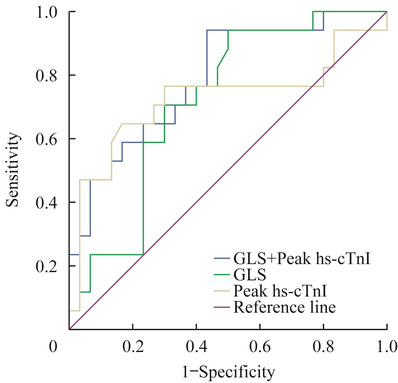
Fig 2 ROC curve analysis of Peak hs-cTnI and GLS diagnostic performance in the model defined as LVESV increased ≥15%
| 1 | Lepper W, Kamp O, Vanoverschelde JL, et al. Intravenous myocardial contrast echocardiography predicts left ventricular remodeling in patients with acute myocardial infarction[J]. J Am Soc Echocardiogr, 2002, 15(9): 849-856. |
| 2 | Ahn KT, Song YB, Choe YH, et al. Impact of transmural necrosis on left ventricular remodeling and clinical outcomes in patients undergoing primary percutaneous coronary intervention for ST-segment elevation myocardial infarction[J]. Int J Cardiovasc Imaging, 2013, 29(4): 835-842. |
| 3 | 邹倩, 周淑丽, 郑甜, 等. 心脏MR组织追踪技术评估心肌梗死后左心室心肌应变的价值[J].中华放射学杂志, 2018, 52(8): 587-592. |
| 4 | Reindl M, Tiller C, Holzknecht M, et al. Prognostic implications of global longitudinal strain by feature-tracking cardiac magnetic resonance in ST-elevation myocardial infarction[J]. Circ Cardiovasc Imaging, 2019, 12(11): e009404. |
| 5 | 沈卫峰, 张奇, 张瑞岩. 2015年急性ST段抬高型心肌梗死诊断和治疗指南解析[J]. 国际心血管病杂志, 2015, 42(4): 217-219. |
| 6 | Legallois D, Hodzic A, Alexandre J, et al. Definition of left ventricular remodelling following ST-elevation myocardial infarction: a systematic review of cardiac magnetic resonance studies in the past decade[J]. Heart Fail Rev, 2020. DOI: 10.1007/s10741-020-09975-3. |
| 7 | Taylor RJ, Moody WE, Umar F, et al. Myocardial strain measurement with feature-tracking cardiovascular magnetic resonance: normal values[J]. Eur Heart J Cardiovasc Imaging, 2015, 16(8): 871-881. |
| 8 | Lombardo A, Niccoli G, Natale L, et al. Impact of microvascular obstruction and infarct size on left ventricular remodeling in reperfused myocardial infarction: a contrast-enhanced cardiac magnetic resonance imaging study[J]. Int J Cardiovasc Imaging, 2012, 28(4): 835-842. |
| 9 | 多国帅, 刘婷, 戴旭. 心脏磁共振组织追踪技术评估冠心病心肌梗死的诊断价值[J]. 磁共振成像, 2018, 9(5): 346-353. |
| 10 | Altiok E, Tiemann S, Becker M, et al. Myocardial deformation imaging by two-dimensional speckle-tracking echocardiography for prediction of global and segmental functional changes after acute myocardial infarction: a comparison with late gadolinium enhancement cardiac magnetic resonance[J]. J Am Soc Echocardiogr, 2014, 27(3): 249-257. |
| 11 | Ben Driss A, Ben Driss Lepage C, Sfaxi A, et al. Strain predicts left ventricular functional recovery after acute myocardial infarction with systolic dysfunction[J]. Int J Cardiol, 2020, 307: 1-7. |
| 12 | Iwahashi N, Kirigaya J, Abe T, et al. Impact of three-dimensional global longitudinal strain for patients with acute myocardial infarction[J]. Eur Heart J Cardiovasc Imaging, 2020. DOI:10.1093/ehjci/jeaa241. |
| 13 | Wang Q, Tan K, Xia H, et al. Left ventricular metabolic remodeling and accompanied dysfunction in type 2 diabetic patients: a 3D speckle tracking analysis[J]. Echocardiography, 2019, 36(3): 486-494. |
| 14 | Neizel M, Korosoglou G, Lossnitzer D, et al. Impact of systolic and diastolic deformation indexes assessed by strain-encoded imaging to predict persistent severe myocardial dysfunction in patients after acute myocardial infarction at follow-up[J]. J Am Coll Cardiol, 2010, 56(13): 1056-1062. |
| 15 | Podlesnikar T, Pizarro G, Fernández-Jiménez R, et al. Left ventricular functional recovery of infarcted and remote myocardium after ST-segment elevation myocardial infarction (METOCARD-CNIC randomized clinical trial substudy)[J]. J Cardiovasc Magn Reson, 2020, 22(1): 44. |
| 16 | Bière L, Donal E, Terrien G, et al. Longitudinal strain is a marker of microvascular obstruction and infarct size in patients with acute ST-segment elevation myocardial infarction[J]. PLoS One, 2014, 9(1): e86959. |
| 17 | Manovel A, Dawson D, Smith B, et al. Assessment of left ventricular function by different speckle-tracking software[J]. Eur J Echocardiogr, 2010, 11(5): 417-421. |
| 18 | Nepper-Christensen L, Lønborg J, Ahtarovski KA, et al. Importance of elevated heart rate in the very early phase of ST-segment elevation myocardial infarction: results from the DANAMI-3 trial[J]. Eur Heart J Acute Cardiovasc Care, 2019, 8(4): 318-328. |
| 19 | HalIdn J, Jensen JK, MwFagerland, et a1. Cardiac troponin Ⅰ for the prediction of functional recovery and left ventricular remodelling following primary percutaneous coronary intervention for ST-elevation myocardial infarction[J].Heart, 2010, 96(23):1892-1897. |
| [1] | Quyang Danzeng, Xiao Huoyuan, Zhang Qingchen, Liu Yuting, Kang Sang, Feng Rui, Pan Jingwei. Diagnostic value of cardiac magnetic resonance for myocardial injury in patients with mild COVID-19 infection [J]. Journal of Shanghai Jiao Tong University (Medical Science), 2026, 46(3): 322-331. |
| [2] | LI Wenli, JIN Lixing, ZHAO Yichao, ZHONG Fangyuan, SHI Yao, LEI Jie, PU Jun, GE Heng. Impact of left ventricular myocardial strain injury on secondary tricuspid regurgitation in acute STEMI assessed by cardiac magnetic resonance [J]. Journal of Shanghai Jiao Tong University (Medical Science), 2025, 45(12): 1578-1588. |
| [3] | LIU Qiming, LU Qifan, CHAI Yezi, JIANG Meng, PU Jun. Short-axis cine cardiac magnetic resonance images-derived radiomics for hypertrophic cardiomyopathy and healthy control classification [J]. Journal of Shanghai Jiao Tong University (Medical Science), 2024, 44(1): 79-86. |
| [4] | LIU Qiming, LU Qifan, CHAI Yezi, JIANG Meng, PU Jun. Radiomics-based left ventricular ejection fraction prediction: a feasibility study [J]. Journal of Shanghai Jiao Tong University (Medical Science), 2023, 43(9): 1162-1168. |
| [5] | Ze-hao FENG, Ye-zi CHAI, Xuan SU, Bao-hang-xing SUN, Qi-ming LIU, Meng JIANG, Jun PU. Association between body mass index and myocardial involvements in patients with systemic lupus erythematosus [J]. JOURNAL OF SHANGHAI JIAOTONG UNIVERSITY (MEDICAL SCIENCE), 2021, 41(2): 180-186. |
| [6] | Pei-kun HU, Jie HE, Lian-ming WU, Heng GE, Jian-rong XU, Jun PU. Effect of microvascular obstruction on left ventricle function and prognosis in patients with ST-segment elevation myocardial infarction [J]. JOURNAL OF SHANGHAI JIAOTONG UNIVERSITY (MEDICAL SCIENCE), 2021, 41(2): 173-179. |
| [7] | Jian-xun DONG, Lai WEI, Jie HE, Ling-cong KONG, Heng GE, Jun PU. Progress of cardiac magnetic resonance in assessment of left ventricular mechanical dyssynchrony [J]. JOURNAL OF SHANGHAI JIAOTONG UNIVERSITY (MEDICAL SCIENCE), 2021, 41(12): 1698-1702. |
| [8] | Ye-zi CHAI, Meng JIANG, Jun PU. Relation between body mass index and left ventricular structure and function in patients with hypertrophic cardiomyopathy: a cardiovascular magnetic resonance imaging study [J]. JOURNAL OF SHANGHAI JIAOTONG UNIVERSITY (MEDICAL SCIENCE), 2021, 41(12): 1636-1641. |
| [9] | FENG Ze-hao1*, ZHANG Qing1*, CHAI Ye-zi1, SU Xuan1, SUN Bao-hang-xing1, LIU Qi-ming1, YAN Fu-hua2, JIANG Meng1#, PU Jun1#. Evaluation of effect of smoking on myocardial injury and prognosis in patients with acute ST-segment elevation myocardial infarction [J]. JOURNAL OF SHANGHAI JIAOTONG UNIVERSITY (MEDICAL SCIENCE), 2020, 40(5): 573-582. |
| [10] | SU Hai-xia, ZHU Ya-qin, ZHANG Tian-kuang, ZHANG Hui-li, GU Jun. Distribution characteristics of fragmented QRS in patients with coronary atherosclerotic heart disease and its relationship with left ventricular remodeling [J]. , 2019, 39(10): 1162-. |
| [11] | TAO Yi-jing1*, XIA Zhi-li1*, GAO Cheng-jie2, GAO Ya-jie1, WU Hao3, WAN Qing1, LI Yong-guang1, LU Zhi-gang1, SHEN Cheng-xing1, PAN Jing-wei1. Analysis of biomarkers for predicting left ventricular remodeling after successful revascularization of acute myocardial infarction [J]. , 2019, 39(1): 60-. |
| [12] | WANG Wei, ZHAO Hang, GE Heng, DING Song, SHEN Xue-dong, PU Jun. Value of two-dimensional speckle-tracking echocardiography in accessing myocardial viability and predicting left ventricular remodeling after acute myocardial infarction [J]. , 2018, 38(12): 1447-. |
| [13] | ZHENG Li, HUANG Guo-qian, WANG Cheng. Evaluation of left ventricular remodeling and function by quantitative real-time three-dimensional echocardiography in patients with acute myocardial infarction [J]. , 2011, 31(2): 194-. |
| [14] | HUANG Yu, LEI Hai-fang, WEI Meng, et al. Changes of plasma NT-ProBNP and MMP-2 levels and relationship with left ventricular remodeling in patients treated by pravastatin with CHF after PCI therapy for AMI [J]. , 2010, 30(3): 336-. |
| Viewed | ||||||
|
Full text |
|
|||||
|
Abstract |
|
|||||
