
Journal of Shanghai Jiao Tong University (Medical Science) ›› 2024, Vol. 44 ›› Issue (12): 1561-1569.doi: 10.3969/j.issn.1674-8115.2024.12.009
• Clinical research • Previous Articles Next Articles
REN Yixuan1,2( ), CHEN Cheng1, CAI Mingci3, CHEN Jiamin1, YANG Xinxin1, WANG Chao2, LIN Xiaozhu2, CHENG Shu3, JIANG Xufeng1,2(), CHEN Dongxu1()
Received:2024-06-18
Accepted:2024-08-27
Online:2024-12-28
Published:2024-12-28
Contact:
JIANG Xufeng,CHEN Dongxu
E-mail:renyixuan1994@163.com;jxf10885@rjh.com.cn;2796372336@qq.com
Supported by:CLC Number:
REN Yixuan, CHEN Cheng, CAI Mingci, CHEN Jiamin, YANG Xinxin, WANG Chao, LIN Xiaozhu, CHENG Shu, JIANG Xufeng, CHEN Dongxu. Research on the characteristics of 18F-FDG PET/CT in mantle cell lymphoma and the discrimination between cellular morphological variants[J]. Journal of Shanghai Jiao Tong University (Medical Science), 2024,(12): 1561-1569.
Add to citation manager EndNote|Ris|BibTeX
URL: https://xuebao.shsmu.edu.cn/EN/10.3969/j.issn.1674-8115.2024.12.009
| Site | Number/n | Percentage/%① | SUVmax |
|---|---|---|---|
| Lymph node | 115 | 99.1 | 8.75±5.47 |
| Extranodal | 99 | 85.3 | 9.52±5.36 |
| Spleen | 62 | 53.4 | 5.84±3.58 |
| Waldeyer's ring | 56 | 48.3 | 9.86±3.58 |
| Bone marrow | 30 | 25.9 | 4.85±2.00 |
| Intestine | 35 | 30.2 | 10.21±5.12 |
| Stomach | 27 | 23.3 | 7.64±3.38 |
| Skin | 11 | 9.5 | 7.65±2.28 |
| Thyroid | 9 | 7.8 | 5.5 (4.2,14.4) |
| Nasal cavity | 2 | 1.7 | 8.10, 38.50 |
| Parotid gland | 2 | 1.7 | 4.6, 8.2 |
| Scrotum | 2 | 1.7 | 3.79, 15.70 |
| Prostate | 2 | 1.7 | 6.0, 10.2 |
| Pancreas | 1 | 0.9 | 3.7 |
| Breast | 1 | 0.9 | 7.0 |
| Eyelid | 1 | 0.9 | 6.7 |
| Lung | 1 | 0.9 | 3.4 |
| Pleura | 1 | 0.9 | 3.1 |
| Gall bladder | 1 | 0.9 | 8.0 |
Tab 1 Distribution of positive lesion and SUVmax in MCL on 18F-FDG PET/CT
| Site | Number/n | Percentage/%① | SUVmax |
|---|---|---|---|
| Lymph node | 115 | 99.1 | 8.75±5.47 |
| Extranodal | 99 | 85.3 | 9.52±5.36 |
| Spleen | 62 | 53.4 | 5.84±3.58 |
| Waldeyer's ring | 56 | 48.3 | 9.86±3.58 |
| Bone marrow | 30 | 25.9 | 4.85±2.00 |
| Intestine | 35 | 30.2 | 10.21±5.12 |
| Stomach | 27 | 23.3 | 7.64±3.38 |
| Skin | 11 | 9.5 | 7.65±2.28 |
| Thyroid | 9 | 7.8 | 5.5 (4.2,14.4) |
| Nasal cavity | 2 | 1.7 | 8.10, 38.50 |
| Parotid gland | 2 | 1.7 | 4.6, 8.2 |
| Scrotum | 2 | 1.7 | 3.79, 15.70 |
| Prostate | 2 | 1.7 | 6.0, 10.2 |
| Pancreas | 1 | 0.9 | 3.7 |
| Breast | 1 | 0.9 | 7.0 |
| Eyelid | 1 | 0.9 | 6.7 |
| Lung | 1 | 0.9 | 3.4 |
| Pleura | 1 | 0.9 | 3.1 |
| Gall bladder | 1 | 0.9 | 8.0 |
| Site | Pathologically confirmed | PET/CT-positive | t value | P value | ||
|---|---|---|---|---|---|---|
| Number/n | SUVmax | Number/n | SUVmax | |||
| Lymph node | 81 | 8.98±5.13 | 115 | 8.75±5.47 | -0.291 | 0.771 |
| Waldeyer's ring | 18 | 9.20±4.36 | 56 | 9.86±3.58 | 0.643 | 0.522 |
| Bone marrow | 53 | 3.81±1.78 | 30 | 4.85±2.00 | 2.449 | 0.016 |
| Stomach | 10 | 9.06±4.71 | 27 | 7.64±3.38 | -1.021 | 0.314 |
| Intestine | 17 | 10.09±6.41 | 35 | 10.21±5.12 | 0.073 | 0.942 |
Tab 2 Comparison of SUVmax values between pathologically diagnosed lesions and 18F-FDG PET/CT-positive lesions
| Site | Pathologically confirmed | PET/CT-positive | t value | P value | ||
|---|---|---|---|---|---|---|
| Number/n | SUVmax | Number/n | SUVmax | |||
| Lymph node | 81 | 8.98±5.13 | 115 | 8.75±5.47 | -0.291 | 0.771 |
| Waldeyer's ring | 18 | 9.20±4.36 | 56 | 9.86±3.58 | 0.643 | 0.522 |
| Bone marrow | 53 | 3.81±1.78 | 30 | 4.85±2.00 | 2.449 | 0.016 |
| Stomach | 10 | 9.06±4.71 | 27 | 7.64±3.38 | -1.021 | 0.314 |
| Intestine | 17 | 10.09±6.41 | 35 | 10.21±5.12 | 0.073 | 0.942 |
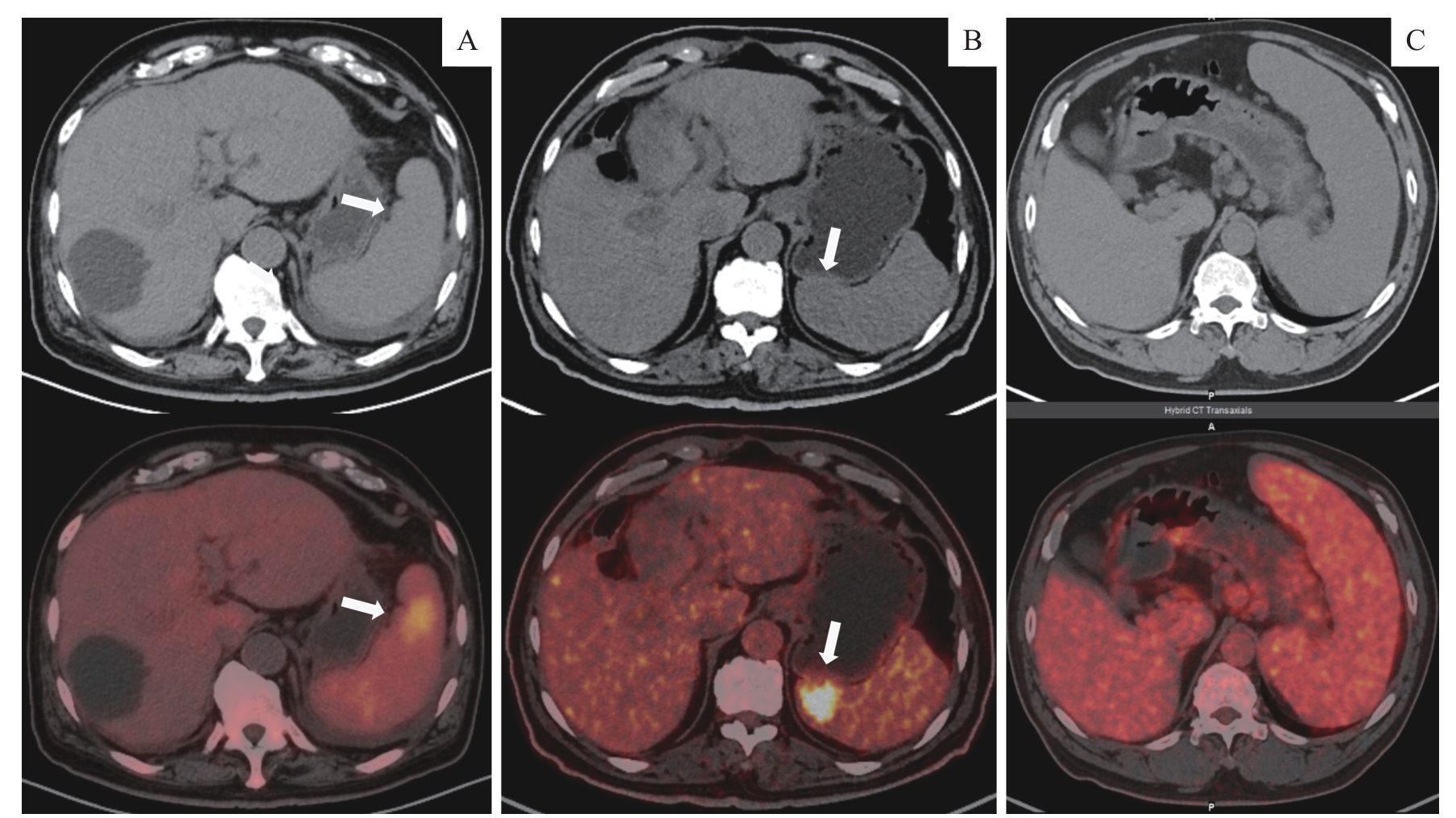
Fig1 18F-FDG PET/CT imaging of the spleens in patients with MCL
| Site | Number/n | 18F-FDG PET/CT | Pathologic diagnosis | Sensitivity/% | Specificity/% | PPV/% | NPV/% | Accuracy/% | |
|---|---|---|---|---|---|---|---|---|---|
| MCL | Not MCL | ||||||||
| Bone marrow | 100 | Positive (27) | 23 | 4 | 43.4 | 91.5 | 85.2 | 58.9 | 66.0 |
| Negative (73) | 30 | 43 | |||||||
| Stomach | 14 | Positive (11) | 10 | 1 | 100.0 | 75.0 | 90.9 | 100.0 | 92.9 |
| Negative (3) | 0 | 3 | |||||||
| Intestine | 19 | Positive (16) | 16 | 0 | 94.1 | 100.0 | 100.0 | 66.7 | 94.7 |
| Negative (3) | 1 | 2 | |||||||
Tab 3 MCL in 18F-FDG PET compared with pathological diagnosis
| Site | Number/n | 18F-FDG PET/CT | Pathologic diagnosis | Sensitivity/% | Specificity/% | PPV/% | NPV/% | Accuracy/% | |
|---|---|---|---|---|---|---|---|---|---|
| MCL | Not MCL | ||||||||
| Bone marrow | 100 | Positive (27) | 23 | 4 | 43.4 | 91.5 | 85.2 | 58.9 | 66.0 |
| Negative (73) | 30 | 43 | |||||||
| Stomach | 14 | Positive (11) | 10 | 1 | 100.0 | 75.0 | 90.9 | 100.0 | 92.9 |
| Negative (3) | 0 | 3 | |||||||
| Intestine | 19 | Positive (16) | 16 | 0 | 94.1 | 100.0 | 100.0 | 66.7 | 94.7 |
| Negative (3) | 1 | 2 | |||||||
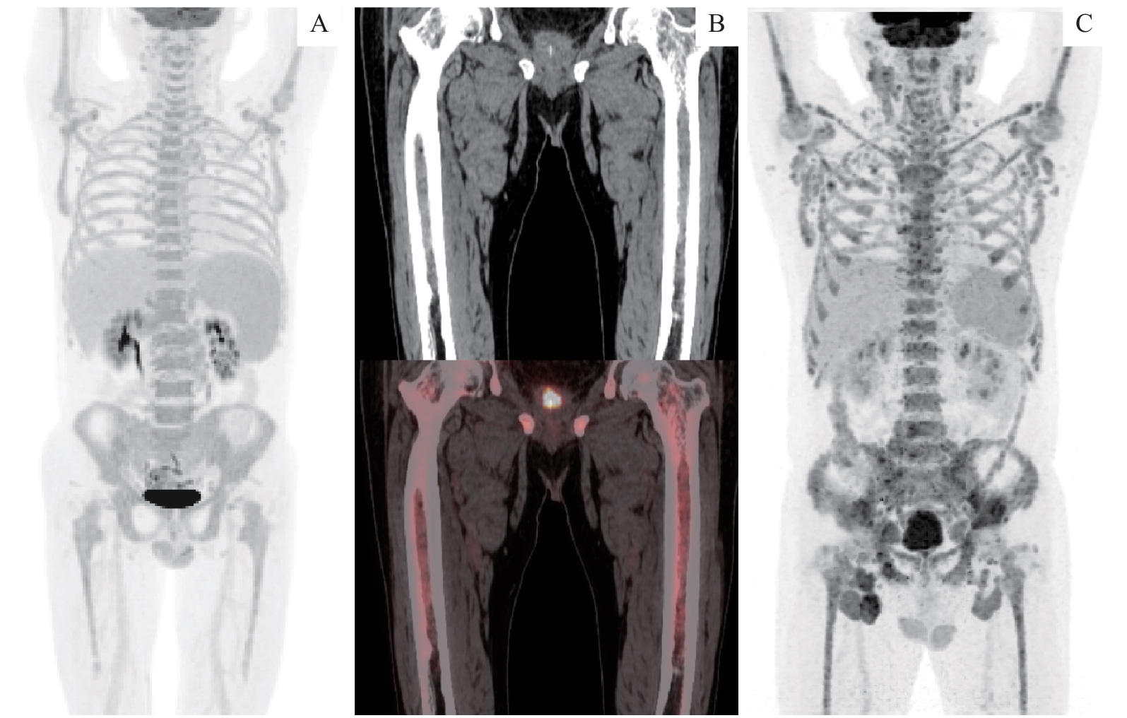
Fig 2 18F-FDG PET/CT imaging of bone marrow in patients with MCL
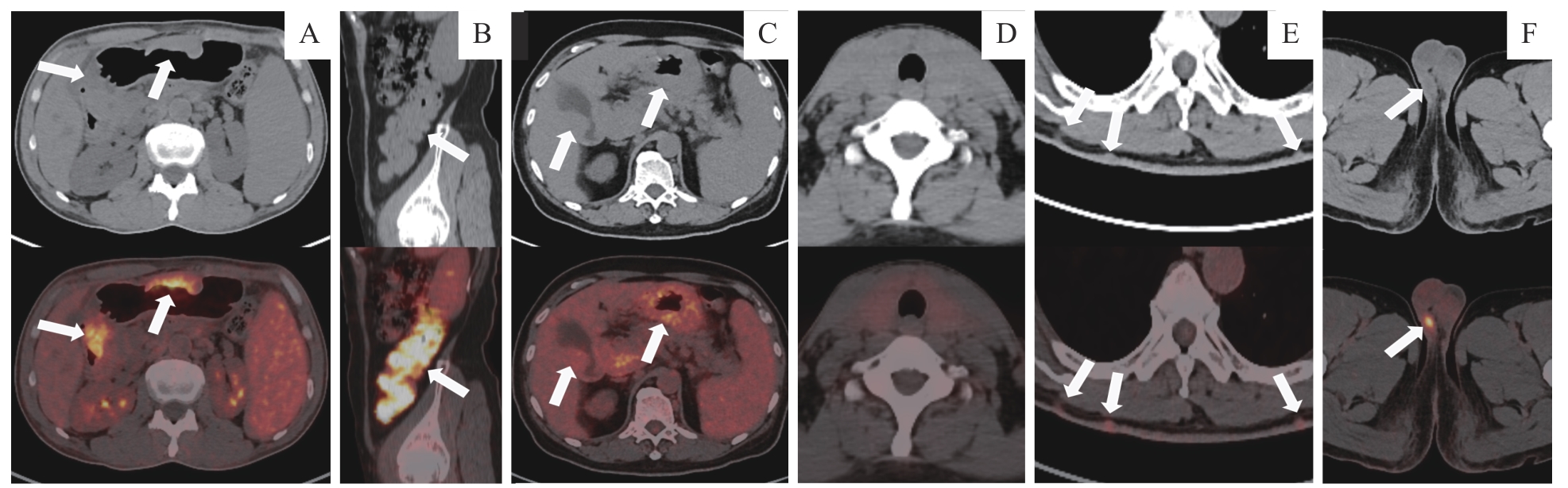
Fig 3 18F-FDG PET/CT images of lesions at different sites in patients with MCL
| Characteristic | Classical type | Aggressive variant | t/χ2 value | P value |
|---|---|---|---|---|
| Number/n(%) | 93 (80.2) | 23 (19.8) | ‒ | ‒ |
| Age/year | 63.71±9.11 | 59.96±10.21 | 1.612 | 0.117 |
| Ki-67/% | 29.98±15.07 | 80.43±15.81 | -14.243 | <0.001 |
| Proportion of Ⅲ‒Ⅳ stage/% | 90.3 | 91.3 | 0.021 | 0.886 |
| Proportion of bone marrow infiltration/% | 51.9 | 57.1 | 0.186 | 0.669 |
| SUVmax | 9.02±4.79 | 15.40±7.90 | -3.708 | 0.001 |
| Minor axis of the largest lymph node/cm | 2.27±1.18 | 2.54±1.99 | -0.641 | 0.527 |
| Proportion of increased spleen metabolism/% | 50.5 | 72.7 | 43.608 | 0.061 |
| Proportion of increased gastrointestinal metabolism/% | 41.9 | 26.1 | 1.951 | 0.163 |
Tab 4 Comparison of differences between the typical type and aggressive variant of MCL in 18F-FDG PET/CT and clinical features
| Characteristic | Classical type | Aggressive variant | t/χ2 value | P value |
|---|---|---|---|---|
| Number/n(%) | 93 (80.2) | 23 (19.8) | ‒ | ‒ |
| Age/year | 63.71±9.11 | 59.96±10.21 | 1.612 | 0.117 |
| Ki-67/% | 29.98±15.07 | 80.43±15.81 | -14.243 | <0.001 |
| Proportion of Ⅲ‒Ⅳ stage/% | 90.3 | 91.3 | 0.021 | 0.886 |
| Proportion of bone marrow infiltration/% | 51.9 | 57.1 | 0.186 | 0.669 |
| SUVmax | 9.02±4.79 | 15.40±7.90 | -3.708 | 0.001 |
| Minor axis of the largest lymph node/cm | 2.27±1.18 | 2.54±1.99 | -0.641 | 0.527 |
| Proportion of increased spleen metabolism/% | 50.5 | 72.7 | 43.608 | 0.061 |
| Proportion of increased gastrointestinal metabolism/% | 41.9 | 26.1 | 1.951 | 0.163 |
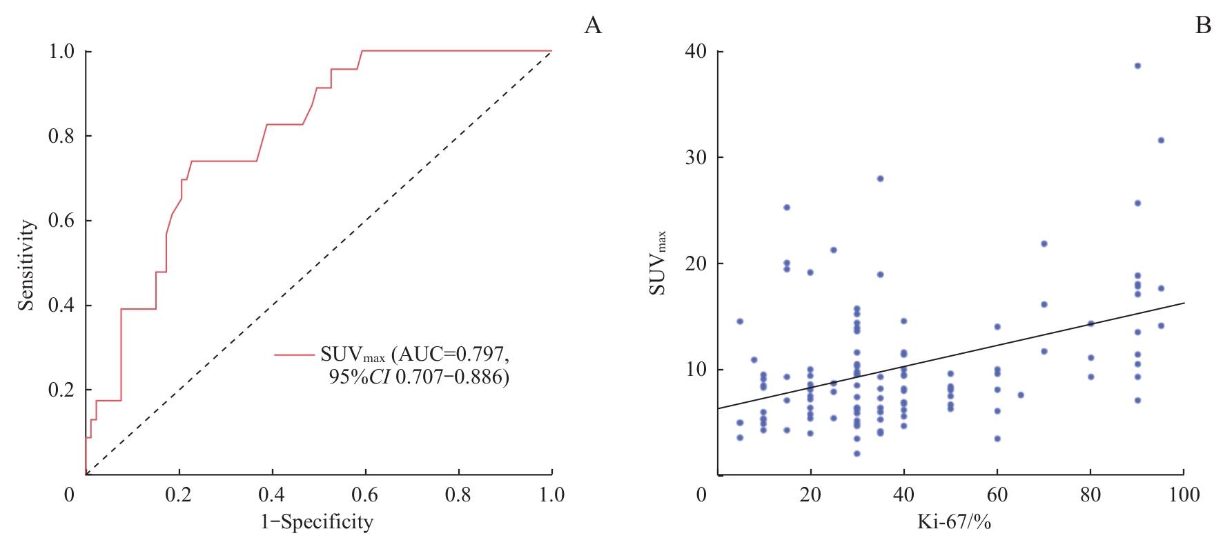
Fig 4 Analysis of the clinical value of SUVmax in MCL
| 1 | ARMITAGE J O, LONGO D L. Mantle-cell lymphoma[J]. N Engl J Med, 2022, 386(26): 2495-2506. |
| 2 | 中国抗癌协会血液肿瘤专业委员会, 中华医学会血液学分会, 中国临床肿瘤学会淋巴瘤专家委员会. 套细胞淋巴瘤诊断与治疗中国指南 (2022年版)[J]. 中华血液学杂志, 2022, 43(7): 529-536. |
| Hematology Oncology Committee of China Anti-cancer Association, The Society of Hematology at Chinese Medical Association, Union for China Lymphoma Investigator at Chinese Society of Clinical Oncology. The guideline of the diagnosis and treatment of mantle cell lymphoma in China (2022) [J].Chinese Journal of Hematology, 2022, 43(7): 529-536. | |
| 3 | 李燕玲, 秦小琪, 马艳萍. 套细胞淋巴瘤发病机制的研究进展[J]. 肿瘤研究与临床, 2024, 36(1): 73-76. |
| LI Y L, QIN X Q, MA Y P. Progress of the pathogenesis in mantle cell lymphoma[J]. Cancer Research and Clinic, 2024, 36(1): 73-76. | |
| 4 | WU M, LI Y, HUANG H Q, et al. Initial treatment patterns and survival outcomes of mantle cell lymphoma patients managed at Chinese academic centers in the rituximab era: a real-world study[J]. Front Oncol, 2021, 11: 770988. |
| 5 | ABRISQUETA P, SCOTT D W, SLACK G W, et al. Observation as the initial management strategy in patients with mantle cell lymphoma[J]. Ann Oncol, 2017, 28(10): 2489-2495. |
| 6 | DREYLING M, KLAPPER W, RULE S. Blastoid and pleomorphic mantle cell lymphoma: still a diagnostic and therapeutic challenge![J]. Blood, 2018, 132(26): 2722-2729. |
| 7 | GERSON J N, HANDORF E, VILLA D, et al. Outcomes of patients with blastoid and pleomorphic variant mantle cell lymphoma[J]. Blood Adv, 2023, 7(24): 7393-7401. |
| 8 | GOUILL S L, DŁUGOSZ-DANECKA M, RULE S, et al. Final results and overall survival data from a phase Ⅱ study of acalabrutinib monotherapy in patients with relapsed/refractory mantle cell lymphoma, including those with poor prognostic factors[J]. Haematologica, 2024, 109(1): 343-350. |
| 9 | ZANONI L, BEZZI D, NANNI C, et al. PET/CT in non-hodgkin lymphoma: an update[J]. Semin Nucl Med, 2023, 53(3): 320-351. |
| 10 | SWERDLOW S H, CAMPO E, PILERI S A, et al. The 2016 revision of the World Health Organization classification of lymphoid neoplasms[J]. Blood, 2016, 127(20): 2375-2390. |
| 11 | LAMBERT L, BURGETOVA A, TRNENY M, et al. The diagnostic performance of whole-body MRI in the staging of lymphomas in adult patients compared to PET/CT and enhanced reference standard-systematic review and meta-analysis[J]. Quant Imaging Med Surg, 2022, 12(2): 1558-1570. |
| 12 | JU S H, LEE S E, YI S, et al. Transcriptomic characteristics according to tumor size and SUVmax in papillary thyroid cancer patients[J]. Sci Rep, 2024, 14(1): 11005. |
| 13 | ALBANO D, TREGLIA G, GAZZILLI M, et al. 18F-FDG PET or PET/CT in mantle cell lymphoma[J]. Clin Lymphoma Myeloma Leuk, 2020, 20(7): 422-430. |
| 14 | ALAVI A, SHRIKANTHAN S, AYDIN A, et al. Fluorodeoxyglucose-positron-emission tomography findings in mantle cell lymphoma[J]. Clin Lymphoma Myeloma Leuk, 2011, 11(3): 261-266. |
| 15 | ALBANO D, FERRO P, BOSIO G, et al. Diagnostic and clinical impact of staging 18F-FDG PET/CT in mantle-cell lymphoma: a two-center experience[J]. Clin Lymphoma Myeloma Leuk, 2019, 19(8): e457-e464. |
| 16 | OÑA-ORTIZ F M, SÁNCHEZ-DEL MONTE J, RAMÍREZ-SOLÍS M E, et al. Mantle cell lymphoma with involvement of the digestive tract[J]. Rev Gastroenterol Mex (Engl Ed), 2019, 84(4): 434-441. |
| 17 | HOSEIN P J, PASTORINI V H, PAES F M, et al. Utility of positron emission tomography scans in mantle cell lymphoma[J]. Am J Hematol, 2011, 86(10): 841-845. |
| 18 | SKRYPETS T, FERRARI C, NASSI L, et al. 18F-FDG PET/CT cannot substitute endoscopy in the staging of gastrointestinal involvement in mantle cell lymphoma. A retrospective multi-center cohort analysis[J]. J Pers Med, 2021, 11(2): 123. |
| 19 | LOVINFOSSE P, HUSTINX R. The role of PET imaging in inflammatory bowel diseases: state-of-the-art review[J]. Q J Nucl Med Mol Imaging, 2022, 66(3): 206-217. |
| 20 | BIEG C, MONGELLI F, PETERLI R, et al. Simplified dual time point FDG-PET/computed tomography for determining dignity of pancreatic lesions[J]. Nucl Med Commun, 2020, 41(7): 682-687. |
| 21 | JIANG H, LI A, JI Z Y, et al. Role of radiomics-based baseline PET/CT imaging in lymphoma: diagnosis, prognosis, and response assessment[J]. Mol Imaging Biol, 2022, 24(4): 537-549. |
| 22 | 吴江, 朱虹, 王新刚, 等. 淋巴瘤脾脏浸润的18F-FDG PET/CT表现[J]. 中国医学影像技术, 2012, 28(6): 1157-1160. |
| WU J, ZHU H, WANG X G, et al. 18F-FDG PET/CT manifestations of spleen infiltration of lymphoma[J]. Chinese Journal of Medical Imaging Technology, 2012, 28(6): 1157-1160. | |
| 23 | LIU Y Y. Clinical significance of diffusely increased splenic uptake on FDG-PET[J]. Nucl Med Commun, 2009, 30(10): 763-769. |
| 24 | PICARDI M, SORICELLI A, GRIMALDI F, et al. Fused FDG-PET/contrast-enhanced CT detects occult subdiaphragmatic involvement of Hodgkin's lymphoma thereby identifying patients requiring six cycles of anthracycline-containing chemotherapy and consolidation radiation of spleen[J]. Ann Oncol, 2011, 22(3): 671-680. |
| 25 | SCHOLTENS A M, VERBERNE H J. FDG uptake in marrow and spleen: what does it mean?: Importance of clinical context in the interpretation of FDG uptake in marrow and spleen in infective endocarditis[J]. J Nucl Cardiol, 2021, 28(6): 2543-2544. |
| 26 | LI H L, WANG X H, ZHANG L F, et al. Correlations between maximum standardized uptake value measured via 18F-fluorodeoxyglucose positron emission tomography/computed tomography and clinical variables and biochemical indicators in adult lymphoma[J]. J Cancer Res Ther, 2019, 15(7): 1581-1588. |
| 27 | NISHIOKA A, URESHINO H, ANDO T, et al. Three coexisting lymphomas in a single patient: composite lymphoma derived from a common germinal center B-cell precursor and unrelated discordant lymphoma[J]. Int J Hematol, 2018, 107(6): 703-708. |
| 28 | PARRY E M, ROULLAND S, OKOSUN J. DLBCL arising from indolent lymphomas: how are they different?[J]. Semin Hematol, 2023, 60(5): 277-284. |
| Viewed | ||||||
|
Full text |
|
|||||
|
Abstract |
|
|||||
