上海交通大学学报(医学版), 2023, 43(5): 540-544 doi: 10.3969/j.issn.1674-8115.2023.05.004

口腔外科专题

磁共振引导的咀嚼肌疼痛放松训练疗效评估

杨海霞,1, 徐丽丽1, 王博成2, 陈敏洁,3

1.上海交通大学医学院附属第九人民医院康复医学科,上海 200011

2.上海交通大学医学院附属第九人民医院放射科,上海 200011

3.上海交通大学医学院附属第九人民医院口腔外科,上海交通大学口腔医学院,国家口腔医学中心,国家口腔疾病临床医学研究中心,上海市口腔医学重点实验室,上海 200011

Evaluation of clinical effect of manipulation on masticatory muscle pain guided by MRI

YANG Haixia,1, XU Lili1, WANG Bocheng2, CHEN Minjie,3

1.Department of Physical Medicine & Rehabilitation, Shanghai Ninth People′s Hospital, Shanghai Jiao Tong University School of Medicine, Shanghai 200011, China

2.Department of Radiology, Shanghai Ninth People′s Hospital, Shanghai Jiao Tong University School of Medicine, Shanghai 200011, China

3.Department of Oral Surgery, Shanghai Ninth People′s Hospital, Shanghai Jiao Tong University School of Medicine; College of Stomatology, Shanghai Jiao Tong University; National Center for Stomatology; National Clinical Research Center for Oral Diseases; Shanghai Key Laboratory of Stomatology, Shanghai 200011, China

通讯作者: 陈敏洁,电子信箱:chenmj_9hospital@126.com

编委: 徐敏

收稿日期: 2023-04-24   接受日期: 2023-05-02  

Corresponding authors: CHEN Minjie, E-mail:chenmj_9hospital@126.com.

Received: 2023-04-24   Accepted: 2023-05-02  

作者简介 About authors

杨海霞(1986—),女,主管治疗师,硕士生;电子信箱:yanghaixia721@sina.com。 E-mail:yanghaixia721@sina.com

摘要

目的·应用磁共振成像(magnetic resonance imaging,MRI)的Dixon技术评估非结构错乱的颞下颌关节紊乱病(temporomandibular joint disorder,TMD)的咀嚼肌疼痛患者的肌肉改变,并评价MRI引导的物理手法治疗咀嚼肌疼痛的临床疗效。方法·连续纳入自2021年6月—2022年9月就诊于上海交通大学医学院附属第九人民医院口腔外科、首次诊断为TMD肌筋膜疼痛(不伴有盘移位)的患者共29例。收集到增强MRI的Dixon序列并进行手法治疗的患者9例。治疗前,利用DICOM Viewer工作站获得9例患者的Dixon图像咀嚼肌疼痛区域值(Z1)、同侧非疼痛区域值(Z2)和对侧对应咀嚼肌区域值(Z3)。依据工作站获得的咀嚼肌异常区域,针对性地进行手法放松治疗。治疗后1~4周进行随访,对比治疗前后主动最大张口度(maximum mouth opening,MMO)和疼痛视觉评分(visual analogue scale,VAS)指标,并评价临床疗效。结果·Dixon序列Z2均值为66.23±32.90,Z3均值为66.27±33.87;而咀嚼肌疼痛区域Z1为131.94±83.99,明显高于Z2和Z3。手法治疗在改善MMO、VAS等方面有明显效果,有效率达88.89%。结论·Dixon序列的影像表现与临床主诉的疼痛点具有很大的相关性,MRI引导的手法放松治疗对改善非结构错乱的颞下颌关节紊乱病的张口度和疼痛有明显疗效。

关键词: 颞下颌关节紊乱病 ; 咀嚼肌 ; 疼痛 ; 磁共振 ; 手法治疗

Abstract

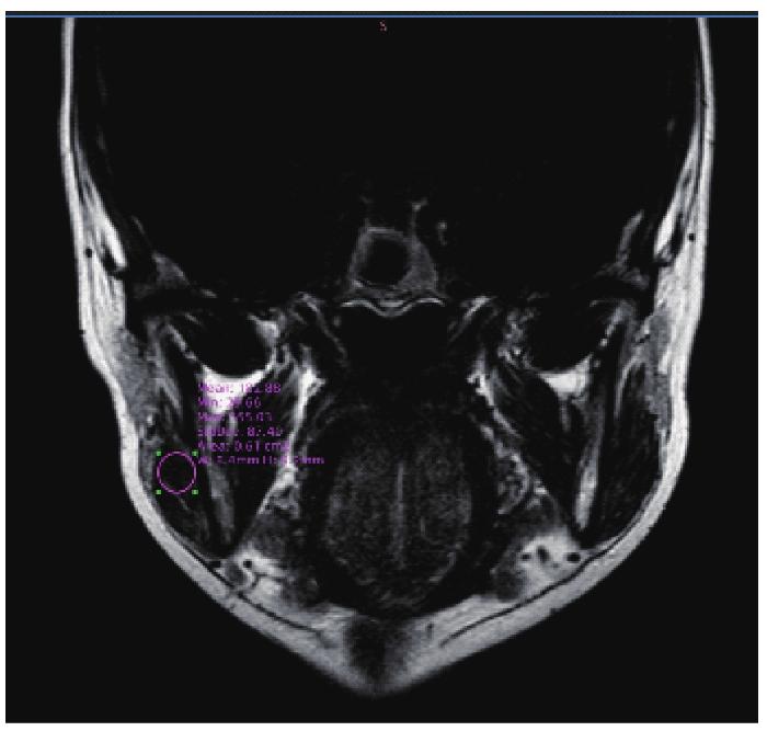
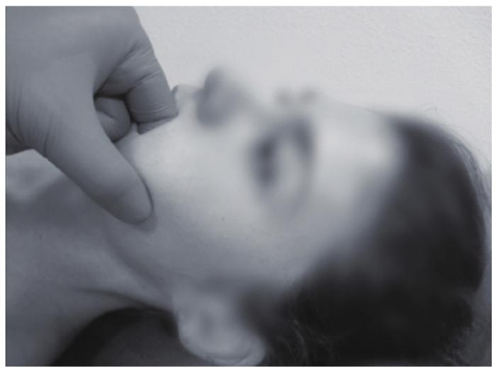
Objective ·To assess muscle changes of patients of temporomandibular joint disorder with nonstructural disorder by using the Dixon technique for MRI of masticatory muscle, and evaluate the clinical effect of manipulation on masticatory muscle pain guided by MRI. Methods ·A total of 29 patients with TMD masticatory muscle pain (without disc displacement) who were diagnosed for the first time in the Department of Oral Surgery, Shanghai Ninth People′s Hospital, Shanghai Jiao Tong University School of Medicine from June 2021 to September 2022 were included. Among them, 9 cases who were assessed with Dixon technique for MRI of masticatory muscle were collected and treated by manipulation. Before treatment, the DICOM Viewer workstation was used to compare the value of masticatory muscle pain area (Z1), the value of ipsilateral non-pain area (Z2) and the value of contralateral corresponding masticatory muscle area (Z3) in Dixon image. Manipulation therapy was performed according to the area of abnormal threshold. Follow-up was performed for 1-4 weeks after treatment, the active maximum mouth opening (MMO) and Visual Analogue Scale (VAS) before and after treatment were compared, and the value of masticatory muscle pain area in Dixon image after treatment was obtained again to evaluate the clinical efficacy. Results ·The mean value of Z2 and Z3 in Dixon was (66.23±32.90) and (66.27±33.87), while Z1 in masticatory muscle pain region was (131.94±83.99), which was significantly higher than Z2 and Z3. Manipulation therapy showed significant improvement in MMO and VAS, and the effective rate was 88.89%. Conclusion ·There is a significant correlation between the imaging findings of Dixon technique for MRI and the pain points of the masticatory muscle reported by the clinical complaints. The manipulation therapy guided by Dixon technique for MRI has a significant effect on improving the degree of mouth opening and pain.

Keywords: temporomandibular joint disorder (TMD) ; masticatory muscle ; pain ; magnetic resonance imaging ; manipulation

PDF (1599KB) 元数据 多维度评价 相关文章 导出 EndNote| Ris| Bibtex  收藏本文

本文引用格式

杨海霞, 徐丽丽, 王博成, 陈敏洁. 磁共振引导的咀嚼肌疼痛放松训练疗效评估. 上海交通大学学报(医学版)[J], 2023, 43(5): 540-544 doi:10.3969/j.issn.1674-8115.2023.05.004

YANG Haixia, XU Lili, WANG Bocheng, CHEN Minjie. Evaluation of clinical effect of manipulation on masticatory muscle pain guided by MRI. Journal of Shanghai Jiao Tong University (Medical Science)[J], 2023, 43(5): 540-544 doi:10.3969/j.issn.1674-8115.2023.05.004

颞下颌关节紊乱病(temporomandibular joint disorder,TMD)是指一系列涉及咀嚼肌组织、关节及其相关结构的疼痛性和非疼痛性疾病。关节杂音、疼痛以及张口受限等症状会显著影响患者的日常生活,其中咀嚼肌疼痛是一类常见的TMD,也是仅次于慢性下腰痛的第二大常见的肌肉骨骼疾病1。针对咀嚼肌疼痛的保守治疗,目前有药物治疗2-3、咬合板治疗4、手法治疗5-6或联合治疗等,但对咀嚼肌疼痛位点的判断多采用触压法,缺乏特定的影像学评估,从而使得手法放松治疗多以医师经验为指导,导致临床疗效不稳定。

磁共振成像(magnetic resonance image,MRI)不仅是确定盘髁关系的金标准,而且可以评估咀嚼肌异常7。MRI Dixon技术又称水-脂分离技术8,即利用脂肪质子和水质子共振频率的差异分离水和脂肪,从而获得同相位、反相位、水像、脂像的图像。有学者将其应用于呼吸肌9和大腿肌群10的评估。Dixon技术在咀嚼肌方面的研究目前较少。本研究纳入正常盘髁关系的咀嚼肌疼痛患者,通过Dixon技术定量评估咀嚼肌疼痛患者的肌肉改变,指导后续物理手法治疗,并进行有效性评估。

1 对象与方法

本研究为前瞻性研究,连续纳入2021年6月—2022年9月就诊于上海交通大学医学院附属第九人民医院口腔外科、首次诊断为TMD肌筋膜疼痛的患者。

纳入标准:① 年龄20~50岁。② 根据2014扩展版Diagnostic Criteria for Temporomandibular Disorders(30),确诊为咀嚼肌疼痛患者。③ 颞下颌关节(temporomandibular joint,TMJ)MRI显示盘髁关系正常。④ 就诊前2周内未经其他治疗。⑤ 身体质量指数(body mass index,BMI)<26。排除标准:① 活动性特异性或非特异性关节炎。② 急性创伤。③ 5年内恶性肿瘤史。④ 精神、神经性疾病史。

1.1 影像学检查

MRI检查采用德国Siemens Aera 1.5T MRI扫描仪,增强剂采用国产马根维显(有效成分为钆喷酸葡胺)注射液15 mL,扫描范围为颌面部。获取Dixon图像,根据主诉咀嚼肌的压痛点,通过DICOM Viewer工作站获得患者的Dixon图像:咀嚼肌疼痛区域值(Z1)、同侧非疼痛区域值(Z2)、对侧对应咀嚼肌区域值(Z3)。工作站可将咀嚼肌的Dixon图像在某区域的最大值、最小值以及均值直观显示(图1)。为了减少误差,取压痛的咀嚼肌的均值作为参考,并多次测量(n=3),取平均值。

图1

图1   咀嚼肌的Dixon图像测量

Note: Getting the the maximum, minimum and average values of the zone at the workstation of DICOM Viewer for 3 times, and then taking the mean values.

Fig 1   Measurement of masticatory muscle in the Dixon image


1.2 手法治疗

参考MRI显示异常区域,医师进行放松手法治疗,方法:① 咬肌:施加压迫手法,并要求患者咬紧牙齿或使用动态的纵向或横向抚摩以进行放松(图2)。② 颞肌:用2个手指从颅(颧)到尾(下颌)方向进行纵向或横向抚摩,进行放松。③ 翼内肌:让患者张口,用示指沿牙弓之间探入末端,与翼内肌的中部接触,并进行静力压迫以进行放松。④ 翼外肌:将示指沿着口腔前庭,与上颌骨牙槽突的上半部分平行的方向,对上颌结节到翼突外侧板施以压迫手法以进行放松。每周治疗3次,隔天1次,共计8~10次。由于疼痛主观感受差异,医师或治疗师手法治疗的力度以轻柔耐受为适度,以不引发新的疼痛为限,治疗的时间为每次15 min左右。

图2

图2   咬肌的手法放松

Fig 2   Manipulation of masseter muscle


1.3 临床疗效评价指标

放松训练4周后评价患者切牙颌间主动最大张口度(maximum mouth opening,MMO)和疼痛视觉模拟评分(visual analogue scale,VAS)。

1.3.1 MMO测量

嘱患者主动张口至最大,用尺测量其上下切牙的距离。

1.3.2 VAS测量

在10 cm的横线上,一端标为0,表示无痛;另一端标为10,表示剧痛;中间部分表示不同程度的疼痛。

1.3.3 疗效评估

根据临床症状对患者生活改善的程度分为3级:① 优,MMO≥35 mm,下颌功能运动与压诊无痛。② 良,治疗后MMO改善≥5 mm,VAS较治疗前的基线水平下降≥2分。③ 差,MMO与VAS无改善、复发或加重。

1.4 统计学方法

采用SPSS 20.0软件进行统计学分析。符合正态分布的定量资料以x±s表示。

2 结果

本研究共纳入29例患者,排除患有空间幽闭恐惧症、对增强剂钆喷酸葡胺过敏、地域偏远未能规律治疗的患者,最终纳入9例,均为女性。入组患者平均年龄(39.78±10.44)岁;平均病程(7.89±10.84)个月(1~36个月),MMO(24.56±5.87)mm,触压诊疼痛位点在咬肌上段2例、咬肌中段1例、咬肌下段2例、颞区1例、冠突区2例和上颌结节区1例。

2.1 患者工作站检查结果

9例患者Z2均值为66.23±32.90,Z3均值为66.27±33.87;咀嚼肌疼痛区域Z1为131.94±83.99,明显高于Z2和Z3。DICOM Viewer工作站测得结果见表1

表1   患者疼痛侧的区域值比较(n=9)

Tab 1  Comparison of the masticatory muscle pain area of patients (n=9)

Case No.Tender area of masticatory muscleZ
Z1Z2Z3
1Left coronoid process124.0082.7083.16
2Right coronoid process323.39112.79115.87
3Left maxillary tuberosity80.6334.0834.62
4Right superior segment of masseter213.55108.51107.27
5Left middle segment of masseter77.0752.8552.85
6Right inferior segment of masseter154.3487.4087.45
7Right temporal region115.1072.4574.36
8Left inferior segment of masseter41.2625.5524.49
9Right superior segment of masseter58.0719.7316.72
Mean value131.94±83.9966.23±32.9066.27±33.87

新窗口打开| 下载CSV


2.2 患者手法治疗结果

9例患者均进行手法治疗,治疗前后的MMO和VAS结果见表2,疗效优为1例,良为7例,差为1例,有效率为88.89%。

表2   手法治疗前后的MMOVAS比较

Table 2  Comparison of MMO and VAS before and after manipulation therapy

Case No.MMO/mmVAS/scoreEfficacy evaluation
BeforeAfterBeforeAfter
1213862good
2153074good
3263574good
4193862good
5243551good
6313942good
7364032poor
8253552good
9243700excellent
Mean value24.56±5.8736.33±2.834.78±2.102.11±1.20

新窗口打开| 下载CSV


3 讨论与结论

TMD咀嚼肌疼痛是由多种因素引起的,如咬合干扰、咀嚼肌活动过度、磨牙症、应激以及损伤、不良姿势以及退化因素等。疼痛通常与受影响肌肉的疲劳、张力、炎症或水肿有关。早期对轻微的压力(触诊)较敏感11-12。并且这种疼痛存在性别差异,女性受影响的频率高于男性13。本研究纳入的9例病例均为女性,有显著的性别倾向。

对咀嚼肌疼痛部位的判断多以患者自述和体检压诊决定,存在一定的主观性。MRI不仅是确定TMD盘髁关系的金标准,也经常用于评估肌肉内水分或脂肪含量的变化14-15。特别是T2加权图像可显示脂肪浸润、水肿或炎症导致的脂肪和水分增加。YUTAKA等16发现主诉有压痛的咬肌,其MRI的T2值明显高于无疼痛侧咬肌,提示压痛患者咬肌的游离水含量较高。在此基础上的Dixon技术利用脂肪和水质子共振频率的差异分离水和脂肪,应用于眼部、头颈部等部位能够获得更高质量的图像。如今Dixon在骨骼肌中的应用越发广泛,有研究17利用Dixon技术量化组织个体中脂肪和水的分布,计算肌肉的脂肪分数,以此评估呼吸肌受累程度9。有研究10用此评估控制性肢端肥大症患者每条大腿肌肉中脂肪分数的百分比,结果显示患者的肌间脂肪分数高于对照组。

本研究首次应用Dixon技术获得咀嚼肌图像,通过工作站软件分析得到压痛侧咀嚼肌病灶区域的平均值,并对比其非病灶区和健侧咀嚼肌,发现其值明显高于后两者,而非病灶区和健侧咀嚼肌均值几乎相当。

针对咀嚼肌疼痛的治疗有药物治疗、咬合板治疗、手法治疗等。其中药物可以改善炎症,减轻疼痛。咬合板能缓解口颌肌肉功能亢进和高张力状态。上述2种方法的缺点是无法针对具体的疼痛肌肉病灶。而手法可以针对疼痛位点进行快速有效的治疗。有研究18表明,手法治疗在治疗TMD方面显示出较好的临床效果。另有报道19称手法治疗和运动干预在改善TMD患者的症状和功能方面有积极作用。针对TMD咀嚼肌疼痛部位,手法可以通过激活低阈值Aβ纤维来调节疼痛20-21。URBAŃSKI等22将60名肌筋膜疼痛患者分为等长收缩后放松治疗组和肌筋膜松解治疗组,2组均进行了10次治疗,评估咀嚼肌疼痛等,结果显示咀嚼肌的疼痛显著下降。然而,以上研究由于治疗前缺乏影像学指导,只能依照患者主诉和临床触压诊获得主观疼痛程度和模糊范围。现针对咀嚼肌疼痛的Dixon技术可显示异常肌肉区域,有利于疼痛区域主诉不明确或压诊无异常的患者,可辅助医师确定异常肌肉的位点,进行临床治疗。

咀嚼肌疼痛会影响日常生活但程度不一。本研究初始就医的29名患者中,仅9名因疼痛导致张口受限的患者愿意选择手法治疗,其他无明显张口受限的患者更愿意选择家庭治疗,如自我热敷或其他治疗,因此无法对比2组的长期预后。希望将来可以开展更进一步的研究。

作者贡献声明

杨海霞、陈敏洁参与了试验设计;杨海霞参与了论文的写作和修改;王博成参与了影像数据分析,徐丽丽参与了临床数据分析。所有作者均阅读并同意了最终稿件的提交。

AUTHOR's CONTRIBUTIONS

The study was designed by YANG Haixia and CHEN Minjie; The manuscript was drafted and revised by YANG Haixia; Image data was analyzed by WANG Bocheng; Clinical data was analyzed by XU Lili. All the authors have read the last version of paper and consented for submission.

利益冲突声明

所有作者声明不存在利益冲突。

COMPETING INTERESTS

All authors disclose no relevant conflict of interests.

参考文献

MAÍSA S G, MARCELE J P, CÉLIA M R. Temporomandibular disorders in fibromyalgia syndrome: a short-communication[J]. Revista Brasileira de Reumatologia, 2015, 55(2): 189-194.

[本文引用: 1]

HARKINS S, LINFORD J, COHEN J, et al. Administration of clonazepam in the treatment of TMD and associated myo-fascial pain: a double-blind pilot study[J]. J Craniomandib Disord, 1991, 5(3): 179-186.

[本文引用: 1]

VENANCIO R A, ALENCAR F J, ZAMPERINI C. Botulinum toxin, lidocaine, and dry-needling injections in patients with myofascial pain and headaches[J]. Cranio, 2009, 27(1): 46-53.

[本文引用: 1]

AL-ANI Z, GRAY R J, DAVIES S J, et al. Stabilization splint therapy for the treatment of temporomandibular myofascial pain: a systematic review[J]. J Dent Educ, 2005, 69(11): 1242-1250.

[本文引用: 1]

VENEZIAN G C, DA S M, MAZZETTO R G, et al. Low level laser effects on pain to palpation and electromyographic activity in TMD patients: a double-blind, randomized, placebo-controlled study[J]. Cranio, 2010, 28(2): 84-91.

[本文引用: 1]

DE L A, STAPPAERTS K, PAPY S. Consueling and physical therapy as treatment for myofascial pain of the masticatory system[J]. J Orofac Pain, 2003, 17(1): 42-49.

[本文引用: 1]

LEE Y H, LEE K M,AUH Q S, et al. Magnetic resonance imaging-based prediction of the relationship between whiplash injury and temporomandibular disorders[J]. Front Neurol, 2017, 8, 725.

[本文引用: 1]

MS S, WALDMAN H, KRINGS B, et al. Effect of curcumin supplementation on exercise-induced oxidative stress, inflammation, muscle damage, and muscle soreness[J]. J Diet Suppl, 2020; 17(4): 401-414.

[本文引用: 1]

VAN D J, PENNATI F, HANSEN H H, et al. Respiratory muscle imaging by ultrasound and MRI in neuromuscular disorders[J]. Eur Respir J, 2021, 58(5): 2100137.

[本文引用: 2]

MARTEL-DUGUECH L, ALONSO-PÉREZ J, BASCUÑANA H, et al. Intramuscular fatty infiltration and physical function in controlled acromegaly[J]. Eur J Endocrinol, 2021, 185(1): 167-177.

[本文引用: 2]

CHANDOLA H C, CHAKRABORTY A. Fibromyalgia and myofascial pain syndrome:a dillema[J]. Indian J Anaesth, 2009, 53(5): 575-581.

[本文引用: 1]

DELGADO E V, ROMERO J C, ESCODA C G. Myofascial pain syndrome associated with trigger points: a literature review. (I): Epidemiology, clinical treatment and etiopathogeny[J]. Med Oral Pathol Oral Cir Bucal, 2009, 14(10):e494-e498.

[本文引用: 1]

VELLY A M, GORNITSKY M, PHILIPPE P. Contributing factors to chronic myofascial pain: a case-control study[J]. Pain, 2003, 104(3): 491-499.

[本文引用: 1]

ARIJI Y, SAKUMA S, IZUMI M, et al. Ultrasonographic features of the masseter muscle in female patients with temporomandibular disorder associated with myofascial pain[J]. Oral Surg Oral Med Oral Pathol Oral Radiol Endod, 2004, 98: 334-41.

[本文引用: 1]

BAKKE M, THOMSEN C E, VILMANN A, et al. Ultrasonographic assessment of the swelling of the human masseter muscle after static and dynamic activity[J]. Arch Oral Biol, 1996, 41: 133-40.

[本文引用: 1]

YUTAKA N, HIDEYOSHI N, TAKAHUMI H I. The relationship between masseter muscle pain and T2 values in temporomandibular joint disorders[J]. Oral Surg Oral Med Oral Pathol Oral Radiol, 2018,126(4): 349-354.

[本文引用: 1]

DIXON W T. Simple proton spectroscopic imaging[J]. Radiology,1984, 153: 189-194.

[本文引用: 1]

MEDLICOTT M S, HARRIS S R. A systematic review of the effectiveness of exercise, manual therapy, electrotherapy, relaxation training, and biofeedback in the management of temporomandibular disorder[J]. Phys Ther, 2006, 86: 955-973.

[本文引用: 1]

SESSLE B J. Neural mechanisms and pathways in craniofacial pain[J]. Can J NeurolSci, 1999, 26(suppl 3): S7-S11.

[本文引用: 1]

GERI T, VICECONTI A, MINACCI M, et al. Manual therapy: exploiting the role of human touch. Musculoskelet Sci Pract[J]. 2019, 44: 102044.

[本文引用: 1]

GYER G, MICHAEL J, INKLEBARGER J, et al. Spinal manipulation therapy: is it all about the brain? A current review of the neurophysiological effects of manipulation[J]. J Integr Med, 2019, 17(5): 328-337.

[本文引用: 1]

URBAŃSKI P, TRYBULEC B, PIHUT M. The Application of Manual Techniques in Masticatory Muscles Relaxation as Adjunctive Therapy in the Treatment of Temporomandibular Joint Disorders[J]. Int J Environ Res Public Health, 2021, 18(24): 12970.

[本文引用: 1]

/