
上海交通大学学报(医学版) ›› 2026, Vol. 46 ›› Issue (7): 875-885.doi: 10.3969/j.issn.1674-8115.2026.07.006
• 论著 · 基础研究 • 上一篇
俸浩然, 刘君君, 王恺纯, 汪年松, 顾思捷( ), 范瑛()
收稿日期:2026-03-31
接受日期:2026-05-08
出版日期:2026-07-28
发布日期:2026-07-28
通讯作者:
范 瑛,主任医师,博士;电子信箱:fanyingsh@126.com。基金资助:
Feng Haoran, Liu Junjun, Wang Kaichun, Wang Niansong, Gu Sijie(), Fan Ying()
Received:2026-03-31
Accepted:2026-05-08
Online:2026-07-28
Published:2026-07-28
Contact:
Fan Ying, E-mail: fanyingsh@126.com.Supported by:摘要:
目的·研究无翅型MMTV整合位点家族成员5A(wingless-type MMTV integration site family member 5A,WNT5A)促进缺血再灌注损伤(ischemia-reperfusion injury,IRI)诱导的急性肾损伤(acute kidney injury,AKI)向慢性肾脏病(chronic kidney disease,CKD)转化的机制。方法·在野生型小鼠中构建IRI诱导的AKI向CKD转化模型,造模第3和第14日时,取肾脏组织行苏木精-伊红(H-E)染色观察组织病理损伤,Masson染色、免疫组织化学染色检测Ⅰ型胶原蛋白α1(collagen type Ⅰ α1 chain,COL1A1)以评估纤维化程度,同时通过转录组测序、实时荧光定量PCR(real-time quantitative PCR,qPCR)、Western blotting定量Wnt5a表达水平。构建Wnt5a杂合敲除(Wnt5a+/- )小鼠,建立IRI诱导的AKI向CKD转化模型,检测血清肌酐及血尿素氮水平以评估肾功能,肾脏损伤和纤维化程度评估方式同上。在转化生长因子-β(transforming growth factor-β,TGF-β)诱导的人肾皮质近端肾小管上皮细胞系HK-2中过表达WNT5A,转录组测序后对差异表达基因行基因本体(Gene Ontology,GO)的生物学过程富集分析,筛选关键靶基因,并通过qPCR和Western blotting验证WNT5A对叉头框蛋白P1(forkhead box protein P1,FOXP1)的调控作用。在HK-2细胞中过表达FOXP1,或在过表达WNT5A时沉默FOXP1,通过qPCR和Western blotting检测NOTCH2(notch receptor 2)通路相关分子NOTCH2、HEY1(hairy/enhancer-of-split related with YRPW motif 1)和HES1(hairy and enhancer of split 1),及纤维化标志物COL1A1和波形蛋白(vimentin,VIM)mRNA和蛋白的表达。结果·IRI术后第3日,小鼠肾组织Wnt5a表达显著升高,伴肾小管明显扩张及炎症细胞浸润;术后第14日,Wnt5a表达进一步升高,肾纤维化标志物COL1A1显著上调。Wnt5a+/- 小鼠与野生型小鼠相比,IRI术后第14日血清肌酐及血尿素氮水平显著下降,肾功能明显改善;同时FOXP1表达下调,肾小管损伤及肾脏纤维化程度减轻。在TGF-β诱导的HK-2细胞中过表达WNT5A可明显上调转录因子FOXP1的表达;转录组测序及GO富集分析提示纤维化及NOTCH通路显著激活。过表达FOXP1可上调HK-2细胞中NOTCH2及其下游靶点HEY1和HES1的表达,COL1A1和VIM的mRNA和蛋白表达也显著增加;反之,在WNT5A过表达的细胞中沉默FOXP1可逆转NOTCH2通路分子及纤维化标志物的上调。结论·WNT5A可上调转录因子FOXP1表达,进而激活NOTCH2信号通路,从而推动IRI诱导的AKI向CKD的转化进程,促进肾纤维化进展。
中图分类号:
俸浩然, 刘君君, 王恺纯, 汪年松, 顾思捷, 范瑛. WNT5A通过FOXP1促进缺血再灌注损伤诱导的急性肾损伤向慢性肾脏病转化的机制研究[J]. 上海交通大学学报(医学版), 2026, 46(7): 875-885.
Feng Haoran, Liu Junjun, Wang Kaichun, Wang Niansong, Gu Sijie, Fan Ying. Mechanism of WNT5A in aggravating the ischemia-reperfusion injury-induced acute kidney injury to chronic kidney disease transition by promoting FOXP1[J]. Journal of Shanghai Jiao Tong University (Medical Science), 2026, 46(7): 875-885.
| Gene | Sequence (5′→3′) |
|---|---|
| FOXP1 (human) | F: GGGGCAGTATGGACAGTGGATGA R: TTGAGAGGTGTGCAGTAGGCGTG |
| NOTCH2 (human) | F: CCTTCCACTGTGAGTGTCTGA R: AGGTAGCATCATTCTGGCAGG |
| HEY1 (human) | F: TCTGCTAAGCTAGAAAAAGCCG R: GTGCGCGTCAAAGTAACCT |
| HES1 (human) | F: TCAACACGACACCGGATAAAC R: GCCGCGAGCTATCTTTCTTCA |
| COL1A1 (human) | F: GTGCGATGACGTGATCTGTGA R: CGGTGGTTTCTTGGTCGGT |
| VIM (human) | F: GACGCCATCAACACCGAGTT R: CTTTGTCGTTGGTTAGCTGGT |
| β-actin (human) | F: AGAGCTACGAGCTGCCTGAC R: AGCACTGTGTTGGCGTACAG |
| Wnt5a (mouse) | F: CAACTGGCAGGACTTTCTCAA R: CATCTCCGATGCCGGAACT |
| Foxp1 (mouse) | F: TCTCGTCCTCGGCACCTT R: GTCACAAACCGCCTCACA |
| β-actin (mouse) | F: AGAGGGAAATCGTGCGTGACA R: CACTGTGTTGGCATAGAGGTC |
表1 qPCR引物序列
Tab 1 Primer sequences for qPCR
| Gene | Sequence (5′→3′) |
|---|---|
| FOXP1 (human) | F: GGGGCAGTATGGACAGTGGATGA R: TTGAGAGGTGTGCAGTAGGCGTG |
| NOTCH2 (human) | F: CCTTCCACTGTGAGTGTCTGA R: AGGTAGCATCATTCTGGCAGG |
| HEY1 (human) | F: TCTGCTAAGCTAGAAAAAGCCG R: GTGCGCGTCAAAGTAACCT |
| HES1 (human) | F: TCAACACGACACCGGATAAAC R: GCCGCGAGCTATCTTTCTTCA |
| COL1A1 (human) | F: GTGCGATGACGTGATCTGTGA R: CGGTGGTTTCTTGGTCGGT |
| VIM (human) | F: GACGCCATCAACACCGAGTT R: CTTTGTCGTTGGTTAGCTGGT |
| β-actin (human) | F: AGAGCTACGAGCTGCCTGAC R: AGCACTGTGTTGGCGTACAG |
| Wnt5a (mouse) | F: CAACTGGCAGGACTTTCTCAA R: CATCTCCGATGCCGGAACT |
| Foxp1 (mouse) | F: TCTCGTCCTCGGCACCTT R: GTCACAAACCGCCTCACA |
| β-actin (mouse) | F: AGAGGGAAATCGTGCGTGACA R: CACTGTGTTGGCATAGAGGTC |
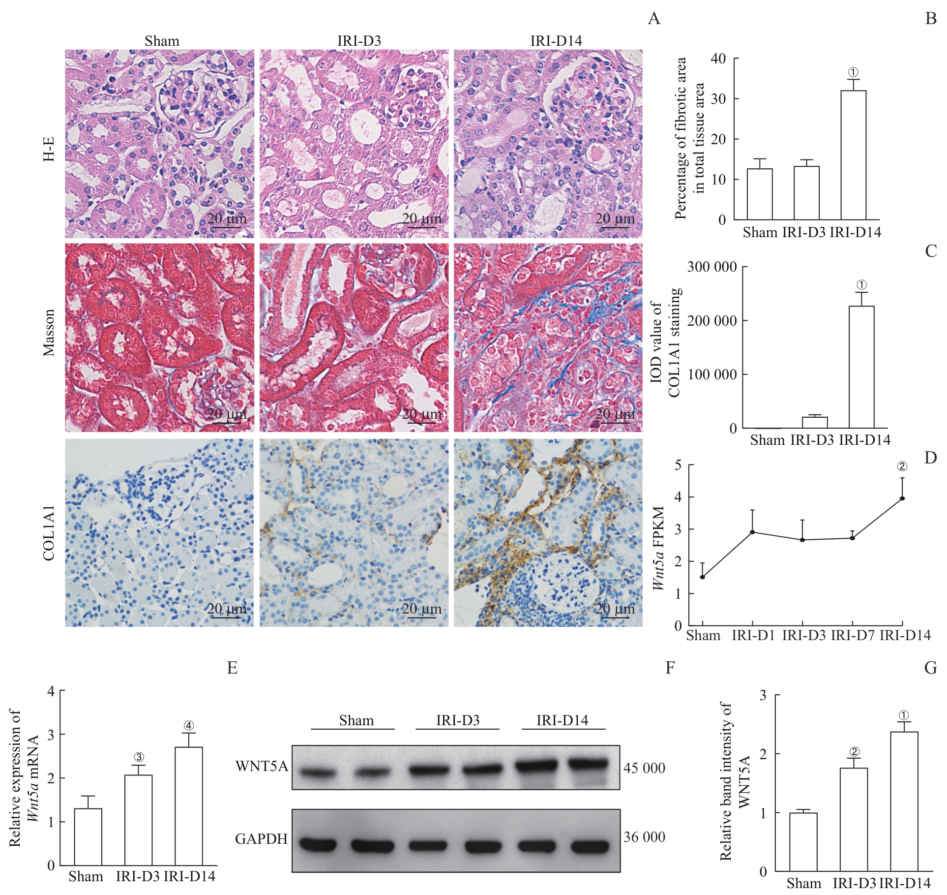
图1 Wnt5a 在IRI诱导的AKI小鼠模型中的表达情况Note: A. Renal histological changes in Sham group and IRI group (D3 and D14) mice evaluated by H-E staining; collagen fiber deposition assessed by Masson′s trichrome staining and immunohistochemical staining of COL1A1 (×400, scale bar=20 μm). B. Quantification of fibrotic area (percentage of total tissue area) based on Masson′s trichrome staining from three non-overlapping independent fields (n=5). C. The average integrated optical density (IOD) of COL1A1 immunohistochemical staining quantified from three non-overlapping independent fields (n=5). D. Temporal changes in Wnt5a gene expression at different time points after IRI in mice (n=5). E. qPCR analysis of Wnt5a expression in Sham group and IRI group (D3 and D14) (n=5). F. Western blotting analysis of WNT5A expression in Sham group and IRI group (D3 and D14). G. Quantification of Western blotting results (n=5). ①P<0.001, ④P=0.024, compared with the IRI-D3 group; ②P<0.001, ③P=0.009, compared with the Sham group.
Fig 1 Expression of Wnt5a in the IRI-induced AKI mouse model
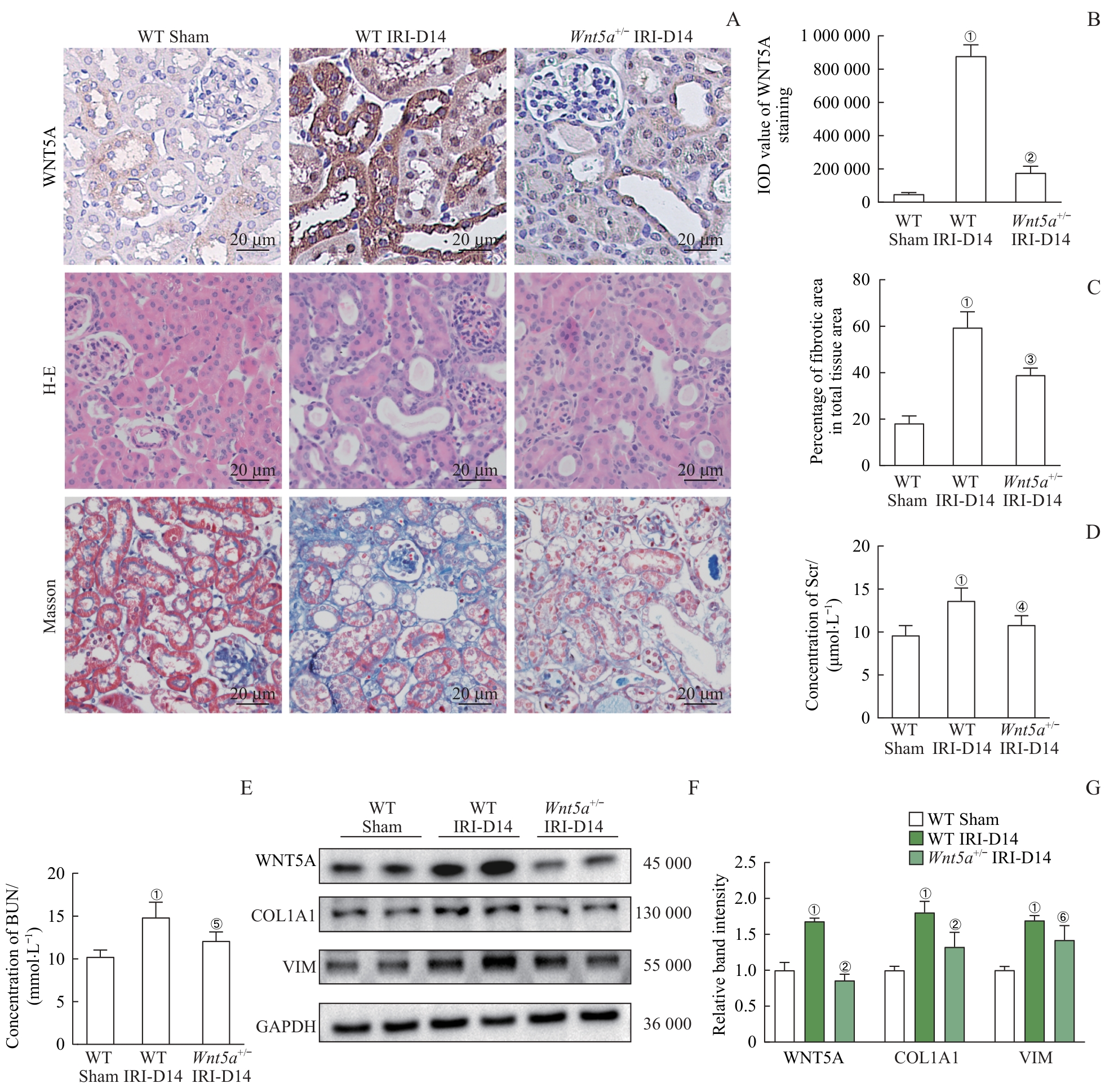
图2 敲除 Wnt5a 显著抑制IRI诱导的AKI小鼠向肾纤维化进展Note: A. Immunohistochemical staining of WNT5A, renal histological changes evaluated by H-E staining, and collagen fiber deposition assessed by Masson′s trichrome staining (×400, scale bar=20 μm). B. The average IOD of WNT5A immunohistochemical staining quantified from three non-overlapping independent fields (n=5). C. Quantification of fibrotic area (percentage of total tissue area) based on Masson′s trichrome staining from three non-overlapping independent fields (n=5). D. Quantification of Scr levels in the three groups (n=5). E. Quantification of BUN levels in the three groups (n=5). F. Western blotting analysis of WNT5A, COL1A1, and VIM expression in the three groups. G. Quantification of the Western blotting results (n=5). ①P<0.001, compared with the WT Sham group; ②P<0.001, ③P=0.004, ④P=0.011, ⑤P=0.015, ⑥P=0.013, compared with the WT IRI-D14 group.
Fig 2 Wnt5a knockout markedly attenuates renal fibrosis progression in IRI-induced AKI mice
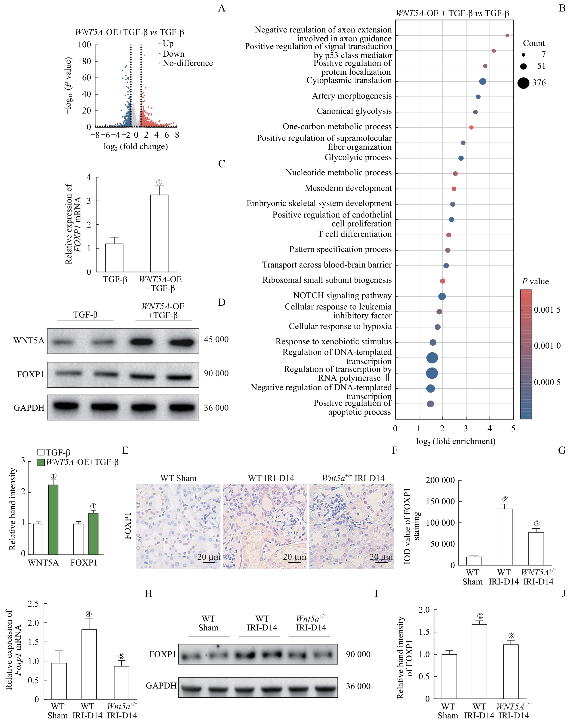
图3 FOXP1是WNT5A介导的肾纤维化下游的关键调控靶点Note: A. Volcano plot of differentially expressed genes between WNT5A-overexpressing (OE) and control HK-2 cells (both treated with TGF-β). B. GO enrichment analysis of differentially expressed genes in the two groups of HK-2 cells. C. qPCR analysis of FOXP1 expression in the two groups of HK-2 cells. D. Western blotting analysis of WNT5A and FOXP1 expression in the two groups of HK-2 cells. E. Quantification of the Western blotting results shown in Figure D. F. Immunohistochemical staining of FOXP1 in WT Sham, WT IRI, and Wnt5a+/- IRI mice (×400, scale bar=20 μm). G. The average IOD of FOXP1 immunohistochemical staining quantified from three non-overlapping independent fields (n=5). H. qPCR analysis of Foxp1 expression in the three groups of mice (n=5). I. Western blotting analysis of FOXP1 expression in the three groups of mice. J. Quantification of the Western blotting results shown in Figure I (n=5). ①P<0.001, compared with the control HK-2 cells; ②P<0.001, ④P=0.003, compared with the WT Sham group; ③P<0.001, ⑤P=0.002, compared with the WT IRI-D14 group.
Fig 3 FOXP1 serves as a key downstream effector of WNT5A-mediated renal fibrosis
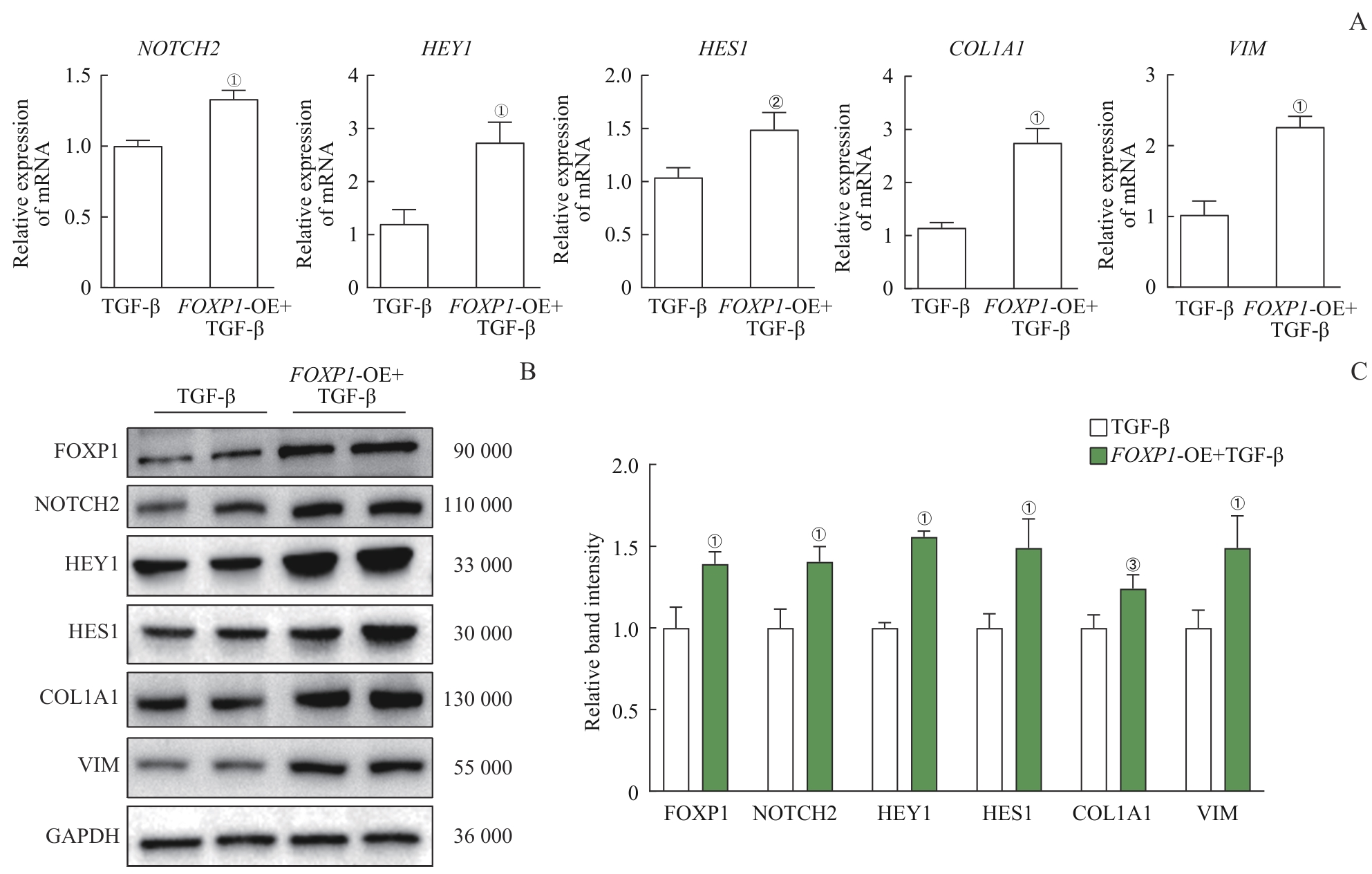
图4 FOXP1 过表达在HK-2纤维化诱导模型中通过NOTCH2通路上调纤维化标志物表达Note: A. qPCR analysis of NOTCH2, HEY1, HES1, COL1A1, and VIM expression in FOXP1-OE and control HK-2 cells (both treated with TGF-β). B. Western blotting analysis of FOXP1, NOTCH2, HEY1, HES1, COL1A1, and VIM expression in the two groups of cells. C. Quantification of the Western blotting results. ①P<0.001, ②P=0.003, ③P=0.004, compared with the control HK-2 cells.
Fig 4 FOXP1 overexpression upregulates the expression of fibrosis markers through the NOTCH2 pathway in TGF-β-treated HK-2 cells
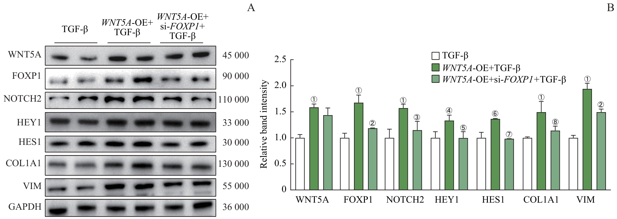
图5 在 WNT5A 过表达的HK-2纤维化诱导细胞模型中沉默 FOXP1 可抑制NOTCH2通路并下调纤维化标志物表达Note: A. Western blotting analysis of WNT5A, FOXP1, NOTCH2, HEY1, HES1, COL1A1, and VIM expression in control, WNT5A-OE, and WNT5A-OE with FOXP1 silencing HK-2 cells (all treated with TGF-β). B. Quantification of the Western blotting results. ①P<0.001, ④P=0.014, ⑥P=0.007, compared with the control HK-2 cells; ②P<0.001, ③P=0.002, ⑤P=0.013, ⑦P=0.005, ⑧P=0.008, compared with the WNT5A-OE HK-2 cells.
Fig 5 Silencing FOXP1 in WNT5A-overexpressing TGF-β-treated HK-2 cells inhibits the NOTCH2 pathway and downregulates the expression of fibrotic markers
| [1] | Ostermann M, Lumlertgul N, Jeong R, et al. Acute kidney injury[J]. Lancet, 2025, 405(10474): 241-256. |
| [2] | Cerda J, Kashani K, Ostermann M, et al. The global epidemiology of acute kidney injury: challenges and opportunities[J]. Nat Rev Nephrol, 2026, 22(3): 179-198. |
| [3] | Hoste E A J, Kellum J A, Selby N M, et al. Global epidemiology and outcomes of acute kidney injury[J]. Nat Rev Nephrol, 2018, 14(10): 607-625. |
| [4] | Dong Y, Zhang Q Z, Wen J J, et al. Ischemic duration and frequency determines AKI-to-CKD progression monitored by dynamic changes of tubular biomarkers in IRI mice[J]. Front Physiol, 2019, 10: 153. |
| [5] | Anders H J, Muruve D A. The inflammasomes in kidney disease[J]. J Am Soc Nephrol, 2011, 22(6): 1007-1018. |
| [6] | Venkatachalam M A, Weinberg J M, Kriz W, et al. Failed tubule recovery, AKI-CKD transition, and kidney disease progression[J]. J Am Soc Nephrol, 2015, 26(8): 1765-1776. |
| [7] | Akoumianakis I, Polkinghorne M, Antoniades C. Non-canonical WNT signalling in cardiovascular disease: mechanisms and therapeutic implications[J]. Nat Rev Cardiol, 2022, 19(12): 783-797. |
| [8] | Schunk S J, Floege J, Fliser D, et al. WNT-β-catenin signalling: a versatile player in kidney injury and repair[J]. Nat Rev Nephrol, 2021, 17(3): 172-184. |
| [9] | Kumawat K, Gosens R. WNT-5A: signaling and functions in health and disease[J]. Cell Mol Life Sci, 2016, 73(3): 567-587. |
| [10] | Trinh-Minh T, Chen C W, Tran Manh C, et al. Noncanonical WNT5A controls the activation of latent TGF-β to drive fibroblast activation and tissue fibrosis[J]. J Clin Invest, 2024, 134(10): e159884. |
| [11] | Gu S J, Feng H R, Li X M, et al. Targeting WNT5A noncanonical signaling attenuates renal fibrosis progression in acute kidney injury[J]. Mol Ther, 2025, 33(10): 5248-5262. |
| [12] | Li S R, Morley M, Lu M M, et al. Foxp transcription factors suppress a non-pulmonary gene expression program to permit proper lung development[J]. Dev Biol, 2016, 416(2): 338-346. |
| [13] | Trink J, Li R Z, Gao B, et al. Modulators of alpha-2 macroglobulin upregulation by high glucose in glomerular mesangial cells[J]. Biomolecules, 2024, 14(11): 1444. |
| [14] | Hsu Y C, Chang P J, Tung C W, et al. De-glycyrrhizinated licorice extract attenuates high glucose-stimulated renal tubular epithelial-mesenchymal transition via suppressing the Notch2 signaling pathway[J]. Cells, 2020, 9(1): 125. |
| [15] | Yu C, Xiong C X, Tang J H, et al. Histone demethylase JMJD3 protects against renal fibrosis by suppressing TGFβ and Notch signaling and preserving PTEN expression[J]. Theranostics, 2021, 11(6): 2706-2721. |
| [16] | Yang L, Xing G L, Wang L, et al. Acute kidney injury in China: a cross-sectional survey[J]. Lancet, 2015, 386(10002): 1465-1471. |
| [17] | Coca S G, Singanamala S, Parikh C R. Chronic kidney disease after acute kidney injury: a systematic review and meta-analysis[J]. Kidney Int, 2012, 81(5): 442-448. |
| [18] | Doi K. The need for disruptive innovation in acute kidney injury[J]. Clin Exp Nephrol, 2020, 24(11): 979-988. |
| [19] | Lin H Y, Liang C J, Yang M Y, et al. Critical roles of tubular mitochondrial ATP synthase dysfunction in maleic acid-induced acute kidney injury[J]. Apoptosis, 2024, 29(5/6): 620-634. |
| [20] | Wang Z W, Zhang C. From AKI to CKD: maladaptive repair and the underlying mechanisms[J]. Int J Mol Sci, 2022, 23(18): 10880. |
| [21] | Huffstater T, Merryman W D, Gewin L S. Wnt/β-catenin in acute kidney injury and progression to chronic kidney disease[J]. Semin Nephrol, 2020, 40(2): 126-137. |
| [22] | Zou Y, Pan L, Shen Y, et al. Cardiac Wnt5a and Wnt11 promote fibrosis by the crosstalk of FZD5 and EGFR signaling under pressure overload[J]. Cell Death Dis, 2021, 12(10): 877. |
| [23] | Li X M, Wen J J, Dong Y, et al. Wnt5a promotes renal tubular inflammation in diabetic nephropathy by binding to CD146 through noncanonical Wnt signaling[J]. Cell Death Dis, 2021, 12(1): 92. |
| [24] | Li G L, Wei W, Suo L G, et al. Low-dose aspirin prevents kidney damage in LPS-induced preeclampsia by inhibiting the WNT5A and NF-κB signaling pathways[J]. Front Endocrinol, 2021, 12: 639592. |
| [25] | Richards T, Modarage K, Dean C, et al. Atmin modulates Pkhd1 expression and may mediate autosomal recessive polycystic kidney disease (ARPKD) through altered non-canonical Wnt/planar cell polarity (PCP) signalling[J]. Biochim Biophys Acta Mol Basis Dis, 2019, 1865(2): 378-390. |
| [26] | Co M, Anderson A G, Konopka G. FOXP transcription factors in vertebrate brain development, function, and disorders[J]. Wiley Interdiscip Rev Dev Biol, 2020, 9(5): e375. |
| [27] | Liu X M, Du S L, Miao R, et al. Targeting the forkhead box protein P1 pathway as a novel therapeutic approach for cardiovascular diseases[J]. Heart Fail Rev, 2022, 27(1): 345-355. |
| [28] | Li H J, Liu P, Xu S Q, et al. FOXP1 controls mesenchymal stem cell commitment and senescence during skeletal aging[J]. J Clin Invest, 2025, 135(4): e191424. |
| [29] | Ling S F, Chen T N, Wang S J, et al. Deacetylation of FOXP1 by HDAC7 potentiates self-renewal of mesenchymal stem cells[J]. Stem Cell Res Ther, 2023, 14(1): 188. |
| [30] | Liu P, Huang S X, Ling S F, et al. Foxp1 controls brown/beige adipocyte differentiation and thermogenesis through regulating β3-AR desensitization[J]. Nat Commun, 2019, 10(1): 5070. |
| [31] | Sasaki Y, Shiozawa E, Watanabe N, et al. t(3;14)(p14.1;q32)/FOXP1-IGH translocation in thyroid extranodal marginal zone lymphoma of mucosa-associated lymphoid tissue (MALT lymphoma)[J]. Leuk Res, 2020, 95: 106399. |
| [32] | Chiang K, Zielinska A E, Shaaban A M, et al. PRMT5 is a critical regulator of breast cancer stem cell function via histone methylation and FOXP1 expression[J]. Cell Rep, 2017, 21(12): 3498-3513. |
| [33] | Shao X, Wei X. FOXP1 enhances fibrosis via activating Wnt/β-catenin signaling pathway in endometriosis[J]. Am J Transl Res, 2018, 10(11): 3610-3618. |
| [34] | Zou Y X, Yiu W H, Lok S W Y, et al. Tubular FoxP2 and kidney fibrosis[J]. J Am Soc Nephrol, 2025, 36(4): 544-558. |
| [35] | Yuan Q, Tang B, Zhang C. Signaling pathways of chronic kidney diseases, implications for therapeutics[J]. Signal Transduct Target Ther, 2022, 7(1): 182. |
| [36] | Xiao M, Bai S J, Chen J, et al. CDKN2B-AS1 participates in high glucose-induced apoptosis and fibrosis via NOTCH2 through functioning as a miR-98-5p decoy in human podocytes and renal tubular cells[J]. Diabetol Metab Syndr, 2021, 13(1): 107. |
| [1] | 商迪华, 陈慧文, 李若琳, 刘英莉, 宋忠臣. 基于代谢组学的伴慢性肾衰竭牙周炎患者唾液代谢物特征分析[J]. 上海交通大学学报(医学版), 2026, 46(5): 576-584. |
| [2] | 应奕雯, 李舒, 张珍, 虞靖然, 张宁, 牟姗. 高危妊娠人群妊娠相关急性肾损伤的临床特征和妊娠结局分析[J]. 上海交通大学学报(医学版), 2026, 46(1): 60-65. |
| [3] | 李玉, 张羽. 慢性肾脏病患者不良妊娠结局的危险因素分析[J]. 上海交通大学学报(医学版), 2024, 44(5): 560-566. |
| [4] | 吴佳晋, 钟晨, 李大伟, 陈若洋, 瞿俊文, 张明. 甲基转移酶3调控pri-miR-21甲基化修饰在糖尿病肾病肾脏纤维化中的作用[J]. 上海交通大学学报(医学版), 2023, 43(1): 1-7. |
| [5] | 谢林, 程烨, 郑琦敏, 张熙, 付莉莉, 陈敏, 王怡, 梅长林, 谢静远, 顾向晨. 淫羊藿苷对急性肾损伤向慢性肾脏病转化小鼠模型的预防性保护作用[J]. 上海交通大学学报(医学版), 2023, 43(1): 8-19. |
| [6] | 赖秀秀, 朱青燕, 谈佳奇, 杨令, 朱琰, 周公民. 小剂量非布司他改善无症状高尿酸血症高龄患者肾功能的临床研究[J]. 上海交通大学学报(医学版), 2022, 42(7): 885-892. |
| [7] | 王亚琨, 许佳瑞, 吴茜茜, 张晓华, 朱迎春, 白寿军. 医养结合综合干预对上海郊区老年慢性肾脏病患者生活质量和精神状态的影响[J]. 上海交通大学学报(医学版), 2022, 42(7): 904-910. |
| [8] | 魏珊, 纪鸥洋, 陈志豪, 黄泽慧, 李璞, 方均燕, 刘英莉. 慢性肾脏病患者安全用药知信行量表的编制及信度、效度分析[J]. 上海交通大学学报(医学版), 2022, 42(12): 1729-1738. |
| [9] | 周丝丝, 巩格言, 马佳莉, 高雯颖, 张莹. 慢性肾脏病患儿-家长过渡期准备与双方生活质量的互依关系分析[J]. 上海交通大学学报(医学版), 2021, 41(12): 1588-1595. |
| [10] | 沈剑箫, 王万鹏, 邵兴华, 吴晶魁, 李舒, 车霞静, 倪兆慧. 顺铂诱导的急性肾损伤中肾脏组织m6A甲基化水平的变化[J]. 上海交通大学学报(医学版), 2021, 41(12): 1603-1611. |
| [11] | 陈胜男,申 燕. 不对称二甲基精氨酸在慢性肾脏病患者动脉粥样硬化发生和发展中的作用[J]. 上海交通大学学报(医学版), 2020, 40(8): 1131-1136. |
| [12] | 周培慧,王丽. 白细胞介素 -27在顺铂所致急性肾损伤中的高表达及对细胞 凋亡的抑制作用[J]. 上海交通大学学报(医学版), 2019, 39(4): 372-. |
| [13] | 廖伟棠1,张文2. 外泌体在急性肾损伤中作用的研究进展[J]. 上海交通大学学报(医学版), 2018, 38(7): 811-. |
| [14] | 平凤,郭勇,刘玉静,曹永梅,李颖川 . 代谢组学在急性肾损伤诊断与治疗中的应用[J]. 上海交通大学学报(医学版), 2017, 37(8): 1174-. |
| [15] | 高磊,吴世勇,蒋丹,王颖,李勇湧,王德伟,常广磊,张鹏,覃铮,张冬颖. 胰岛素样因子6对单侧输尿管梗阻小鼠肾脏纤维化和钙化的影响[J]. 上海交通大学学报(医学版), 2017, 37(12): 1616-. |
| 阅读次数 | ||||||
|
全文 |
|
|||||
|
摘要 |
|
|||||
