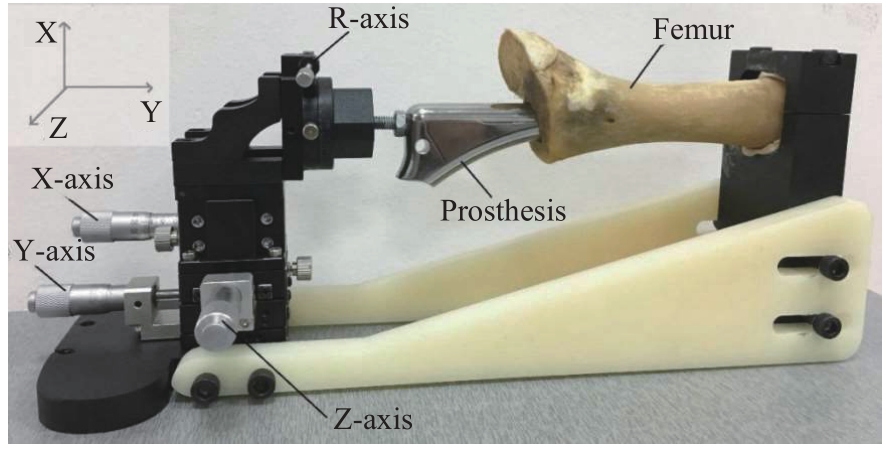
| 1 |
边焱焱, 程开源, 常晓, 等. 2011至2019年中国人工髋膝关节置换手术量的初步统计与分析[J]. 中华骨科杂志, 2020, 40(21): 1453-1460.
|
|
BIAN Y Y, CHENG K Y, CHANG X, et al. Reports and analysis of amount of hip and knee arthroplasty in China from 2011 to 2019[J]. Chinese Journal of Orthopaedics, 2020, 40(21): 1453-1460.
|
| 2 |
LEWIS P L, GRAVES S E, CUTHBERT A, et al. What is the risk of repeat revision when patellofemoral replacement is revised to TKA? An analysis of 482 cases from a large national arthroplasty registry[J]. Clin Orthop Relat Res, 2019, 477(6): 1402-1410.
|
| 3 |
ROOF M A, NARAYANAN S, LORENTZ N, et al. Impact of time to revision total knee arthroplasty on outcomes following aseptic failure[J]. Knee Surg Relat Res, 2023, 35(1): 15.
|
| 4 |
OLTEAN-DAN D, APOSTU D, TOMOAIA G, et al. Causes of revision after total hip arthroplasty in an orthopedics and traumatology regional center[J]. Med Pharm Rep, 2022, 95(2): 179-184.
|
| 5 |
FONTALIS A, HADDAD F S. Roentgen stereophotogrammetric analysis: still a very valuable tool in the orthopaedic research armamentarium[J]. Bone Joint Res, 2022, 11(4): 210-213.
|
| 6 |
XU J, SONNTAG R, KRETZER J P, et al. Model-based roentgen stereophotogrammetric analysis to monitor the head-taper junction in total hip arthroplasty in vivo: and they do move[J]. Materials, 2020, 13(7): 1543.
|
| 7 |
DAMMERER D, BLUM P, PUTZER D, et al. Subsidence of a metaphyseal-anchored press-fit stem after 4-year follow-up: an EBRA-FCA analysis[J]. Arch Orthop Trauma Surg, 2022, 142(8): 2075-2082.
|
| 8 |
DAMMERER D, BLUM P, PUTZER D, et al. Good mid-term results with the trident peripheral self-locking cup: a clinical evaluation and migration measurement with EBRA[J]. Arch Orthop Trauma Surg, 2021, 141(2): 327-332.
|
| 9 |
CLARKE S G, LOGISHETTY K, HALEWOOD C, et al. Low dose CT-based spatial analysis (CTSA) to measure implant migration after ceramic hip resurfacing arthroplasty (HRA): a phantom study[J]. Proc Inst Mech Eng H, 2023, 237(3): 359-367.
|
| 10 |
LI G A, VAN DE VELDE S K, BINGHAM J T. Validation of a non-invasive fluoroscopic imaging technique for the measurement of dynamic knee joint motion[J]. J Biomech, 2008, 41(7): 1616-1622.
|
| 11 |
ZOU D Y, TAN J Q, ZHENG N, et al. Larger medial contact area and more anterior contact position in medial-pivot than posterior-stabilized total knee arthroplasty during in-vivo lunge activity[J]. Bioengineering, 2023, 10(3): 290.
|
| 12 |
HU Y, ZOU D Y, JIANG M D, et al. Postoperative hip center position is associated with gait symmetry in range of axial rotation in dysplasia patients after THA[J]. Front Surg, 2023, 10: 1135327.
|
| 13 |
廖广姗.基于模型和图像的动态Fluoroscopic Stereophotogrammetric Analysis(FSA)技术检测人工髋关节无菌性松动的研究[D]. 上海:上海交通大学, 2013.
|
|
LIAO G S. A study on the detection of aseptic loosening of artificial hip joints using dynamic fluoroscopic stereographic analysis (FSA) technology based on models and images [D]. Shanghai: Shanghai Jiao Tong University, 2013.
|
| 14 |
廖广姗, 李慧武, 王金武, 等. 人工髋关节无菌性松动失效的生物力学分析与诊断推理[J]. 医用生物力学, 2012, 27(3): 251-257.
|
|
LIAO G S, LI H W, WANG J W, et al. Biomechanical analysis and reasoning on aseptic loosening failure after total hip arthroplasty[J]. Journal of Medical Biomechanics, 2012, 27(3): 251-257.
|
| 15 |
PRINS A H, KAPTEIN B L, STOEL B C, et al. Performance of local optimization in single-plane fluoroscopic analysis for total knee arthroplasty[J]. J Biomech, 2015, 48(14): 3837-3845.
|
| 16 |
FONSECA ULLOA C A, SEEGER A, HAGEDORN F S, et al. Development and validation of an algorithm to determine the minimal factors needed for non-invasive measurement of the in vivo primary stability of cementless hip implants[J]. Med Eng Phys, 2023, 111: 103932.
|
| 17 |
KIEVIT A J, BUIJS G S, DOBBE J G G, et al. Promising results of an non-invasive measurement of knee implant loosening using a loading device, CT-scans and 3D image analysis[J]. Clin Biomech, 2023, 104: 105930.
|
| 18 |
HEILEMANN M, WENDLER T, MÜNST P, et al. A novel micromotion measurement method to gain instructive insight into the acetabular bone-implant interface[J]. Med Eng Phys, 2020, 86: 138-145.
|
 ), 雷浩2(