上海交通大学学报(医学版) ›› 2024, Vol. 44 ›› Issue (9): 1094-1103.doi: 10.3969/j.issn.1674-8115.2024.09.004
• 论著 · 基础研究 • 上一篇
收稿日期:
2024-05-03
接受日期:
2024-07-18
出版日期:
2024-09-28
发布日期:
2024-10-09
通讯作者:
鞠强
E-mail:2894791478@qq.com;jqedison@163.com;qiangju401@sina.com
作者简介:
梁梦晨(1999—),女,博士生;电子信箱:2894791478@qq.com基金资助:
LIANG Mengchen(), LI Jiaqi(
), WU Xinyi, MO Xiaohui, JU Qiang(
)
Received:
2024-05-03
Accepted:
2024-07-18
Online:
2024-09-28
Published:
2024-10-09
Contact:
JU Qiang
E-mail:2894791478@qq.com;jqedison@163.com;qiangju401@sina.com
Supported by:
摘要:
目的·研究中度、重度寻常痤疮患者与健康者非皮损区毛囊菌群群落结构和载量的差异,探讨微生物与寻常痤疮及其严重程度之间的关系。方法·采用横断面研究,选取2022年8月—2023年8月在上海交通大学医学院附属仁济医院皮肤科就诊的中度、重度寻常痤疮(简称痤疮)患者和健康志愿者。取中度、重度痤疮患者及健康志愿者面部非皮损区毛囊内容物进行16S rRNA高通量测序以及实时荧光定量聚合酶链反应(quantitative real-time polymerase chain reaction,qPCR)检测,分析不同严重程度痤疮患者毛囊细菌菌群多样性、物种组成差异和菌群载量差异。结果·纳入10例中度痤疮患者、11例重度痤疮患者及11例健康志愿者,3组间年龄、性别等一般资料差异均无统计学意义。中度痤疮组和重度痤疮组相对于健康组细菌α多样性均显著下降(P=0.020,P=0.013)。主坐标分析(principal coordinates analysis,PCoA)图表明健康组人群样本分布较为集中,组内差异较小,而中度痤疮组及重度痤疮组人群样本分布有一定的趋势但较为离散,组内之间存在较大差异。3组样本趋势分布差异明显,组间微生物群落结构差异较大。相似性分析结果显示健康组与中度痤疮组(P=0.027)、健康组与重度痤疮组(P=0.017)组间β多样性有显著差异,物种组成相似度低。中度痤疮组与重度痤疮组(P=0.160)组间物种相似度较高。门水平上3组的优势菌群均为放线菌门(Actinobacteria)、厚壁菌门(Firmicutes)、变形菌门(Proteobacteria)以及拟杆菌门(Bacteroidetes)。属水平上健康组优势菌为丙酸杆菌属(Propionibacterium)、未分类的放线菌属(unclassified Actinomycetales),2组痤疮组优势菌均为葡萄球菌属(Staphylococcus)、丙酸杆菌属。相对于健康组,中度痤疮组和重度痤疮组非皮损区毛囊中的葡萄球菌属相对丰度均显著增加(P=0.010,P=0.019),毛囊菌群载量均显著增加(均P=0.001)。与中度痤疮组相比,重度痤疮组毛囊样本中细菌载量显著升高(P=0.017)。结论·中度、重度痤疮患者与健康者非皮损区毛囊微生物群落结构不同,痤疮组微生物多样性明显下降。中度痤疮组及重度痤疮组非皮损区毛囊中的葡萄球菌属相对丰度较健康组均显著增加。随着痤疮严重程度的增加,非皮损区毛囊内细菌载量显著增加。研究提示痤疮的发生以及严重程度可能与毛囊菌群的群落结构和载量有关。
中图分类号:
梁梦晨, 李嘉祺, 吴心怡, 莫小辉, 鞠强. 中度与重度寻常痤疮患者非皮损区毛囊菌群分析:一项单中心横断面研究[J]. 上海交通大学学报(医学版), 2024, 44(9): 1094-1103.
LIANG Mengchen, LI Jiaqi, WU Xinyi, MO Xiaohui, JU Qiang. Analysis of hair follicle microbiota in non-lesional areas of patients with moderate-to-severe acne vulgaris: a single-center cross-sectional study[J]. Journal of Shanghai Jiao Tong University (Medical Science), 2024, 44(9): 1094-1103.
Primer name | Primer sequence (5′→3′) | Amplified region |
---|---|---|
341F | CCTACGGGNGGCWGCAG | V3‒4[ |
805R | GACTACHVGGGTATCTAATCC |
表1 引物序列
Tab 1 Primer sequences
Primer name | Primer sequence (5′→3′) | Amplified region |
---|---|---|
341F | CCTACGGGNGGCWGCAG | V3‒4[ |
805R | GACTACHVGGGTATCTAATCC |
Step | Program |
---|---|
Step 1 | 95 ℃ 30 s |
Step 2 (40 cycles) | 95 ℃ 5 s; 55 ℃ 30 s; 72 ℃ 30 s |
Step 3 (dissociation stage) | 94 ℃ 30 s; 70 ℃ 1.5 min; 94 ℃ 10 s |
表2 qPCR绝对定量程序
Tab 2 qPCR absolute quantification program
Step | Program |
---|---|
Step 1 | 95 ℃ 30 s |
Step 2 (40 cycles) | 95 ℃ 5 s; 55 ℃ 30 s; 72 ℃ 30 s |
Step 3 (dissociation stage) | 94 ℃ 30 s; 70 ℃ 1.5 min; 94 ℃ 10 s |
Characteristic | C group (n=11) | M group (n=10) | S group (n=11) | H/χ² value | P value |
---|---|---|---|---|---|
Gender/n(%) | 0.182 | 0.913 | |||
Male | 5 (45.5) | 5 (50.0) | 6 (54.5) | ||
Female | 6 (54.5) | 5 (50.0) | 5 (45.5) | ||
Age/year | 24.64±1.63 | 21.90±2.88 | 23.18±5.42 | 3.827 | 0.252 |
Ethnic group/n(%) | 1.971 | 0.373 | |||
Han | 10 (90.9) | 10 (100.0) | 11 (100.0) | ||
Bai | 1 (9.1) | 0 (0) | 0 (0) | ||
Education/n(%) | 2.676 | 0.262 | |||
Senior high school/technical secondary school | 4 (36.4) | 2 (20.0) | 6 (54.5) | ||
Junior college and above | 7 (63.6) | 8 (80.0) | 5 (45.5) | ||
Work/n(%) | 1.703 | 0.427 | |||
In service | 3 (27.3) | 2 (20.0) | 5 (45.5) | ||
Not in service | 8 (72.7) | 8 (80.0) | 6 (54.5) | ||
Habitual residence/n(%) | 2.147 | 0.342 | |||
Shanghai | 11 (100.0) | 9 (90.0) | 9 (81.8) | ||
Others | 0 (0) | 1 (10.0) | 2 (18.2) |
表3 3组人群一般资料
Tab 3 General characteristics of the three groups
Characteristic | C group (n=11) | M group (n=10) | S group (n=11) | H/χ² value | P value |
---|---|---|---|---|---|
Gender/n(%) | 0.182 | 0.913 | |||
Male | 5 (45.5) | 5 (50.0) | 6 (54.5) | ||
Female | 6 (54.5) | 5 (50.0) | 5 (45.5) | ||
Age/year | 24.64±1.63 | 21.90±2.88 | 23.18±5.42 | 3.827 | 0.252 |
Ethnic group/n(%) | 1.971 | 0.373 | |||
Han | 10 (90.9) | 10 (100.0) | 11 (100.0) | ||
Bai | 1 (9.1) | 0 (0) | 0 (0) | ||
Education/n(%) | 2.676 | 0.262 | |||
Senior high school/technical secondary school | 4 (36.4) | 2 (20.0) | 6 (54.5) | ||
Junior college and above | 7 (63.6) | 8 (80.0) | 5 (45.5) | ||
Work/n(%) | 1.703 | 0.427 | |||
In service | 3 (27.3) | 2 (20.0) | 5 (45.5) | ||
Not in service | 8 (72.7) | 8 (80.0) | 6 (54.5) | ||
Habitual residence/n(%) | 2.147 | 0.342 | |||
Shanghai | 11 (100.0) | 9 (90.0) | 9 (81.8) | ||
Others | 0 (0) | 1 (10.0) | 2 (18.2) |
图1 3组人群毛囊菌群的α多样性分析Note: A. Species accumulation curve. B. Venn diagrams of the OTU numbers of the three groups. C. Box plots of Chao1 index of the three groups.
Fig 1 α diversity analysis of follicular microbiome of the three groups
图2 3组人群毛囊菌群的β多样性分析Note: PCoA scatter plot based on β diversity index between samples.
Fig 2 β-diversity analysis of hair follicle microbiome of the three groups
Phylum | Abundance/% | ||
---|---|---|---|
C group (n=11) | M group (n=10) | S group (n=11) | |
Actinobacteria | 61.48±19.39 | 58.48±17.59 | 49.64±28.26 |
Firmicutes | 21.94±16.10 | 35.42±18.37 | 43.90±26.42 |
Proteobacteria | 14.84±15.23 | 5.27±5.79 | 5.51±5.41 |
Bacteroidetes | 0.86±0.88 | 0.27±0.29 | 0.59±1.13 |
Others | 0.88±0.55 | 0.56±0.57 | 0.36±0.49 |
表4 3组样本细菌菌群在门水平上的丰度(%)
Tab 4 Abundance of bacterial flora at the phylum level in three sample groups (%)
Phylum | Abundance/% | ||
---|---|---|---|
C group (n=11) | M group (n=10) | S group (n=11) | |
Actinobacteria | 61.48±19.39 | 58.48±17.59 | 49.64±28.26 |
Firmicutes | 21.94±16.10 | 35.42±18.37 | 43.90±26.42 |
Proteobacteria | 14.84±15.23 | 5.27±5.79 | 5.51±5.41 |
Bacteroidetes | 0.86±0.88 | 0.27±0.29 | 0.59±1.13 |
Others | 0.88±0.55 | 0.56±0.57 | 0.36±0.49 |
Genus | Abundance/% | ||
---|---|---|---|
C group (n=11) | M group (n=10) | S group (n=11) | |
Staphylococcus | 13.05±11.54 | 31.54±17.84 | 37.30±27.09 |
Propionibacterium | 36.99±17.67 | 25.94±19.63 | 23.78±16.30 |
Unclassified Actinomycetales | 18.78±19.20 | 23.38±15.96 | 19.58±17.88 |
Corynebacterium | 2.09±2.77 | 8.28±11.28 | 5.47±4.74 |
Unclassified Neisseriaceae | 4.36±8.64 | 0.06±0.08 | 0.86±2.48 |
Others | 24.73±22.90 | 10.80±9.32 | 13.01±22.05 |
表5 3组样本细菌菌群在属水平上的丰度(%)
Tab 5 Abundance of bacterial flora at the genus level in three sample groups (%)
Genus | Abundance/% | ||
---|---|---|---|
C group (n=11) | M group (n=10) | S group (n=11) | |
Staphylococcus | 13.05±11.54 | 31.54±17.84 | 37.30±27.09 |
Propionibacterium | 36.99±17.67 | 25.94±19.63 | 23.78±16.30 |
Unclassified Actinomycetales | 18.78±19.20 | 23.38±15.96 | 19.58±17.88 |
Corynebacterium | 2.09±2.77 | 8.28±11.28 | 5.47±4.74 |
Unclassified Neisseriaceae | 4.36±8.64 | 0.06±0.08 | 0.86±2.48 |
Others | 24.73±22.90 | 10.80±9.32 | 13.01±22.05 |
图5 C组和M组人群毛囊菌群LEfSe分析进化分支图Note: The circles radiating from the inside out represent the classification levels from phylum to genus. Each circle and its shaded range at different classification levels represent the classification at that level, and the diameter of the circle is proportional to the relative abundance of the species. The species represented by the English letters next to the circles are shown in the legend. The red nodes represent microbial communities with high abundance and important roles in the red group, the green nodes represent microbial communities with important roles in the green group, and the yellow nodes represent species with no differences between groups.
Fig 5 LEfSe cladogram of analysis of follicular microbiome in C group and M group
图6 C组和S组人群毛囊菌群LEfSe分析进化分支图Note: The circles radiating from the inside out represent the classification levels from phylum to genus. Each circle and its shaded range at different classification levels represent the classifications at those levels, and the diameter of the circle is proportional to the relative abundance of the species. The species represented by the English letters next to the circles are shown in the legend. The red nodes represent microbial communities with high abundance and important roles in the red group, the green nodes represent microbial communities with important roles in the green group, and the yellow nodes represent species with no differences between groups.
Fig 6 LEfSe cladogram of analysis of hair follicle microbiome in Group C and Group S
图7 M组和S组人群毛囊菌群LEfSe分析进化分支图Note: The circles radiating from the inside out represent the classification level from phylum to genus. Each circle and its shaded range at different classification levels represent the classification at that level, and the diameter of the circle is proportional to the relative abundance of the species. The species represented by the English letters next to the circle are shown in the legend. The red nodes represent microbial communities with high abundance and important roles in the red group, the green nodes represent microbial communities with important roles in the green group, and the yellow nodes represent species with no differences between groups.
Fig 7 LEfSe cladogram of analysis of hair follicle microbiome in Group M and Group S
1 | CHEN Y, KNIGHT R, GALLO R L. Evolving approaches to profiling the microbiome in skin disease[J]. Front Immunol, 2023, 14: 1151527. |
2 | LAYTON A M, RAVENSCROFT J. Adolescent acne Vulgaris: current and emerging treatments[J]. Lancet Child Adolesc Health, 2023, 7(2): 136-144. |
3 | BERRY K, LIM J, ZAENGLEIN A L. Acne Vulgaris: treatment made easy for the primary care physician[J]. Pediatr Ann, 2020, 49(3): e109-e115. |
4 | CHUA W, POH S E, LI H. Secretory proteases of the human skin microbiome[J]. Infect Immun, 2022, 90(1): e0039721. |
5 | SHI J, CHENG J W, ZHANG Q, et al. Comparison of the skin microbiota of patients with acne Vulgaris and healthy controls[J]. Ann Palliat Med, 2021, 10(7): 7933-7941. |
6 | NUMATA S, AKAMATSU H, AKAZA N, et al. Analysis of facial skin-resident microbiota in Japanese acne patients[J]. Dermatology, 2014, 228(1): 86-92. |
7 | XU X X, RAN X, TANG J Q, et al. Skin microbiota in non-inflammatory and inflammatory lesions of acne Vulgaris: the underlying changes within the pilosebaceous unit[J]. Mycopathologia, 2021, 186(6): 863-869. |
8 | WITKOWSKI J A, PARISH L C. The assessment of acne: an evaluation of grading and lesion counting in the measurement of acne[J]. Clin Dermatol, 2004, 22(5): 394-397. |
9 | 
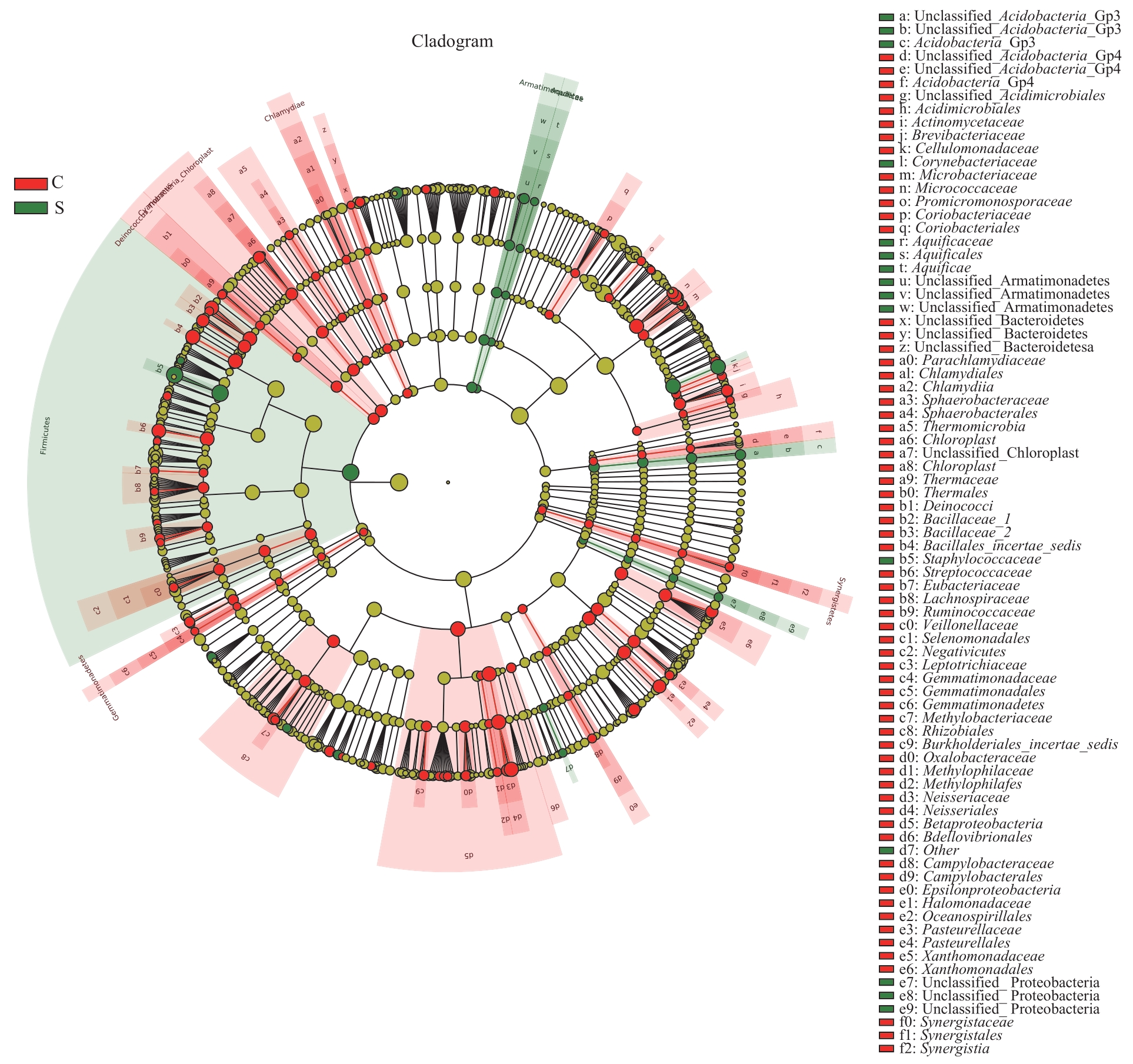
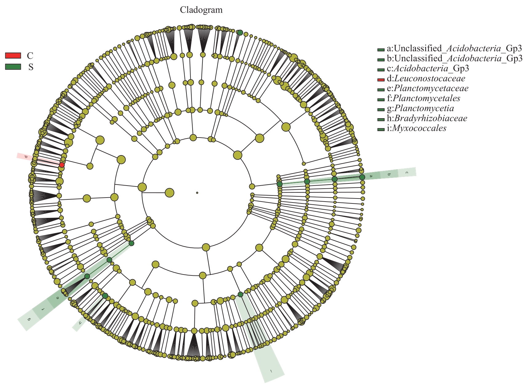
SPESHOCK J L, BRADY J A, EASTMAN J, et al. Impact of manure storage time and temperature on microbial composition and stable fly (Diptera: Muscidae) development[J]. AiM, 2019, 9(3): 248-265. |
10 | LEE Y B, BYUN E J, KIM H S. Potential role of the microbiome in acne: a comprehensive review[J]. J Clin Med, 2019, 8(7): 987. |
11 | BYRD A L, BELKAID Y, SEGRE J A. The human skin microbiome[J]. Nat Rev Microbiol, 2018, 16(3): 143-155. |
12 | KOH L F, ONG R Y, COMMON J E. Skin microbiome of atopic dermatitis[J]. Allergol Int, 2022, 71(1): 31-39. |
13 | SUNG K H. Microbiota in Rosacea[J]. Am J Clin Dermatol, 2020, 21(Suppl 1): 1-11. |
14 | SKOWRON K, BAUZA-KASZEWSKA J, KRASZEWSKA Z, et al. Human skin microbiome: impact of intrinsic and extrinsic factors on skin microbiota[J]. Microorganisms, 2021, 9(3): 543. |
15 | ISARD O, KNOL A C, ARIÈS M F, et al. Propionibacterium acnes activates the IGF-1/IGF-1R system in the epidermis and induces keratinocyte proliferation[J]. J Invest Dermatol, 2011, 131(1): 59-66. |
16 | AKAZA N, AKAMATSU H, KISHI M, et al. Effects of Propionibacterium acnes on various mRNA expression levels in normal human epidermal keratinocytes in vitro[J]. J Dermatol, 2009, 36(4): 213-223. |
17 | FITZ-GIBBON S, TOMIDA S, CHIU B H, et al. Propionibacterium acnes strain populations in the human skin microbiome associated with acne[J]. J Invest Dermatol, 2013, 133(9): 2152-2160. |
18 | DRENO B, MARTIN R, MOYAL D, et al. Skin microbiome and acne Vulgaris: Staphylococcus, a new actor in acne[J]. Exp Dermatol, 2017, 26(9): 798-803. |
19 | O'NEILL A M, NAKATSUJI T, HAYACHI A, et al. Identification of a human skin commensal bacterium that selectively kills cutibacteriumacnes[J]. J Invest Dermatol, 2020, 140(8): 1619-1628.e2. |
20 | XIA X L, LI Z H, LIU K W, et al. Staphylococcal LTA-induced miR-143 inhibits Propionibacterium acnes-mediated inflammatory response in skin[J]. J Invest Dermatol, 2016, 136(3): 621-630. |
21 | COTTER P D, HILL C, ROSS R P. Bacterial lantibiotics: strategies to improve therapeutic potential[J]. Curr Protein Pept Sci, 2005, 6(1): 61-75. |
22 | MARITO S, KESHARI S, TRAISAENG S, et al. Electricity-producing Staphylococcus epidermidis counteracts Cutibacterium acnes[J]. Sci Rep, 2021, 11(1): 12001. |
23 | DAGNELIE M A, CORVEC S, TIMON-DAVID E, et al. Cutibacterium acnes and Staphylococcus epidermidis: the unmissable modulators of skin inflammatory response[J]. Exp Dermatol, 2022, 31(3): 406-412. |
24 | LI D H, CHEN Q, LIU Y, et al. The prevalence of acne in Mainland China: a systematic review and meta-analysis[J]. BMJ Open, 2017, 7(4): e015354. |
25 | LI C X, YOU Z X, LIN Y X, et al. Skin microbiome differences relate to the grade of acne Vulgaris[J]. J Dermatol, 2019, 46(9): 787-790. |
26 | KÕLJALG U, NILSSON R H, ABARENKOV K, et al. Towards a unified paradigm for sequence-based identification of fungi[J]. Mol Ecol, 2013, 22(21): 5271-5277. |
27 | SU T T, LAI S C, LEE A, et al. Meta-analysis: proton pump inhibitors moderately increase the risk of small intestinal bacterial overgrowth[J]. J Gastroenterol, 2018, 53(1): 27-36. |
28 | PIMENTEL M, SAAD R J, LONG M D, et al. ACG clinical guideline: small intestinal bacterial overgrowth[J]. Am J Gastroenterol, 2020, 115(2): 165-178. |
29 | EFREMOVA I, MASLENNIKOV R, POLUEKTOVA E, et al. Epidemiology of small intestinal bacterial overgrowth[J]. World J Gastroenterol, 2023, 29(22): 3400-3421. |
[1] | . 隆突性皮肤纤维肉瘤多学科诊治实施规范——上海交通大学医学院附属第九人民医院专家共识(2020年版)[J]. 上海交通大学学报(医学版), 2021, 41(12): 1669-1675. |
[2] | 庄昊俊, 郭美亮, 刘婉雯, 邓辉. Janus激酶抑制剂在特应性皮炎治疗中的临床应用研究进展[J]. 上海交通大学学报(医学版), 2021, 41(7): 963-966. |
阅读次数 | ||||||||||||||||||||||||||||||||||||||||||||||||||
全文 1017
|
|
|||||||||||||||||||||||||||||||||||||||||||||||||
摘要 256
|
|
|||||||||||||||||||||||||||||||||||||||||||||||||