上海交通大学学报(医学版) ›› 2026, Vol. 46 ›› Issue (10): 1313-1322.doi: 10.3969/j.issn.1674-8115.2026.10.001

• 创新团队成果专栏 •    

单细胞测序探索Otoferlin基因敲除后耳聋小鼠耳蜗内细胞变化

马晓彤1,2,3,4, 蔡瑞捷1,2,3,4, 赖艺洋1,2,3,4, 汪子荃1,2,3,4, 刘辉辉1,2,3,4(), 吴皓1,2,3,4()   

  1. 1.上海交通大学医学院附属第九人民医院耳鼻咽喉头颈外科,上海 200011
    2.上海交通大学医学院耳科学研究所,上海 200125
    3.上海市耳鼻疾病转化医学重点实验室,上海 200125
    4.上海交通大学医学院医学技术学院听力与言语康复系,上海 200125
  • 收稿日期:2026-01-20 接受日期:2026-04-22 出版日期:2026-10-28 发布日期:2026-10-28
  • 通讯作者: 刘辉辉,电子信箱: liuhent9th@163.com。
    吴 皓,电子信箱:wuhao@shsmu.edu.cn。
  • 基金资助:
    上海市2024年度“科技创新行动计划”(24J12800300);国家自然科学基金青年科学基金项目(82101211);中国博士后科学基金(2024M752052)

Changes in cochlear cell subsets in Otoferlin-knockout deaf mice revealed by single-cell sequencing

Ma Xiaotong1,2,3,4, Cai Ruijie1,2,3,4, Lai Yiyang1,2,3,4, Wang Ziquan1,2,3,4, Liu Huihui1,2,3,4(), Wu Hao1,2,3,4()   

  1. 1.Department of Otorhinolaryngology Head and Neck Surgery, Shanghai Ninth People's Hospital, Shanghai Jiao Tong University School of Medicine, Shanghai 200011, China
    2.Ear Institute, Shanghai Jiao Tong University School of Medicine, Shanghai 200125, China
    3.Shanghai Key Laboratory of Translational Medicine on Ear and Nose Diseases, Shanghai 200125, China
    4.Department of Audiology and Speech-Language Pathology, School of Medical Technology, Shanghai Jiao Tong University School of Medicine, Shanghai 200125, China
  • Received:2026-01-20 Accepted:2026-04-22 Online:2026-10-28 Published:2026-10-28
  • Contact: Liu Huihui, E-mail: liuhent9th@163.com.
    Wu Hao, E-mail: wuhao@shsmu.edu.cn.
  • Supported by:
    Shanghai Science and Technology Innovation Action Plan(24J12800300);National Natural Science Foundation of China for Young Scientists(82101211);China Postdoctoral Science Foundation(2024M752052)

摘要:

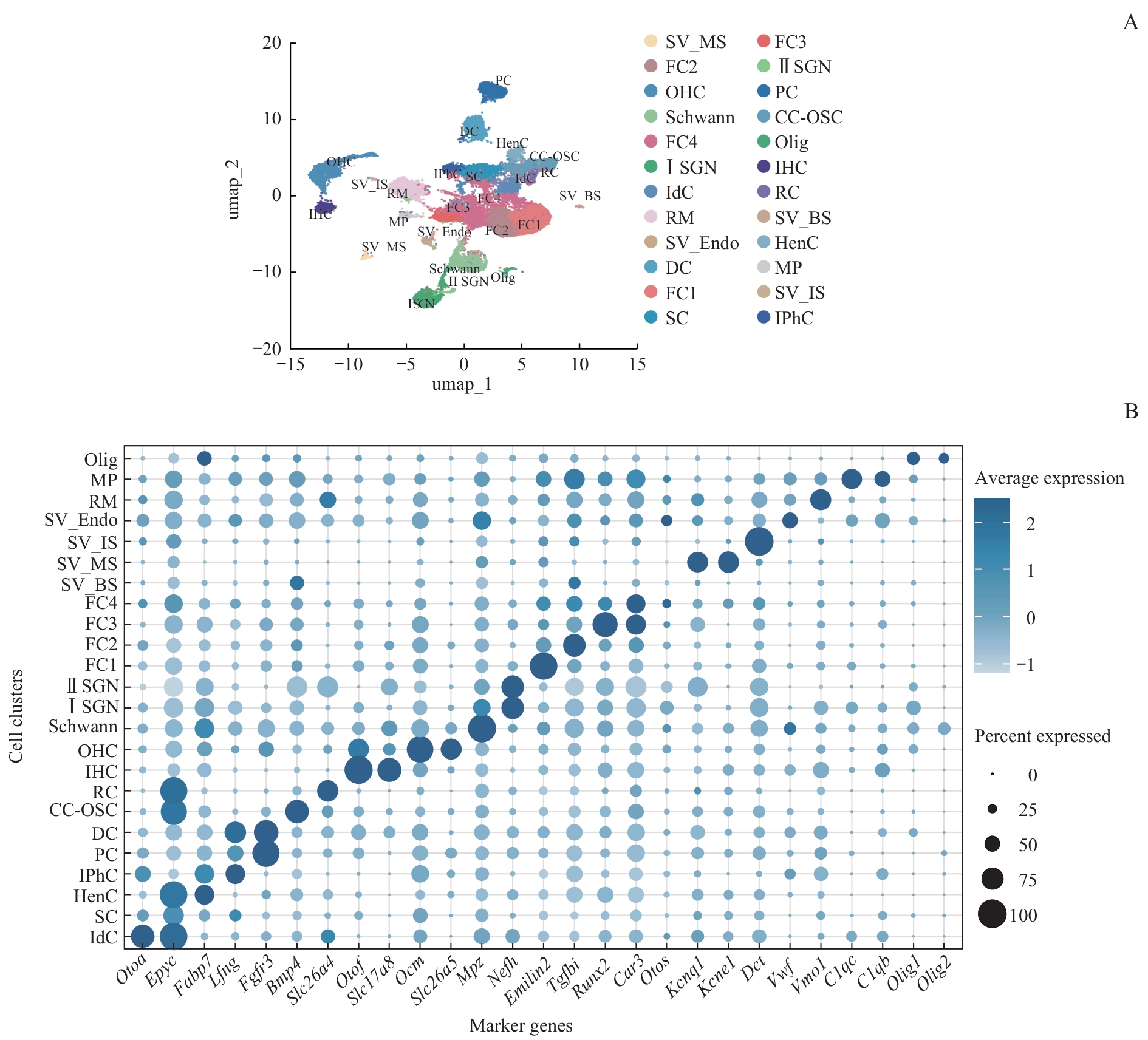
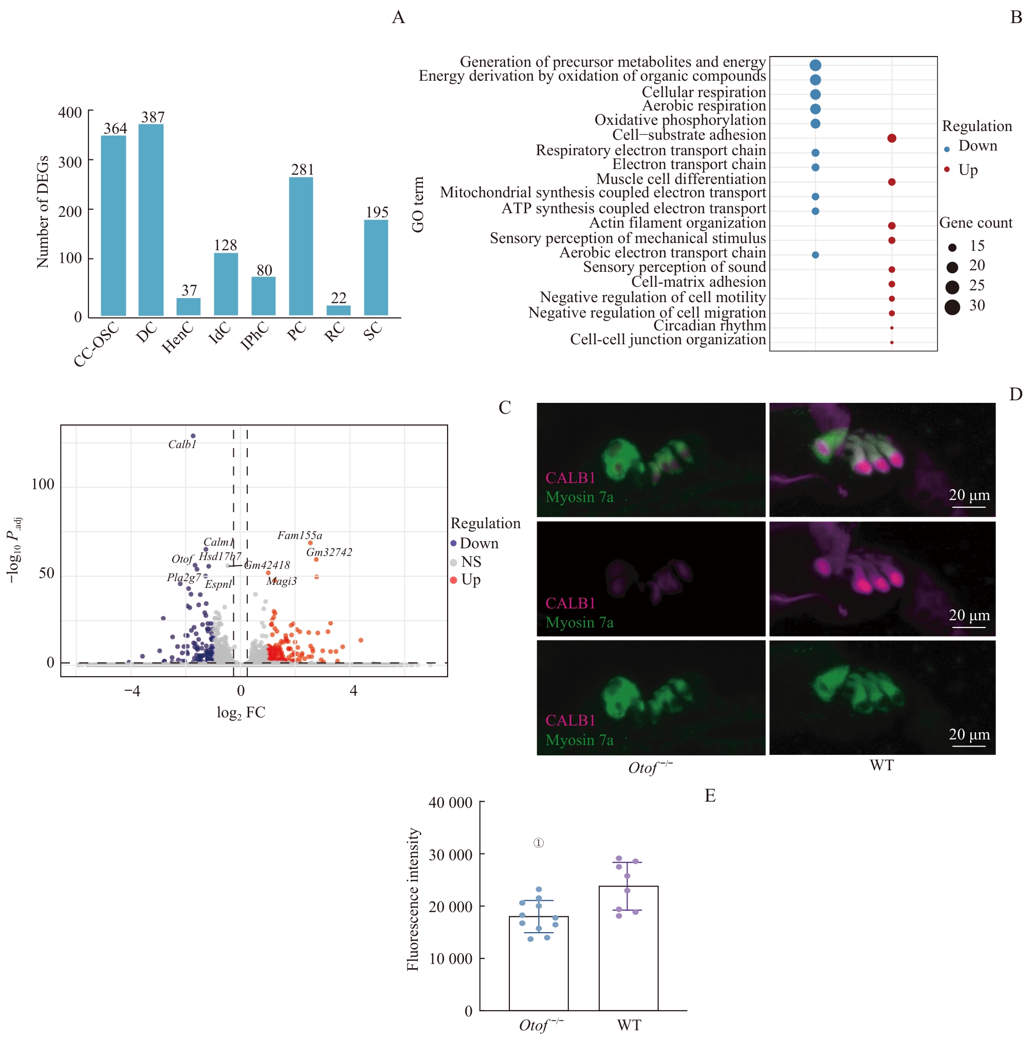
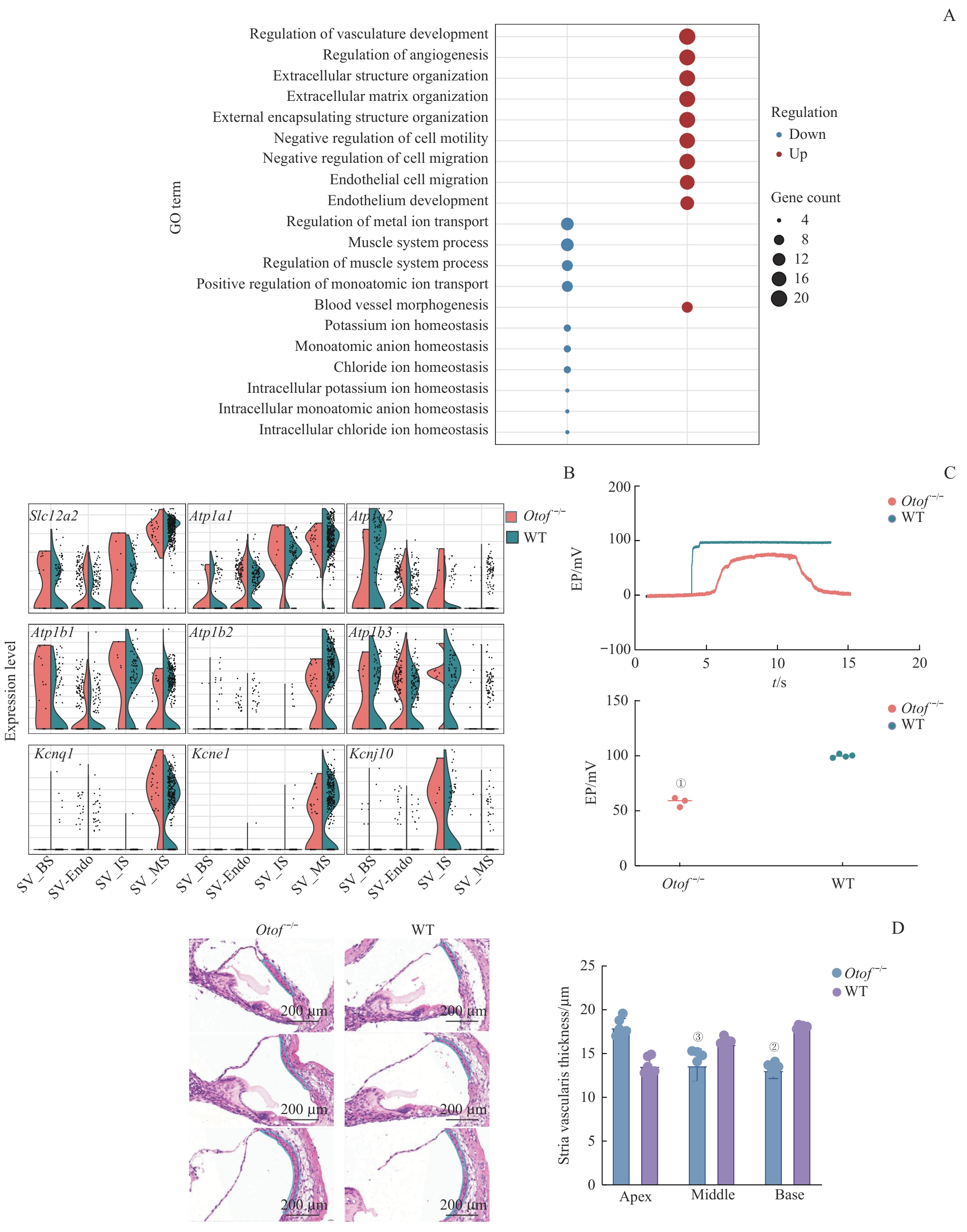
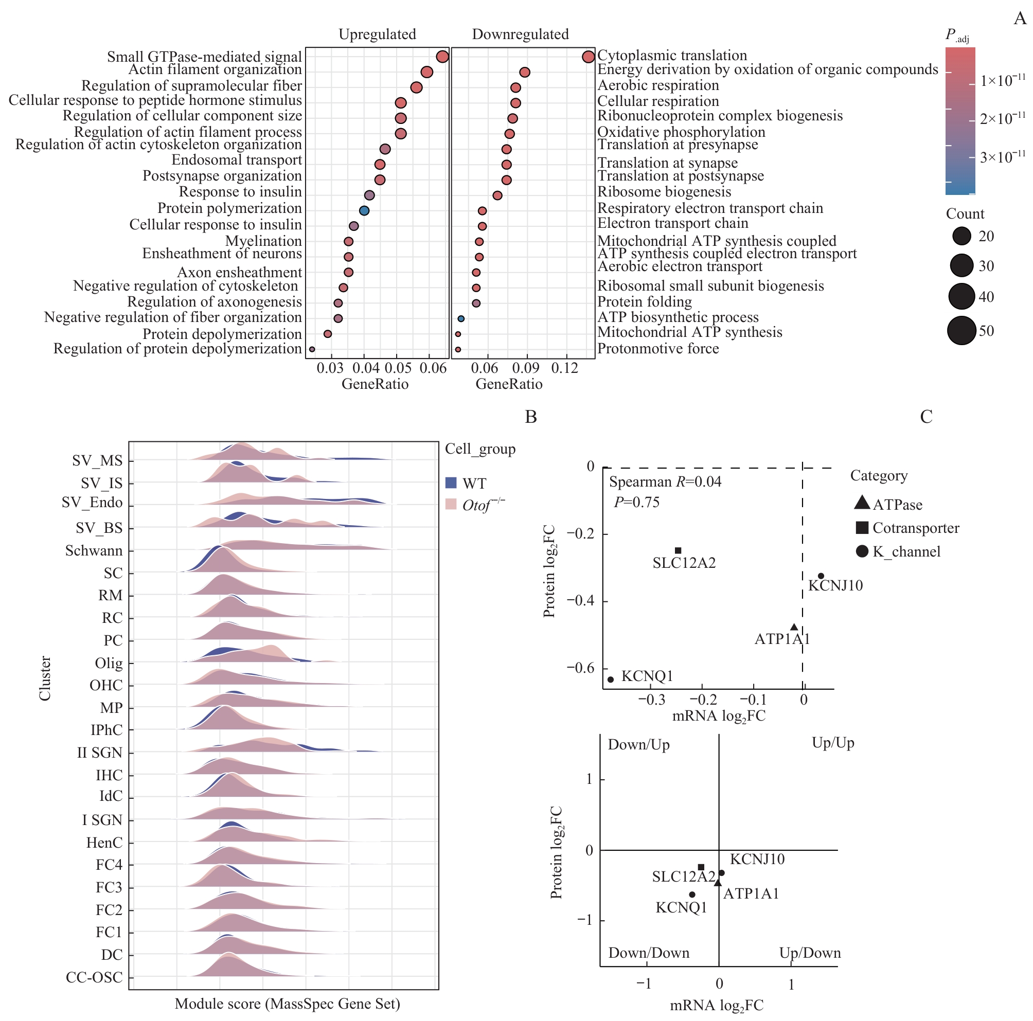
目的·观察Otoferlin基因敲除对小鼠耳蜗不同细胞亚群转录特征及细胞稳态的影响,探讨其在遗传性耳聋发生中的作用。方法·选取Otoferlin基因敲除小鼠(Otof-/-组)及同背景野生型小鼠(WT组),采用单细胞转录组测序技术获取耳蜗细胞转录谱,结合特异性标记基因鉴定耳蜗细胞亚群。筛选差异表达基因(differentially expressed genes,DEGs),并进行基因本体(Gene Ontology,GO)功能富集分析。整合免疫荧光染色、内淋巴电位(endolymphatic potential,EP)检测及血管纹形态学测量进行功能验证,并结合蛋白质组学与基因集模块打分分析技术揭示Otoferlin基因敲除对耳蜗细胞的影响。结果·通过单细胞测序分析,成功鉴定出 24 个耳蜗细胞亚群。2组细胞亚群整体构成比例的差异无统计学意义,但DEGs则呈现明显的细胞类型特异性分布特征。支持细胞中的外沟细胞、柱细胞及Deiters细胞的DEGs富集程度较高,主要参与细胞外基质组织、耳蜗发育及核糖体代谢等生物学过程;外毛细胞DEGs则显著富集于线粒体氧化呼吸链相关通路;血管纹细胞中,与离子稳态维持及上皮发育密切相关的基因表达模式发生显著改变,关键离子转运基因Slc12a2和Kcnj10表达异常。蛋白质组学分析进一步显示Otoferlin基因敲除小鼠Corti器的内质网-高尔基体囊泡运输及线粒体翻译等通路相较于WT小鼠存在明显功能异常,且该异常在血管纹细胞及部分神经胶质细胞中表现尤为突出。结论·Otoferlin 基因敲除耳聋小鼠耳蜗细胞亚群的整体组成未发生显著变化,但血管纹离子转运功能发生障碍,内淋巴电位显著降低,耳聋小鼠表现出血管纹等非神经元细胞功能异常。

关键词: 单细胞转录组测序, Otoferlin基因, 遗传性耳聋, 耳蜗细胞

Abstract:

Objective ·To investigate the effects of Otoferlin gene-knockout on transcriptional profiles and cellular homeostasis of different cochlear cell subsets in mice, and to explore its role in the pathogenesis of hereditary deafness. Methods ·Otoferlin-knockout mice (Otof-/- group) and wild-type mice (WT group) with the same genetic background were used. Single-cell RNA sequencing was performed to obtain transcriptional profiles of cochlear cells. Cell-specific marker genes were used to identify cochlear cell subsets. Differentially expressed genes (DEGs) were screened, followed by Gene Ontology (GO) functional enrichment analysis. Immunofluorescence staining, endolymphatic potential (EP) measurement, and morphological assessment of the stria vascularis were applied for functional validation. Proteomics and gene set module scoring were further used to reveal the impacts of Otoferlin knockout on cochlear cells. Results ·A total of 24 cochlear cell subsets were identified via single-cell sequencing. No significant difference was observed in the overall proportions of cell subsets between the two groups, while DEGs exhibited obvious cell-type-specific distribution patterns. DEGs were most enriched in supporting cells, including outer sulcus cells, pillar cells, and Deiters' cells, and were mainly involved in extracellular matrix organization, cochlear development, and ribosome metabolism. DEGs in outer hair cells were significantly enriched in mitochondrial oxidative respiratory chain pathways. In stria vascularis cells, the expression patterns of genes related to ion homeostasis and epithelial development were markedly altered, with abnormal expression of the key ion transport genes Slc12a2 and Kcnj10. Proteomic analysis further revealed marked abnormalities in endoplasmic reticulum-Golgi vesicular transport and mitochondrial translation pathways in the organ of Corti of Otoferlin-knockout mice compared with WT mice, and such abnormalities were most prominent in stria vascularis cells and some glial cells. Conclusion ·Although the overall composition of cochlear cell subsets did not change significantly in deaf mice with Otoferlin-knockout, ion transport dysfunction in the stria vascularis and a marked reduction in EP can be detected. Deaf mice present functional abnormalities in non-neuronal cells, including those of the stria vascularis.

Key words: single-cell RNA sequencing, Otoferlin gene, hereditary hearing loss, cochlear cell

中图分类号: