上海交通大学学报(医学版) ›› 2022, Vol. 42 ›› Issue (7): 875-884.doi: 10.3969/j.issn.1674-8115.2022.07.005
王雨心( ), 孙瑞琪, 刘坚华(), 何伟娜()
收稿日期:2022-03-19
接受日期:2022-06-25
出版日期:2022-07-28
发布日期:2022-09-04
通讯作者:
何伟娜,电子信箱:hewn0319@sjtu.edu.cn。作者简介:王雨心(1995—),女,硕士生;电子信箱yuxinwang@sjtu.edu.cn。
基金资助:
WANG Yuxin(), SUN Ruiqi, LIU Jianhua(), HE Weina()
Received:2022-03-19
Accepted:2022-06-25
Online:2022-07-28
Published:2022-09-04
Contact:
HE Weina, E-mail: hewn0319@sjtu.edu.cn.Supported by:摘要:
目的·合成水溶性pH响应性花菁类近红外荧光探针,评估其光学性能,并将其应用于模拟肿瘤微环境的在体成像。方法·应用两步经典的化学反应,合成水溶性pH响应性近红外荧光探针R2S,同步合成脂溶性探针R2Z作为对照。应用磁共振氢谱、质谱及高效液相色谱验证探针的化学结构及纯度。利用紫外-可见、光致发光光谱学测试评价探针的pH响应性、响应可逆性、光稳定性及结构稳定性。利用细胞荧光共定位成像评价探针的细胞膜通透性,使用HCT-116细胞、HeLa细胞进行细胞毒性实验,使用BALB/c健康雌性小鼠进行小动物在体成像实验,评估探针的安全性。通过在小鼠背部左侧和右侧分别皮下注射pH 6.50和pH 7.40的PBS溶液模拟肿瘤微酸性环境和正常的细胞环境,随后注射探针R2S进行在体成像实验,比较R2S在pH 6.50侧和pH 7.40侧的荧光强度。结果·成功合成了水溶性pH响应性近红外荧光探针R2S及其脂溶性类似物R2Z。随着溶液酸性逐渐增强(从pH 11.10到pH 3.47),R2S的最大吸收波长由642 nm红移至774 nm,最大发射波长亦由794 nm红移至808 nm。探针R2S的酸解离常数(pKa)值约为6.88。与脂溶性探针R2Z相比,R2S在保留了较好细胞膜通透性的前提下,展现出更大的斯托克斯位移、更好的响应可逆性、更高的稳定性。R2Z在12.5 μmol/L浓度下,细胞相对存活率低于80%;而R2S在100 μmol/L浓度下,仍未表现出明显的细胞生长抑制现象,表明其具有更高的生物安全性。R2S在模拟肿瘤微酸性环境的低pH组织和正常pH组织中的成像具有明显的区分度。结论·水溶性pH响应性花菁类近红外荧光探针R2S对pH变化响应灵敏、稳定,最佳响应范围与肿瘤所处微环境的pH相符,在肿瘤成像领域展现出了较强的在体成像应用潜力。
中图分类号:
王雨心, 孙瑞琪, 刘坚华, 何伟娜. 开发用于肿瘤微环境成像的pH敏感荧光探针[J]. 上海交通大学学报(医学版), 2022, 42(7): 875-884.
WANG Yuxin, SUN Ruiqi, LIU Jianhua, HE Weina. Development of pH-responsive fluorescent probe for tumor microenvironment imaging[J]. Journal of Shanghai Jiao Tong University (Medical Science), 2022, 42(7): 875-884.
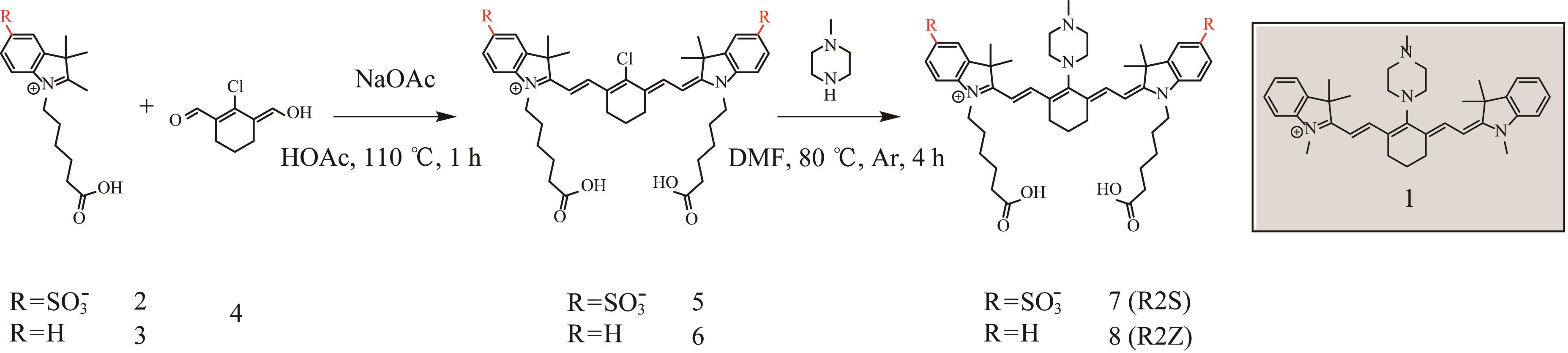
图1 探针R2S与R2Z的合成路线Note: NaOAc—sodium acetate; HOAc—acetic acid; DMF—N, N-dimethylformamide; Ar—argon.
Fig 1 Synthetic routes of the target probes R2S and R2Z
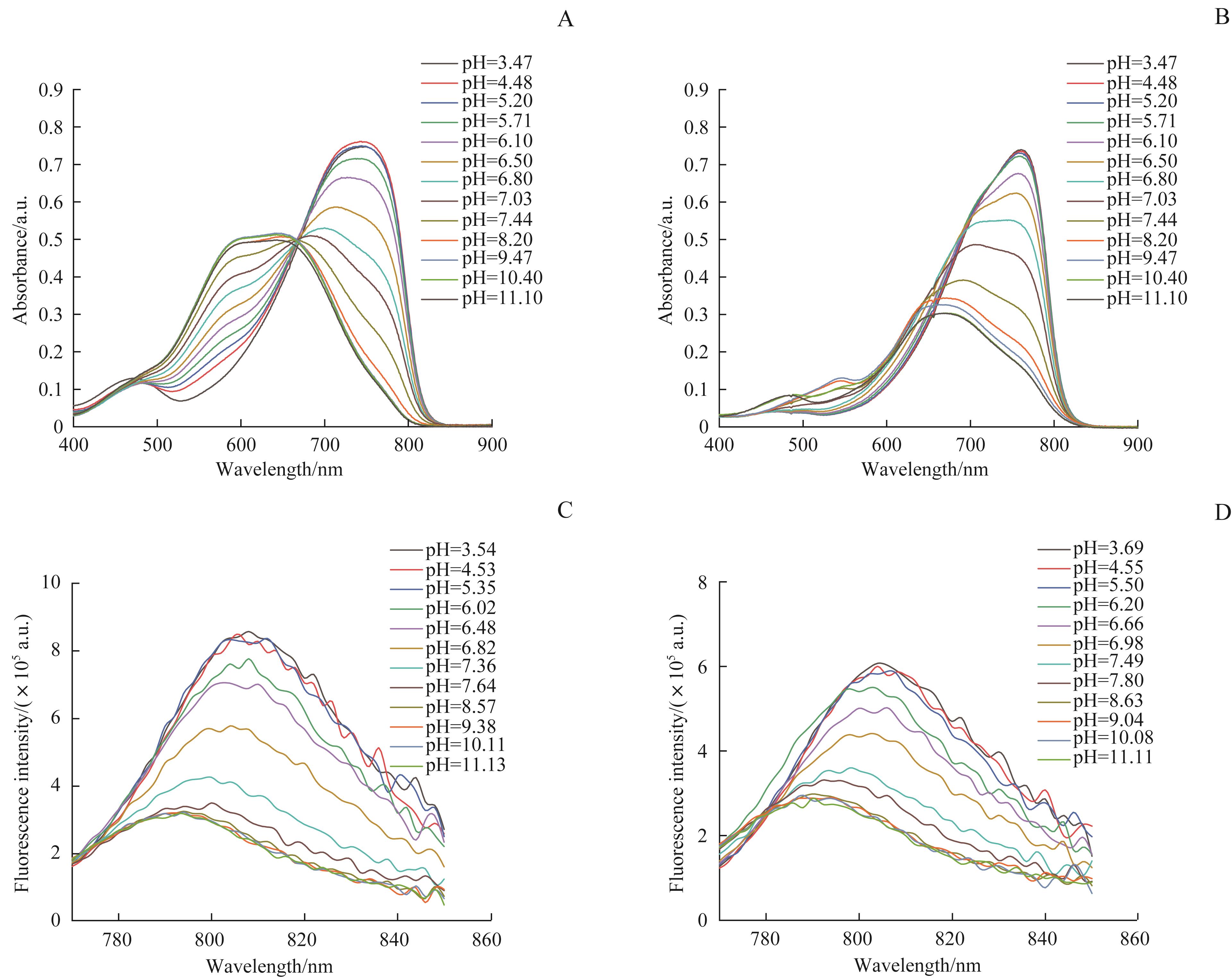
图2 R2S与R2Z在不同pH值下的吸收光谱及荧光光谱Note: A/B. Absorption spectra of 20 μmol/L R2S (A) and 20 μmol/L R2Z (B). C/D. Fluorescence spectra of 1 μmol/L R2S (C) and 20 μmol/L R2Z (D). All samples were measured in PBS at pH 3.47, 4.48, 5.20, 5.71, 6.10, 6.50, 6.80, 7.03, 7.44, 8.20, 9.47, 10.40 and 11.10 (A/B) or pH 3.54, 4.53, 5.35, 6.02, 6.48, 6.82, 7.36, 7.64, 8.57, 9.58, 10.11 and 11.13 (C) or pH 3.69, 4.55, 5.50, 6.20, 6.66, 6.98, 7.49, 7.80, 8.63, 9.04, 10.08 and 11.11 (D).
Fig 2 Absorption spectra and fluorescence spectra of the probes R2S and R2Z at different pH values
| Parameter | R2S/nm | R2Z/nm |
|---|---|---|
| λAbsmax (protonated) | 744 | 760 |
| λAbsmax (deprotonated) | 642 | 662 |
| λEmmax (protonated) | 808 | 804 |
| λEmmax (deprotonated) | 794 | 790 |
| Stokes shift (protonated) | 64 | 44 |
表1 R2S与R2Z的光学参数
Tab 1 Spectroscopic parameters of probes R2S and R2Z
| Parameter | R2S/nm | R2Z/nm |
|---|---|---|
| λAbsmax (protonated) | 744 | 760 |
| λAbsmax (deprotonated) | 642 | 662 |
| λEmmax (protonated) | 808 | 804 |
| λEmmax (deprotonated) | 794 | 790 |
| Stokes shift (protonated) | 64 | 44 |
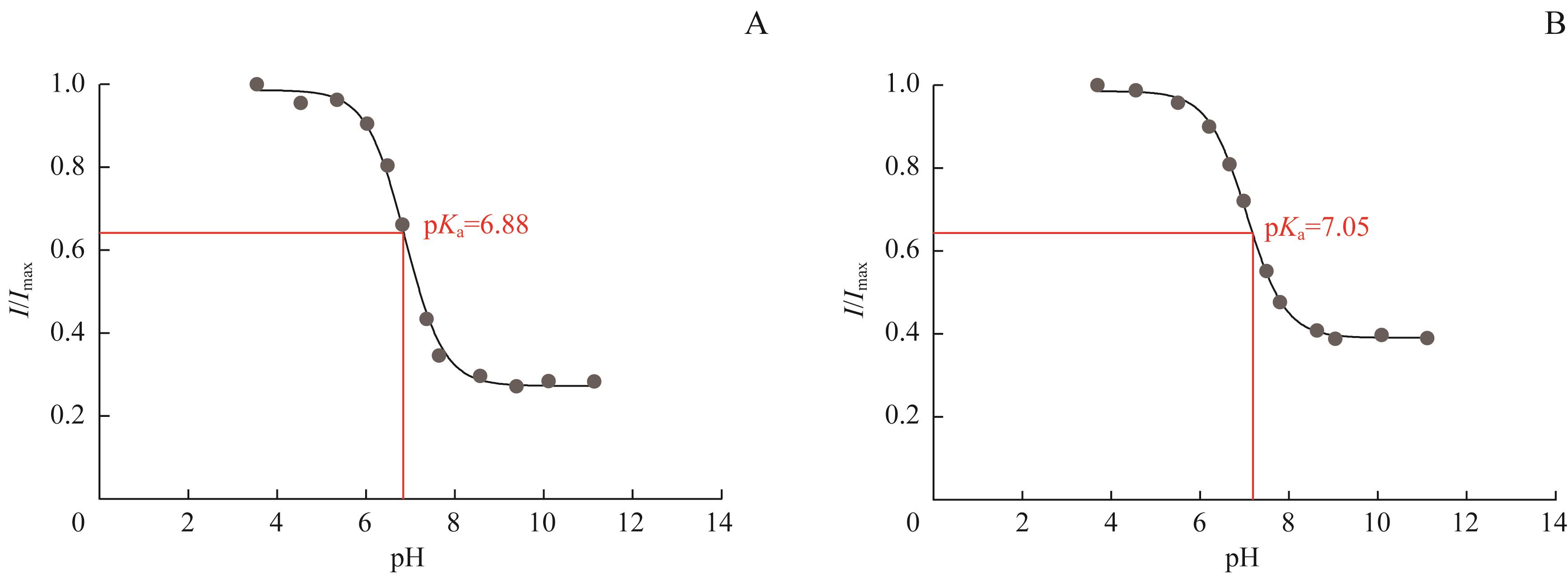
图3 R2S与R2Z在不同pH值下的相对荧光强度Note: A. Changes in fluorescence ratio I/Imax of R2S at 750 nm excitation, 808 nm emission in PBS at the pH values of Fig. 2C. B. Changes in fluorescence ratio I/Imax of R2Z at 750 nm excitation, 804 nm emission in PBS at the pH values of Fig. 2D.
Fig 3 Relative fluorescence intensity of probes R2S and R2Z upon different pH values
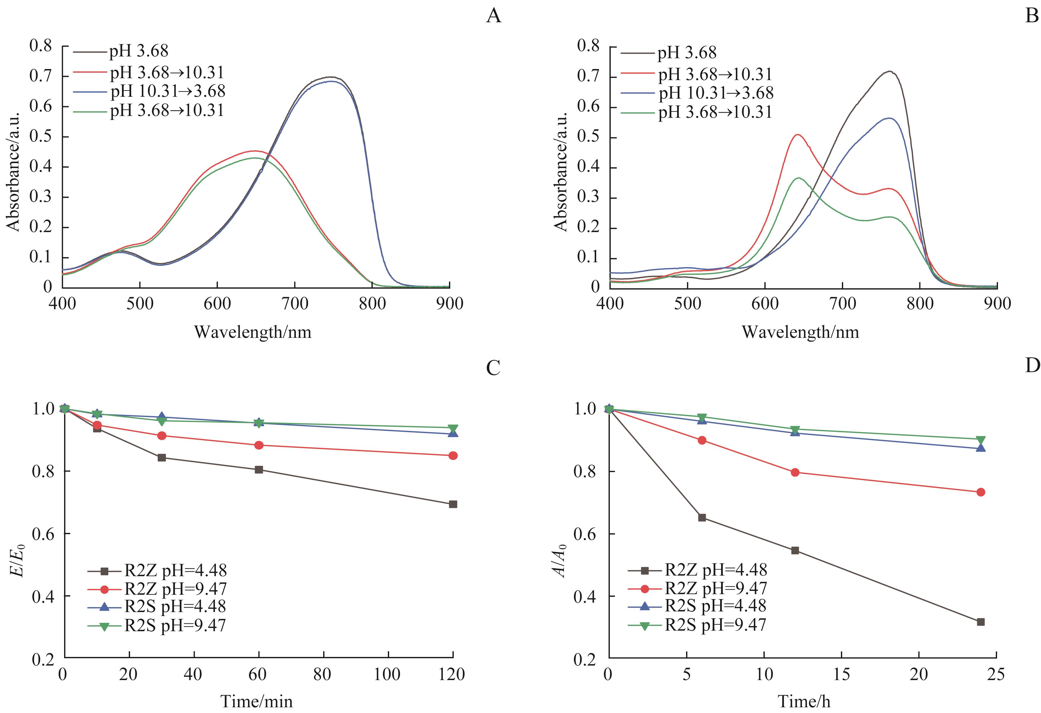
图4 R2S与R2Z的pH响应可逆性和稳定性Note: A/B. Changes of absorption spectra of probes R2S (A) and R2Z (B) toward cyclically adjusted pH between 3.68 and 10.31. C. Time-dependent changes of fluorescence intensity at 805 nm of probes R2S and R2Z under UV light irradiation with a wavelength of 254 nm. D. Time-dependent changes of absorbance at 750 nm of probes R2S and R2Z under 37 ℃. E—emission intensity of probes at different time; A—absorbance of probes at different time.
Fig 4 Reversible changes toward cyclically adjusted pH and stabilities of probes R2S and R2Z
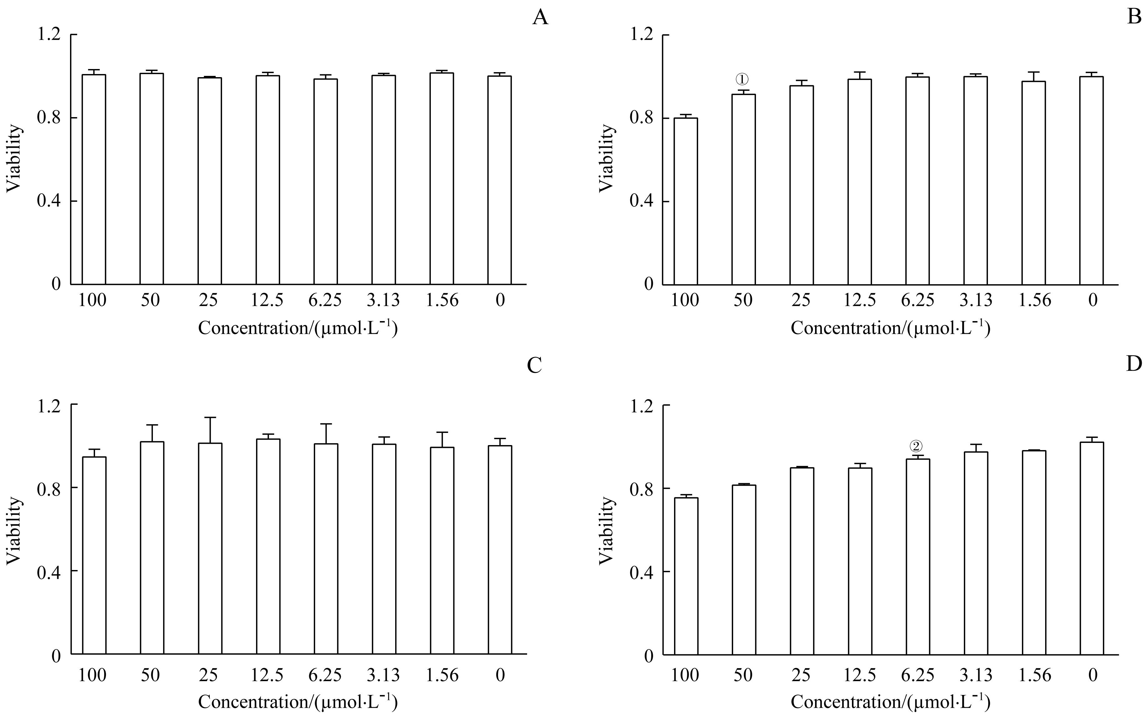
图5 R2S与R2Z对HCT-116细胞和HeLa细胞的毒性Note: A/B. Cytotoxicity of HCT-116 cells after being treated with R2S (A) and R2Z (B) at different concentrations for 24 h. C/D. Cytotoxicity of HeLa cells after being treated with R2S (C) and R2Z (D) at different concentrations for 24 h. ①P=0.007, ②P=0.009, compared with the control group.
Fig 5 Cytotoxicity evaluation in HCT-116 and HeLa cells of probes R2S and R2Z
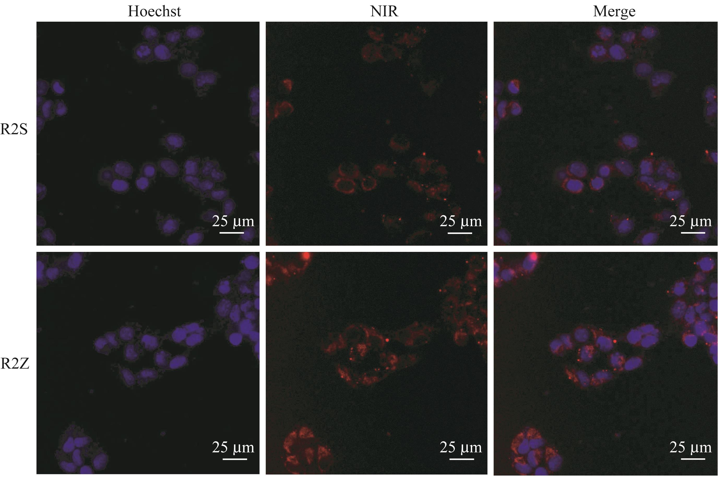
图6 R2S与R2Z的HeLa细胞成像Note: Confocal fluorescence imaging for 10 μg/mL Hoechst (blue channel, 430?470 nm) and 10 μmol/L R2S or R2Z (red channel, 700?800 nm) in HeLa cells.
Fig 6 Imaging of probes R2S and R2Z in HeLa cells
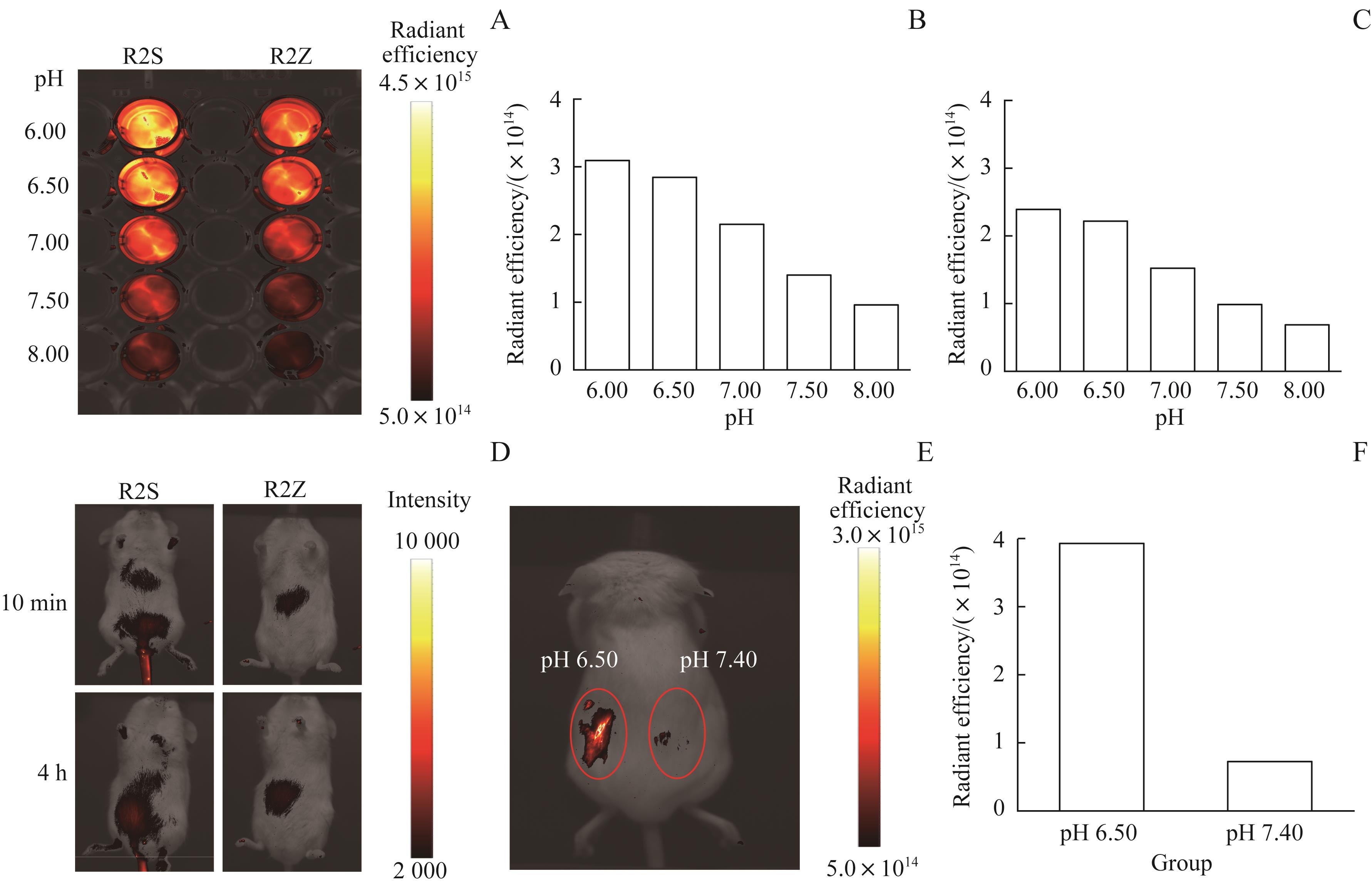
图7 R2S与R2Z的体外与体内NIR成像Note: A. NIR images of 20 μmol/L R2S and R2Z in PBS at pH 6.00, 6.50, 7.00, 7.50 and 8.00. B/C. Radiant efficiency of R2S (B) and R2Z (C) at above pH values. D. In vivo NIR images of mice after intravenous injection of R2S and R2Z (10 μmol/L, 200 μL). E. In vivo NIR images of a mouse labeled with R2S. PBS solution (50 μL, pH 6.50) and PBS solution (50 μL, pH 7.40) were injected into two adjacent spots on the back of the mouse. Then, R2S (5 μL, 50 μmol/L) was subcutaneously injected into these two different locations. F. Radiant efficiency of the spot at pH 6.50 and spot at pH 7.40.
Fig 7 In vitro and in vivo NIR imaging of probes R2S and R2Z
| 1 | CAO M M, LI H, SUN D Q, et al. Cancer burden of major cancers in China: a need for sustainable actions[J]. Cancer Commun (Lond), 2020, 40(5): 205-210. |
| 2 | SIEGEL R L, MILLER K D, JEMAL A. Cancer statistics, 2020[J]. CA Cancer J Clin, 2020, 70(1): 7-30. |
| 3 | PERUMAL V, SIVAKUMAR P M, ZARRABI A, et al. Near infra-red polymeric nanoparticle based optical imaging in cancer diagnosis[J]. J Photochem Photobiol B, 2019, 199: 111630. |
| 4 | LUO S L, ZHANG E L, SU Y P, et al. A review of NIR dyes in cancer targeting and imaging[J]. Biomaterials, 2011, 32(29): 7127-7138. |
| 5 | LI H D, KIM D, YAO Q C, et al. Activity-based NIR enzyme fluorescent probes for the diagnosis of tumors and image-guided surgery[J]. Angew Chem Int Ed Engl, 2021, 60(32): 17268-17289. |
| 6 | ZHOU Z X, LU Z R. Molecular imaging of the tumor microenvironment[J]. Adv Drug Deliv Rev, 2017, 113: 24-48. |
| 7 | WEBB B A, CHIMENTI M, JACOBSON M P, et al. Dysregulated pH: a perfect storm for cancer progression[J]. Nat Rev Cancer, 2011, 11(9): 671-677. |
| 8 | BOEDTKJER E, BUNCH L, PEDERSEN S F. Physiology, pharmacology and pathophysiology of the pH regulatory transport proteins NHE1 and NBCn1: similarities, differences, and implications for cancer therapy[J]. Curr Pharm Des, 2012, 18(10): 1345-1371. |
| 9 | ANDERSEN A P, MOREIRA J M A, PEDERSEN S F. Interactions of ion transporters and channels with cancer cell metabolism and the tumour microenvironment[J]. Philos Trans R Soc Lond B Biol Sci, 2014, 369(1638): 20130098. |
| 10 | BOEDTKJER E, PEDERSEN S F. The acidic tumor microenvironment as a driver of cancer[J]. Annu Rev Physiol, 2020, 82: 103-126. |
| 11 | FRY D R, BOBBITT D R. Investigation of dynamically modified optical-fiber sensors for pH sensing at the extremes of the pH scale[J]. Microchem J, 2001, 69(2): 25-33. |
| 12 | TANG X, ZHU Z, WANG Y, et al. A dual site controlled probe for fluorescent monitoring of intracellular pH and colorimetric monitoring of Cu2+[J]. Sens Actuat B Chem, 2018, 270: 35-44. |
| 13 | ZHU H, FAN J L, DU J J, et al. Fluorescent probes for sensing and imaging within specific cellular organelles[J]. Acc Chem Res, 2016, 49(10): 2115-2126. |
| 14 | LEE M H, PARK N, YI C, et al. Mitochondria-immobilized pH-sensitive off-on fluorescent probe[J]. J Am Chem Soc, 2014, 136(40): 14136-14142. |
| 15 | JIN D, WANG B W, HOU Y Q, et al. Novel near-infrared pH-sensitive cyanine-based fluorescent probes for intracellular pH monitoring[J]. Dyes Pigments, 2019, 170: 107612. |
| 16 | ZHENG X H, XING D, ZHOU F F, et al. Indocyanine green-containing nanostructure as near infrared dual-functional targeting probes for optical imaging and photothermal therapy[J]. Mol Pharm, 2011, 8(2): 447-456. |
| 17 | MA Y, TONG S, BAO G, et al. Indocyanine green loaded SPIO nanoparticles with phospholipid-PEG coating for dual-modal imaging and photothermal therapy[J]. Biomaterials, 2013, 34(31): 7706-7714. |
| 18 | TANG B, LIU X, XU K, et al. A dual near-infrared pH fluorescent probe and its application in imaging of HepG2 cells[J]. Chem Commun (Camb), 2007(36): 3726-3728. |
| 19 | ZHAO X, WEI R S, CHEN L G, et al. Glucosamine modified near-infrared cyanine as a sensitive colorimetric fluorescent chemosensor for aspartic and glutamic acid and its applications[J]. New J Chem, 2014, 38(10): 4791-4798. |
| 20 | MU H Y, MIKI K, HARADA H, et al. pH-activatable cyanine dyes for selective tumor imaging using near-infrared fluorescence and photoacoustic modalities[J]. ACS Sens, 2021, 6(1): 123-129. |
| 21 | ZHANG Z, ACHILEFU S. Design, synthesis and evaluation of near-infrared fluorescent pH indicators in a physiologically relevant range[J]. Chem Commun (Camb), 2005(47): 5887-5889. |
| 22 | YU H, SUN M T, ZHANG K, et al. A reversible near-infrared pH probes for optical measurements of pH in complete water system and living cells[J]. Sens Actuat B Chem, 2015, 219: 294-300. |
| 23 | TANG B, YU F B, LI P, et al. A near-infrared neutral pH fluorescent probe for monitoring minor pH changes: imaging in living HepG2 and HL-7702 cells[J]. J Am Chem Soc, 2009, 131(8): 3016-3023. |
| 24 | MYOCHIN T, KIYOSE K, HANAOKA K, et al. Rational design of ratiometric near-infrared fluorescent pH probes with various pKa values, based on aminocyanine[J]. J Am Chem Soc, 2011, 133(10): 3401-3409. |
| 25 | SHI Y X, MENG X C, YANG H R, et al. Lysosome-specific sensing and imaging of pH variations in vitro and in vivo utilizing a near-infrared boron complex[J]. J Mater Chem B, 2019, 7(22): 3569-3575. |
| 26 | VAN DER WAL S, KUIL J, VALENTIJN A R P M, et al. Synthesis and systematic evaluation of symmetric sulfonated centrally CC bonded cyanine near-infrared dyes for protein labelling[J]. Dyes Pigments, 2016, 132: 7-19. |
| 27 | MASKASKY J E. Molecular orientation of individual J aggregates on gelatin-grown AgBr tabular microcrystals[J]. Langmuir, 1991, 7(2): 407-421. |
| 28 | 王伟, 姚祖光. 含N-烷基吲哚环方酸菁染料的合成及性能[J]. 感光科学与光化学, 1997, 15(4): 321-326. |
| WANG W, YAO Z G. Synthesis and properties of squarylium cyanine dyes containing N-alkyl indole[J]. Photogaraphic Sci Photoch, 1997, 15(4): 321-326. | |
| 29 | CAO J, WAN S N, TIAN J M, et al. Fast clearing RGD-based near-infrared fluorescent probes for in vivo tumor diagnosis[J]. Contrast Media Mol Imaging, 2012, 7(4): 390-402. |
| 30 | JEONG C, NOH I, REJINOLD N S, et al. Self-assembled supramolecular bilayer nanoparticles composed of near-infrared dye as a theranostic nanoplatform to encapsulate hydrophilic drugs effectively[J]. ACS Biomater Sci Eng, 2020, 6(1): 474-484. |
| [1] | 缪可言, 贾浩, 杨溪. 花生四烯酸代谢通路重塑肿瘤免疫微环境的研究进展[J]. 上海交通大学学报(医学版), 2026, 46(4): 521-528. |
| [2] | 章玥, 陈青建. 基于质谱流式细胞技术的乳腺癌免疫微环境预后特征分析[J]. 上海交通大学学报(医学版), 2026, 46(1): 34-42. |
| [3] | 汤开然, 冯成领, 韩邦旻. 基于单细胞测序与转录组测序构建M2巨噬细胞基因相关的前列腺癌预后模型[J]. 上海交通大学学报(医学版), 2025, 45(5): 549-561. |
| [4] | 张烨晟, 杨易静, 黄依雯, 施珑玙, 王曼媛, 陈思思. 肿瘤微环境免疫细胞调节肿瘤细胞耐药性的研究进展[J]. 上海交通大学学报(医学版), 2024, 44(7): 830-838. |
| [5] | 刘林楠, 冯莉, 王龙, 刘嘉寅, 范志松. 多能蛋白聚糖在恶性肿瘤中的表达及生物学作用的研究进展[J]. 上海交通大学学报(医学版), 2024, 44(4): 525-530. |
| [6] | 李钰, 姜艺凡, 童荣亮, 陈迪宇, 吴健. FOXM1与肿瘤代谢关系的研究进展[J]. 上海交通大学学报(医学版), 2024, 44(10): 1323-1329. |
| [7] | 魏兰懿, 薛晓川, 陈君君, 杨全军, 王梦月, 韩永龙. 骨肉瘤免疫微环境中肿瘤相关巨噬细胞及其靶向治疗的研究进展[J]. 上海交通大学学报(医学版), 2023, 43(5): 624-630. |
| [8] | 吴瑞芳, 冯明, 孟健. 脂肪酸结合蛋白4在肥胖相关肿瘤中的作用综述[J]. 上海交通大学学报(医学版), 2023, 43(10): 1311-1316. |
| [9] | 马芳芳, 秦洁洁, 任灵杰, 唐笑梅, 刘佳, 施敏敏, 蒋玲曦. 基于水凝胶微球建立胰腺癌原代细胞的3D培养模型[J]. 上海交通大学学报(医学版), 2023, 43(1): 79-87. |
| [10] | 林家俞, 秦洁洁, 蒋玲曦. 肿瘤微环境中免疫细胞的代谢研究进展[J]. 上海交通大学学报(医学版), 2022, 42(8): 1122-1130. |
| [11] | 李静威, 王俐文, 蒋玲曦, 詹茜, 陈皓, 沈柏用. 胰腺癌免疫抑制性肿瘤微环境研究综述[J]. 上海交通大学学报(医学版), 2021, 41(8): 1103-1108. |
| [12] | 那迪娜·帕尔哈提null, 严妍, 车千纪, 罗菁, 刘鑫男, 李斌. 嵌合抗原受体T细胞疗法在胶质母细胞瘤中的应用与展望[J]. 上海交通大学学报(医学版), 2021, 41(7): 982-986. |
| [13] | 顾琦晟, 张米粒, 曹灿, 李继坤. 基于TCGA数据库分析胃癌可变剪接与肿瘤免疫的关系[J]. 上海交通大学学报(医学版), 2021, 41(4): 448-458. |
| [14] | 刘梦珂, 纪濛濛, 程林, 黄金艳, 孙晓建, 赵维莅, 王黎. 黄芩苷抗肿瘤作用机制的研究进展[J]. 上海交通大学学报(医学版), 2021, 41(2): 246-250. |
| [15] | 赵伟光,刘志宏. 肿瘤相关成纤维细胞调控肿瘤免疫炎症微环境的研究进展[J]. 上海交通大学学报(医学版), 2020, 40(9): 1288-1293. |
| 阅读次数 | ||||||
|
全文 |
|
|||||
|
摘要 |
|
|||||