Journal of Shanghai Jiao Tong University (Medical Science) ›› 2025, Vol. 45 ›› Issue (4): 434-442.doi: 10.3969/j.issn.1674-8115.2025.04.005
• Clinical research • Previous Articles Next Articles
WANG Sirui1,2, KONG Gai2, LI Hui2, QIAN Zhenying2, CUI Huiru2, TANG Yingying1,2( )
)
Received:2024-12-10
Accepted:2024-12-31
Online:2025-04-28
Published:2025-04-28
Contact:
TANG Yingying
E-mail:yytang@smhc.org.cn
Supported by:CLC Number:
WANG Sirui, KONG Gai, LI Hui, QIAN Zhenying, CUI Huiru, TANG Yingying. Impact of transcranial magnetic stimulation therapy on the volumes of amygdala and hippocampal subfields in patients with major depressive disorder[J]. Journal of Shanghai Jiao Tong University (Medical Science), 2025, 45(4): 434-442.
Add to citation manager EndNote|Ris|BibTeX
URL: https://xuebao.shsmu.edu.cn/EN/10.3969/j.issn.1674-8115.2025.04.005
| Clinical scale | Before TMS | After TMS | T value | P value |
|---|---|---|---|---|
| HAMD-24 | 27 (12, 40) | 10 (0, 27) | 3.0 | <0.001 |
| MADRS | 27 (16, 36) | 7 (0, 26) | 1.5 | <0.001 |
| HAMA | 19 (9, 39) | 10 (0, 27) | 28.0 | <0.001 |
Tab 1 Clinical assessment scores of depressive and anxiety symptoms before and after TMS treatment
| Clinical scale | Before TMS | After TMS | T value | P value |
|---|---|---|---|---|
| HAMD-24 | 27 (12, 40) | 10 (0, 27) | 3.0 | <0.001 |
| MADRS | 27 (16, 36) | 7 (0, 26) | 1.5 | <0.001 |
| HAMA | 19 (9, 39) | 10 (0, 27) | 28.0 | <0.001 |
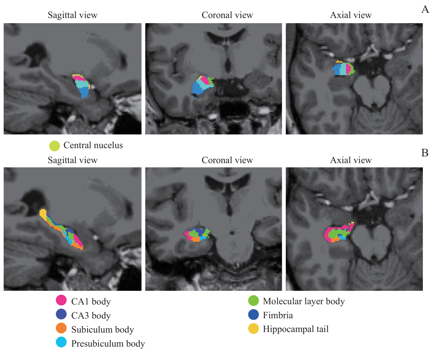
Fig 1 Illustrations of the segmentation of the right amygdala and hippocampal subfields
| Subfield | Time | Laterality | Time × Laterality | ||||||
|---|---|---|---|---|---|---|---|---|---|
| F value | P value | η2 | F value | P value | η2 | F value | P value | η2 | |
| La | 0.897 | 0.347 | 0.015 | 18.151 | <0.001 | 0.242 | 5.533 | 0.022 | 0.088 |
| Ba | 0.352 | 0.556 | 0.006 | 59.562 | <0.001 | 0.511 | 3.425 | 0.069 | 0.057 |
| AB | 0.075 | 0.785 | 0.001 | 108.26 | <0.001 | 0.655 | 2.702 | 0.106 | 0.045 |
| AAA | 3.796 | 0.056 | 0.062 | 89.549 | <0.001 | 0.611 | 1.767 | 0.189 | 0.030 |
| Ce | 4.204 | 0.045 | 0.069 | 44.796 | <0.001 | 0.440 | 2.440 | 0.124 | 0.041 |
| Me | 2.307 | 0.134 | 0.390 | 29.001 | <0.001 | 0.337 | 3.143 | 0.082 | 0.052 |
| Co | 0.242 | 0.625 | 0.004 | 102.340 | <0.001 | 0.642 | 0.914 | 0.343 | 0.016 |
| CAT | 0.410 | 0.525 | 0.007 | 59.627 | <0.001 | 0.511 | 0.281 | 0.598 | 0.005 |
| PL | 0.446 | 0.507 | 0.008 | 6.263 | 0.015 | 0.099 | 0.030 | 0.863 | 0.001 |
| Whole amygdala | 0.170 | 0.681 | 0.003 | 83.962 | <0.001 | 0.596 | 4.825 | 0.032 | 0.078 |
Tab 2 Two-way ANOVA of bilateral amygdala subfield volumes before and after TMS treatment
| Subfield | Time | Laterality | Time × Laterality | ||||||
|---|---|---|---|---|---|---|---|---|---|
| F value | P value | η2 | F value | P value | η2 | F value | P value | η2 | |
| La | 0.897 | 0.347 | 0.015 | 18.151 | <0.001 | 0.242 | 5.533 | 0.022 | 0.088 |
| Ba | 0.352 | 0.556 | 0.006 | 59.562 | <0.001 | 0.511 | 3.425 | 0.069 | 0.057 |
| AB | 0.075 | 0.785 | 0.001 | 108.26 | <0.001 | 0.655 | 2.702 | 0.106 | 0.045 |
| AAA | 3.796 | 0.056 | 0.062 | 89.549 | <0.001 | 0.611 | 1.767 | 0.189 | 0.030 |
| Ce | 4.204 | 0.045 | 0.069 | 44.796 | <0.001 | 0.440 | 2.440 | 0.124 | 0.041 |
| Me | 2.307 | 0.134 | 0.390 | 29.001 | <0.001 | 0.337 | 3.143 | 0.082 | 0.052 |
| Co | 0.242 | 0.625 | 0.004 | 102.340 | <0.001 | 0.642 | 0.914 | 0.343 | 0.016 |
| CAT | 0.410 | 0.525 | 0.007 | 59.627 | <0.001 | 0.511 | 0.281 | 0.598 | 0.005 |
| PL | 0.446 | 0.507 | 0.008 | 6.263 | 0.015 | 0.099 | 0.030 | 0.863 | 0.001 |
| Whole amygdala | 0.170 | 0.681 | 0.003 | 83.962 | <0.001 | 0.596 | 4.825 | 0.032 | 0.078 |
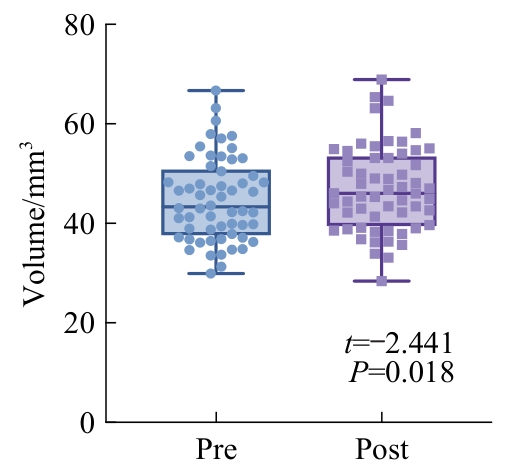
Fig 2 Change in the volumes of the right amygdala central nucleus after TMS treatment
| Subfield | Time | Laterality | Time × Laterality | ||||||
|---|---|---|---|---|---|---|---|---|---|
| F value | P value | η2 | F value | P value | η2 | F value | P value | η2 | |
| CA1-body | 3.910 | 0.053 | 0.064 | 67.705 | <0.001 | 0.543 | 4.673 | 0.035 | 0.067 |
| CA1-head | 0.148 | 0.702 | 0.003 | 86.886 | <0.001 | 0.604 | 1.217 | 0.275 | 0.021 |
| CA3-body | 12.769 | <0.001 | 0.183 | 82.281 | <0.001 | 0.591 | 7.449 | 0.008 | 0.166 |
| CA3-head | 0.261 | 0.611 | 0.005 | 21.449 | <0.001 | 0.273 | 1.851 | 0.179 | 0.031 |
| CA4-body | 6.293 | 0.015 | 0.099 | 1.844 | 0.180 | 0.031 | 0.002 | 0.968 | <0.001 |
| CA4-head | 0.026 | 0.872 | 0.0005 | 20.487 | <0.001 | 0.264 | 3.107 | 0.083 | 0.052 |
| Fimbria | 15.254 | <0.001 | 0.211 | 7.824 | 0.007 | 0.121 | 1.837 | 0.181 | 0.031 |
| Fissure | 0.874 | 0.354 | 0.015 | 1.970 | 0.166 | 0.033 | 0.247 | 0.621 | 0.004 |
| GC-DG-body | 6.720 | 0.012 | 0.105 | 1.966 | 0.166 | 0.033 | 0.125 | 0.725 | 0.002 |
| GC-DG-head | 0.001 | 0.973 | <0.001 | 25.200 | <0.001 | 0.307 | 2.654 | 0.109 | 0.044 |
| HATA | 0.370 | 0.546 | 0.006 | 6.398 | 0.014 | 0.101 | 0.684 | 0.412 | 0.012 |
| Hippocampus tail | 26.765 | <0.001 | 0.320 | 65.029 | <0.001 | 0.533 | 0.267 | 0.608 | 0.005 |
| ML-body | 20.469 | <0.001 | 0.264 | 44.278 | <0.001 | 0.437 | 7.873 | 0.007 | 0.121 |
| ML-head | 0.007 | 0.936 | <0.001 | 34.724 | <0.001 | 0.379 | 2.142 | 0.149 | 0.036 |
| Parasubiculum | 0.064 | 0.801 | 0.001 | 13.377 | <0.001 | 0.190 | 0.279 | 0.599 | 0.005 |
| Presubiculum-body | 6.277 | 0.015 | 0.099 | 18.069 | <0.001 | 0.241 | 1.514 | 0.224 | 0.026 |
| Presubiculum-head | 3.017 | 0.083 | 0.052 | 15.834 | <0.001 | 0.217 | 0.637 | 0.428 | 0.011 |
| SUB-body | 17.011 | <0.001 | 0.230 | 0.010 | 0.921 | <0.001 | 1.019 | 0.317 | 0.018 |
| SUB-head | 0.278 | 0.600 | 0.005 | 1.118 | 0.295 | 0.019 | 0.166 | 0.685 | 0.003 |
| Whole hippocampus | 10.728 | 0.002 | 0.158 | 65.042 | <0.001 | 0.533 | 2.941 | 0.092 | 0.049 |
| Whole hippocampus-body | 14.750 | <0.001 | 0.206 | 12.169 | <0.001 | 0.176 | 3.168 | 0.080 | 0.053 |
| Whole hippocampus-head | 0.034 | 0.855 | 0.001 | 33.231 | <0.001 | 0.368 | 1.459 | 0.232 | 0.025 |
Tab 3 Two-way ANOVA of bilateral hippocampal subfield volumes before and after TMS treatment
| Subfield | Time | Laterality | Time × Laterality | ||||||
|---|---|---|---|---|---|---|---|---|---|
| F value | P value | η2 | F value | P value | η2 | F value | P value | η2 | |
| CA1-body | 3.910 | 0.053 | 0.064 | 67.705 | <0.001 | 0.543 | 4.673 | 0.035 | 0.067 |
| CA1-head | 0.148 | 0.702 | 0.003 | 86.886 | <0.001 | 0.604 | 1.217 | 0.275 | 0.021 |
| CA3-body | 12.769 | <0.001 | 0.183 | 82.281 | <0.001 | 0.591 | 7.449 | 0.008 | 0.166 |
| CA3-head | 0.261 | 0.611 | 0.005 | 21.449 | <0.001 | 0.273 | 1.851 | 0.179 | 0.031 |
| CA4-body | 6.293 | 0.015 | 0.099 | 1.844 | 0.180 | 0.031 | 0.002 | 0.968 | <0.001 |
| CA4-head | 0.026 | 0.872 | 0.0005 | 20.487 | <0.001 | 0.264 | 3.107 | 0.083 | 0.052 |
| Fimbria | 15.254 | <0.001 | 0.211 | 7.824 | 0.007 | 0.121 | 1.837 | 0.181 | 0.031 |
| Fissure | 0.874 | 0.354 | 0.015 | 1.970 | 0.166 | 0.033 | 0.247 | 0.621 | 0.004 |
| GC-DG-body | 6.720 | 0.012 | 0.105 | 1.966 | 0.166 | 0.033 | 0.125 | 0.725 | 0.002 |
| GC-DG-head | 0.001 | 0.973 | <0.001 | 25.200 | <0.001 | 0.307 | 2.654 | 0.109 | 0.044 |
| HATA | 0.370 | 0.546 | 0.006 | 6.398 | 0.014 | 0.101 | 0.684 | 0.412 | 0.012 |
| Hippocampus tail | 26.765 | <0.001 | 0.320 | 65.029 | <0.001 | 0.533 | 0.267 | 0.608 | 0.005 |
| ML-body | 20.469 | <0.001 | 0.264 | 44.278 | <0.001 | 0.437 | 7.873 | 0.007 | 0.121 |
| ML-head | 0.007 | 0.936 | <0.001 | 34.724 | <0.001 | 0.379 | 2.142 | 0.149 | 0.036 |
| Parasubiculum | 0.064 | 0.801 | 0.001 | 13.377 | <0.001 | 0.190 | 0.279 | 0.599 | 0.005 |
| Presubiculum-body | 6.277 | 0.015 | 0.099 | 18.069 | <0.001 | 0.241 | 1.514 | 0.224 | 0.026 |
| Presubiculum-head | 3.017 | 0.083 | 0.052 | 15.834 | <0.001 | 0.217 | 0.637 | 0.428 | 0.011 |
| SUB-body | 17.011 | <0.001 | 0.230 | 0.010 | 0.921 | <0.001 | 1.019 | 0.317 | 0.018 |
| SUB-head | 0.278 | 0.600 | 0.005 | 1.118 | 0.295 | 0.019 | 0.166 | 0.685 | 0.003 |
| Whole hippocampus | 10.728 | 0.002 | 0.158 | 65.042 | <0.001 | 0.533 | 2.941 | 0.092 | 0.049 |
| Whole hippocampus-body | 14.750 | <0.001 | 0.206 | 12.169 | <0.001 | 0.176 | 3.168 | 0.080 | 0.053 |
| Whole hippocampus-head | 0.034 | 0.855 | 0.001 | 33.231 | <0.001 | 0.368 | 1.459 | 0.232 | 0.025 |
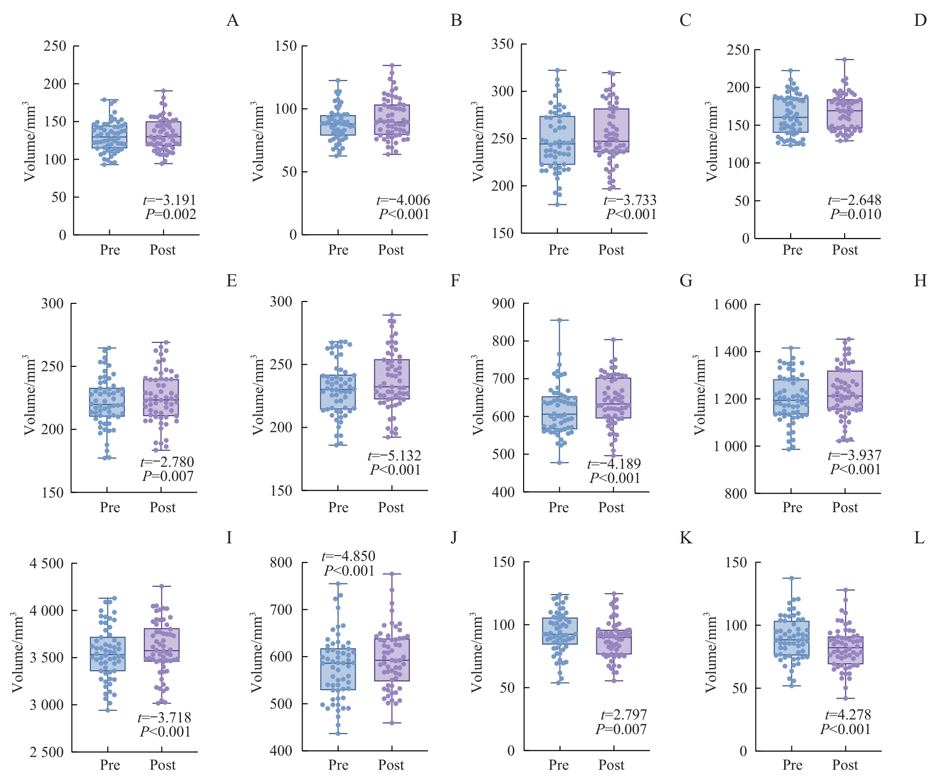
Fig 3 Comparison of volumes of bilateral hippocampal subfields before and after TMS treatment
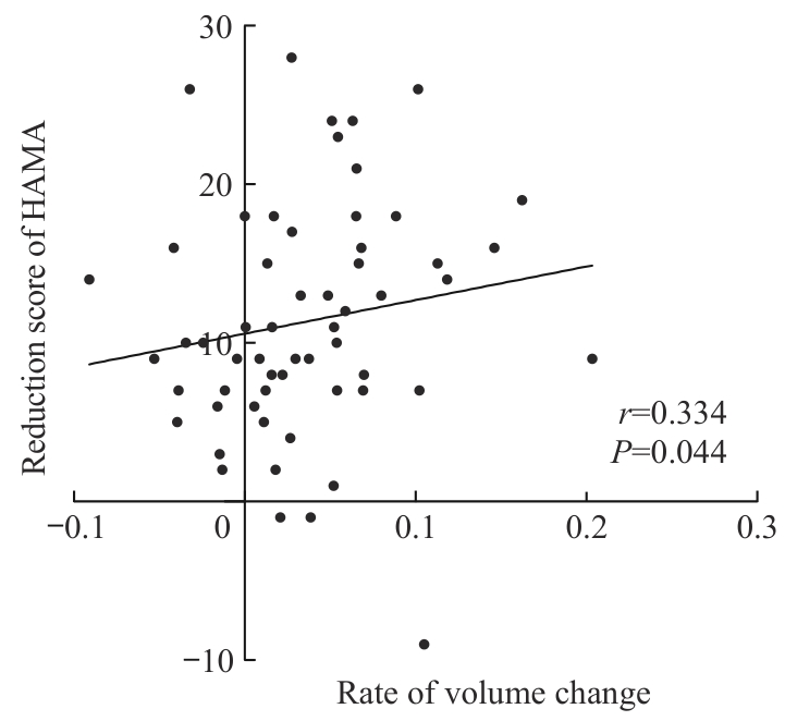
Fig 4 Correlation between the rate of left hippocampal tail volume change and reduction scores in anxiety symptoms in TMS responders
| 1 | LU J, XU X F, HUANG Y Q, et al. Prevalence of depressive disorders and treatment in China: a cross-sectional epidemiological study[J]. Lancet Psychiatry, 2021, 8(11): 981-990. |
| 2 | JACOB Y, MORRIS L S, VERMA G, et al. Altered hippocampus and amygdala subregion connectome hierarchy in major depressive disorder[J]. Transl Psychiatry, 2022, 12(1): 209. |
| 3 | PLOSKI J E, VAIDYA V A. The neurocircuitry of posttraumatic stress disorder and major depression: insights into overlapping and distinct circuit dysfunction-a tribute to ron duman[J]. Biol Psychiatry, 2021, 90(2): 109-117. |
| 4 | MCKINNON M C, YUCEL K, NAZAROV A, et al. A meta-analysis examining clinical predictors of hippocampal volume in patients with major depressive disorder[J]. J Psychiatry Neurosci, 2009, 34(1): 41-54. |
| 5 | SCHMAAL L, POZZI E, HO T C, et al. ENIGMA MDD: seven years of global neuroimaging studies of major depression through worldwide data sharing[J]. Transl Psychiatry, 2020, 10(1): 172. |
| 6 | FRODL T, MEISENZAHL E, ZETZSCHE T, et al. Enlargement of the amygdala in patients with a first episode of major depression[J]. Biol Psychiatry, 2002, 51(9): 708-714. |
| 7 | KRONENBERG G, TEBARTZ VAN ELST L, REGEN F, et al. Reduced amygdala volume in newly admitted psychiatric in-patients with unipolar major depression[J]. J Psychiatr Res, 2009, 43(13): 1112-1117. |
| 8 | KIM H, HAN K M, CHOI K W, et al. Volumetric alterations in subregions of the amygdala in adults with major depressive disorder[J]. J Affect Disord, 2021, 295: 108-115. |
| 9 | CONG E Z, LI Q F, CHEN H Y, et al. Association between the volume of subregions of the amygdala and major depression with suicidal thoughts and anxiety in a Chinese cohort[J]. J Affect Disord, 2022, 312: 39-45. |
| 10 | 储召松. 抑郁症患者杏仁核亚区结构磁共振成像研究[D]. 昆明: 昆明医科大学, 2021. |
| CHU Z S. Structural magnetic resonance imaging study of amygdala subregions in patients with major depressive disorder[D]. Kunming: Kunming Medical University, 2021. | |
| 11 | RODDY D, KELLY J R, FARRELL C, et al. Amygdala substructure volumes in major depressive disorder[J]. Neuroimage Clin, 2021, 31: 102781. |
| 12 | ZAVALIANGOS-PETROPULU A, MCCLINTOCK S M, JOSHI S H, et al. Hippocampal subfield volumes in treatment resistant depression and serial ketamine treatment[J]. Front Psychiatry, 2023, 14: 1227879. |
| 13 | WU C C, JIA L L, MU Q L, et al. Altered hippocampal subfield volumes in major depressive disorder with and without anhedonia[J]. BMC Psychiatry, 2023, 23(1): 540. |
| 14 | HAN K M, KIM A, KANG W, et al. Hippocampal subfield volumes in major depressive disorder and bipolar disorder[J]. Eur Psychiatry, 2019, 57: 70-77. |
| 15 | RODDY D W, FARRELL C, DOOLIN K, et al. The hippocampus in depression: more than the sum of its parts? Advanced hippocampal substructure segmentation in depression[J]. Biol Psychiatry, 2019, 85(6): 487-497. |
| 16 | LEFAUCHEUR J P, ALEMAN A, BAEKEN C, et al. Evidence-based guidelines on the therapeutic use of repetitive transcranial magnetic stimulation (rTMS): an update (2014‒2018)[J]. Clin Neurophysiol, 2020, 131(2): 474-528. |
| 17 | CASH R F H, ZALESKY A. Personalized and circuit-based transcranial magnetic stimulation: evidence, controversies, and opportunities[J]. Biol Psychiatry, 2024, 95(6): 510-522. |
| 18 | SYDNOR V J, CIESLAK M, DUPRAT R, et al. Cortical-subcortical structural connections support transcranial magnetic stimulation engagement of the amygdala[J]. Sci Adv, 2022, 8(25): eabn5803. |
| 19 | DALHUISEN I, ACKERMANS E, MARTENS L, et al. Longitudinal effects of rTMS on neuroplasticity in chronic treatment-resistant depression[J]. Eur Arch Psychiatry Clin Neurosci, 2021, 271(1): 39-47. |
| 20 | HAYASAKA S, NAKAMURA M, NODA Y, et al. Lateralized hippocampal volume increase following high-frequency left prefrontal repetitive transcranial magnetic stimulation in patients with major depression[J]. Psychiatry Clin Neurosci, 2017, 71(11): 747-758. |
| 21 | SEEWOO B J, RODGER J, DEMITRACK M A, et al. Neurostructural differences in adolescents with treatment-resistant depression and treatment effects of transcranial magnetic stimulation[J]. Int J Neuropsychopharmacol, 2022, 25(8): 619-630. |
| 22 | SAYGIN Z M, KLIEMANN D, IGLESIAS J E, et al. High-resolution magnetic resonance imaging reveals nuclei of the human amygdala: manual segmentation to automatic atlas[J]. Neuroimage, 2017, 155: 370-382. |
| 23 | IGLESIAS J E, AUGUSTINACK J C, NGUYEN K, et al. A computational atlas of the hippocampal formation using ex vivo, ultra-high resolution MRI: application to adaptive segmentation of in vivo MRI[J]. Neuroimage, 2015, 115: 117-137. |
| 24 | KALIN N H, SHELTON S E, DAVIDSON R J. The role of the central nucleus of the amygdala in mediating fear and anxiety in the primate[J]. J Neurosci, 2004, 24(24): 5506-5515. |
| 25 | AMARAL D G, PRICE J L, PITKANEN A, et al. The amygdala: neurobiological aspects of emotion, memory, and mental dysfunction[M]. New York: Wiley-Liss, 1992: 1-66. |
| 26 | GILPIN N W, HERMAN M A, ROBERTO M. The central amygdala as an integrative hub for anxiety and alcohol use disorders[J]. Biol Psychiatry, 2015, 77(10): 859-869. |
| 27 | TAKAMIYA A, CHUNG J K, LIANG K C, et al. Effect of electroconvulsive therapy on hippocampal and amygdala volumes: systematic review and meta-analysis[J]. Br J Psychiatry, 2018, 212(1): 19-26. |
| 28 | ENNEKING V, LEEHR E J, DANNLOWSKI U, et al. Brain structural effects of treatments for depression and biomarkers of response: a systematic review of neuroimaging studies[J]. Psychol Med, 2020, 50(2): 187-209. |
| 29 | TATU L, VUILLIER F. Structure and vascularization of the human hippocampus[J]. Front Neurol Neurosci, 2014, 34: 18-25. |
| 30 | TESEN H, WATANABE K, OKAMOTO N, et al. Volume of amygdala subregions and clinical manifestations in patients with first-episode, drug-naïve major depression[J]. Front Hum Neurosci, 2022, 15: 780884. |
| 31 | BROWN S G, RUTLAND J W, VERMA G, et al. Structural MRI at 7T reveals amygdala nuclei and hippocampal subfield volumetric association with Major Depressive Disorder symptom severity[J]. Sci Rep, 2019, 9(1): 10166. |
| 32 | CAO B, PASSOS I C, MWANGI B, et al. Hippocampal subfield volumes in mood disorders[J]. Mol Psychiatry, 2017, 22(9): 1352-1358. |
| 33 | XU Y W, CUI D, ZHAO Y, et al. Volumetric alterations of the hippocampal subfields in major depressive disorder with and without suicidal ideation[J]. Behav Brain Res, 2023: 114733. |
| 34 | FRODL T, SCHAUB A, BANAC S, et al. Reduced hippocampal volume correlates with executive dysfunctioning in major depression[J]. J Psychiatry Neurosci, 2006, 31(5): 316-323. |
| 35 | ZHANG Y X, LIU X, YANG Y, et al. Revealing complexity: segmentation of hippocampal subfields in adolescents with major depressive disorder reveals specific links to cognitive dysfunctions[J]. Eur Psychiatry, 2024, 68(1): e5. |
| 36 | GBYL K, VIDEBECH P. Electroconvulsive therapy increases brain volume in major depression: a systematic review and meta-analysis[J]. Acta Psychiatr Scand, 2018, 138(3): 180-195. |
| 37 | MALLER J J, JEFF DASKALAKIS Z, FITZGERALD P B. Hippocampal volumetrics in depression: the importance of the posterior tail[J]. Hippocampus, 2007, 17(11): 1023-1027. |
| 38 | CHU Z S, YUAN L J, LIAN K, et al. Reduced gray matter volume of the hippocampal tail in melancholic depression: evidence from an MRI study[J]. BMC Psychiatry, 2024, 24(1): 183. |
| 39 | MALLER J J, BROADHOUSE K, RUSH A J, et al. Increased hippocampal tail volume predicts depression status and remission to anti-depressant medications in major depression[J]. Mol Psychiatry, 2018, 23(8): 1737-1744. |
| 40 | NOGOVITSYN N, MULLER M, SOUZA R, et al. Hippocampal tail volume as a predictive biomarker of antidepressant treatment outcomes in patients with major depressive disorder: a CAN-BIND report[J]. Neuropsychopharmacology, 2020, 45(2): 283-291. |
| [1] | WANG Haihong, YUAN Chenxin, GAN Hong, JIANG Haifeng, ZHAO Yan, DU Jiang, ZHANG Yi. A randomized controlled study on the effect of intermittent theta burst stimulation on craving, mood, and cognitive function in alcohol-dependent patients during the withdrawal period [J]. Journal of Shanghai Jiao Tong University (Medical Science), 2025, 45(3): 349-356. |
| [2] | CHEN Shentse, CHEN Yiming, WANG Fan, ZHANG Mengke, YANG Weichieh, LÜ Dongbin, HONG Wu. Advances in dietary interventions for the treatment of depression-related symptoms [J]. Journal of Shanghai Jiao Tong University (Medical Science), 2024, 44(8): 1050-1055. |
| [3] | HU Canfang, ZHONG Chuanyu, CAO Li. Research progress of neuromodulation in the treatment of Parkinson's disease [J]. Journal of Shanghai Jiao Tong University (Medical Science), 2024, 44(2): 258-263. |
| [4] | LI Siyuan, HE Shen, LI Huafang. Recent advance in autophagy-related pathways and key biomarkers in major depressive disorder [J]. Journal of Shanghai Jiao Tong University (Medical Science), 2023, 43(10): 1324-1331. |
| [5] | Xin LI, Qing FAN. Application progress of machine learning in the study of facial features of patients with depression [J]. JOURNAL OF SHANGHAI JIAOTONG UNIVERSITY (MEDICAL SCIENCE), 2022, 42(1): 124-129. |
| [6] | Yi WANG, Cheng CHENG, Hong-yan SHEN, Hong-yan GAO, Yue-ning DAI, Zheng-hui YI. Meta-analysis of efficacy of transcranial magnetic stimulation for the treatment of cognitive function and behavioral and psychological symptoms of dementia in patients with Alzheimer′s disease [J]. JOURNAL OF SHANGHAI JIAOTONG UNIVERSITY (MEDICAL SCIENCE), 2021, 41(7): 931-941. |
| [7] | Rui-jie GENG, Lin YAO, Xin-xin HUANG, Shun-ying YU, Cheng-mei YUAN, Wu HONG, Qin-yu LÜ, Qing-zhong WANG, Zheng-hui YI, Yi-ru FANG. Identification of differentially expressed gene modules in major depressive disorder based on weighted gene co-expression network analysis [J]. JOURNAL OF SHANGHAI JIAOTONG UNIVERSITY (MEDICAL SCIENCE), 2021, 41(6): 724-731. |
| [8] | Lin-jie SHEN, Yu-xin HUANG, Yong WANG, Hua JIN. Review of non-invasive brain stimulation for the treatment of somatic symptoms in major depressive disorder [J]. JOURNAL OF SHANGHAI JIAOTONG UNIVERSITY (MEDICAL SCIENCE), 2021, 41(11): 1535-1539. |
| [9] | LIN Qing-qing, QIU Jian-yin. Social and interpersonal dysfunction of patients with major depressive disorder [J]. JOURNAL OF SHANGHAI JIAOTONG UNIVERSITY (MEDICAL SCIENCE), 2020, 40(7): 985-989. |
| [10] | HUANG Yan, ZHANG Xian-gao, LIN Heng, CHENG Shu-ying. Meta-analysis of effect and safety of repetitive transcranial magnetic stimulation on patients with schizophrenia [J]. , 2020, 40(1): 81-. |
| [11] | ZHAO Dong-mei1, 2*, DING Lei1*, LIU Hong-ye1, 3, PENG Dai-hui1. Advanced research of kynurenine pathway mechanism in suicide of major depressive disorder [J]. , 2019, 39(7): 805-. |
| [12] | XU Xiao-min, CHEN Tian-zhen, JIANG Hai-feng. Modulating neural circuits in substance addiction with repetitive transcranial magnetic stimulation [J]. , 2019, 39(6): 655-. |
| [13] | ZHU Jun-juan,ZHANG Tian-hong,ZHANG Ling,XU Li-hua,WEI Yan-yan,TANG Ying-ying,WANG Ji-jun. Effect of accelerated repetitive transcranial magnetic stimulation on suicide ideation in depressive patients [J]. , 2019, 39(5): 534-. |
| [14] | ZHU Li-na1, ZHANG Qiong2, CAI Jun1, ZHANG Wei-bo1, ZHU Hong-xia1. Effect of cerebellar vermal repetitive transcranial magnetic stimulation with theta burst stimulation paradigm on cognitive functions of patients with schizophrenia [J]. , 2019, 39(3): 282-. |
| [15] | LI Pu-yu, WANG Zhen. Application of repetitive transcranial magnetic stimulation in the treatment of obsessive-compulsive disorder [J]. , 2019, 39(12): 1477-. |
| Viewed | ||||||
|
Full text |
|
|||||
|
Abstract |
|
|||||