Journal of Shanghai Jiao Tong University (Medical Science) ›› 2025, Vol. 45 ›› Issue (9): 1202-1213.doi: 10.3969/j.issn.1674-8115.2025.09.012
• Clinical research • Previous Articles Next Articles
LI Siyu1, CHEN Ya1, HU Wentao2, DAI Yongming3, WU Yingwei1( )
)
Received:2025-03-06
Accepted:2025-03-25
Online:2025-09-28
Published:2025-09-30
Contact:
WU Yingwei
E-mail:wuyw0103@hotmail.com
Supported by:CLC Number:
LI Siyu, CHEN Ya, HU Wentao, DAI Yongming, WU Yingwei. Using diffusion-relaxation correlation spectroscopic imaging to assess the heterogeneity of head and neck tumors and identify occult lymph node metastasis[J]. Journal of Shanghai Jiao Tong University (Medical Science), 2025, 45(9): 1202-1213.
Add to citation manager EndNote|Ris|BibTeX
URL: https://xuebao.shsmu.edu.cn/EN/10.3969/j.issn.1674-8115.2025.09.012
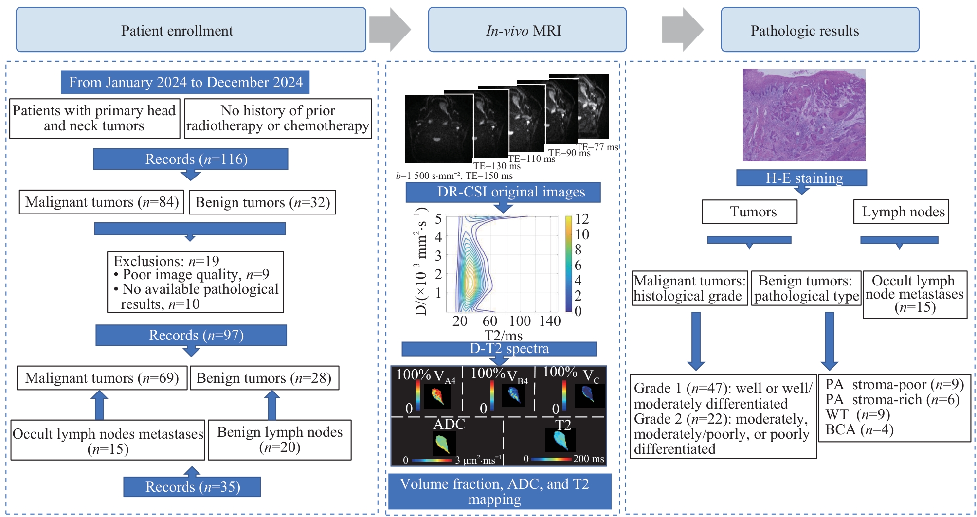
Fig 1 Diagram of this study
| Item | PA stroma-poor (n=9) | PA stroma-rich (n=6) | WT (n=9) | BCA (n=4) | P value |
|---|---|---|---|---|---|
| Age/year | 45.4±13.9 | 40.8±11.0 | 51.7±10.1 | 41.8±9.0 | 0.101 |
| Sex/n (%) | 0.423 | ||||
| Male | 5 (55.6) | 2 (33.3) | 7 (77.8) | 2(50.0) | |
| Female | 4 (44.4) | 4 (66.7) | 2 (22.2) | 2 (50.0) | |
| MD/cm | 2.56±1.04 | 2.00±0.88 | 3.33±0.97 | 1.98±0.64 | 0.040 |
| ADC/(μm2·ms-1) | 1.35±0.13 | 2.04±0.10 | 0.87±0.21 | 1.72±0.18 | <0.001 |
| T2/ms | 80.32±19.89 | 155.85±23.84 | 67.12±5.56 | 124.29±19.16 | <0.001 |
Tab 1 Clinical and imaging data of patients with benign head and neck tumors
| Item | PA stroma-poor (n=9) | PA stroma-rich (n=6) | WT (n=9) | BCA (n=4) | P value |
|---|---|---|---|---|---|
| Age/year | 45.4±13.9 | 40.8±11.0 | 51.7±10.1 | 41.8±9.0 | 0.101 |
| Sex/n (%) | 0.423 | ||||
| Male | 5 (55.6) | 2 (33.3) | 7 (77.8) | 2(50.0) | |
| Female | 4 (44.4) | 4 (66.7) | 2 (22.2) | 2 (50.0) | |
| MD/cm | 2.56±1.04 | 2.00±0.88 | 3.33±0.97 | 1.98±0.64 | 0.040 |
| ADC/(μm2·ms-1) | 1.35±0.13 | 2.04±0.10 | 0.87±0.21 | 1.72±0.18 | <0.001 |
| T2/ms | 80.32±19.89 | 155.85±23.84 | 67.12±5.56 | 124.29±19.16 | <0.001 |
| Item | Grade 1 (n=47) | Grade 2 (n=22) | P value |
|---|---|---|---|
| Age/year | 51.8±14.6 | 59.6±10.1 | 0.043 |
| Sex/n (%) | 0.715 | ||
| Male | 32 (68.1) | 14 (63.6) | |
| Female | 14 (31.9) | 8 (36.4) | |
| Subsite/n (%) | 0.599 | ||
| Oral tongue | 39 (83.0) | 18 (81.8) | |
| Oral floor | 1 (2.1) | 1 (4.5) | |
| Gingiva | 5 (10.6) | 1 (4.5) | |
| Oropharynx | 2 (4.3) | 2 (9.1) | |
| MD/cm | 2.85 (1.98, 3.75) | 3.00 (2.05, 3.73) | 0.605 |
| DOI/cm | 1.30 (0.90, 1.68) | 1.30 (1.00, 2.00) | 0.506 |
| cT stage/n (%) | 0.973 | ||
| T1‒2 | 13 (27.7) | 6 (27.3) | |
| T3‒4 | 34 (72.3) | 16 (72.7) | |
| LNM/n (%) | 0.044 | ||
| Negative | 25 (53.2) | 6 (27.3) | |
| Positive | 22 (46.8) | 16 (72.1) | |
| ADC/(μm2·ms-1) | 0.98 (0.92, 1.12) | 0.92 (0.87, 1.04) | 0.053 |
| T2/ms | 65.46 (52.28, 70.40) | 62.75 (56.91, 69.13) | 0.827 |
| DR-CSI VA4/% | 40.2 (25.7, 54.1) | 22.9 (17.9, 33.5) | 0.002 |
| DR-CSI VB4/% | 41.3 (28.6, 58.4) | 59.3 (52.8, 67.8) | 0.002 |
| DR-CSI VC4/% | 14.9 (12.1, 20.6) | 14.2 (12.7, 19.4) | 0.718 |
Tab 2 Clinical and imaging data of patients with HNSCC of different histological grades
| Item | Grade 1 (n=47) | Grade 2 (n=22) | P value |
|---|---|---|---|
| Age/year | 51.8±14.6 | 59.6±10.1 | 0.043 |
| Sex/n (%) | 0.715 | ||
| Male | 32 (68.1) | 14 (63.6) | |
| Female | 14 (31.9) | 8 (36.4) | |
| Subsite/n (%) | 0.599 | ||
| Oral tongue | 39 (83.0) | 18 (81.8) | |
| Oral floor | 1 (2.1) | 1 (4.5) | |
| Gingiva | 5 (10.6) | 1 (4.5) | |
| Oropharynx | 2 (4.3) | 2 (9.1) | |
| MD/cm | 2.85 (1.98, 3.75) | 3.00 (2.05, 3.73) | 0.605 |
| DOI/cm | 1.30 (0.90, 1.68) | 1.30 (1.00, 2.00) | 0.506 |
| cT stage/n (%) | 0.973 | ||
| T1‒2 | 13 (27.7) | 6 (27.3) | |
| T3‒4 | 34 (72.3) | 16 (72.7) | |
| LNM/n (%) | 0.044 | ||
| Negative | 25 (53.2) | 6 (27.3) | |
| Positive | 22 (46.8) | 16 (72.1) | |
| ADC/(μm2·ms-1) | 0.98 (0.92, 1.12) | 0.92 (0.87, 1.04) | 0.053 |
| T2/ms | 65.46 (52.28, 70.40) | 62.75 (56.91, 69.13) | 0.827 |
| DR-CSI VA4/% | 40.2 (25.7, 54.1) | 22.9 (17.9, 33.5) | 0.002 |
| DR-CSI VB4/% | 41.3 (28.6, 58.4) | 59.3 (52.8, 67.8) | 0.002 |
| DR-CSI VC4/% | 14.9 (12.1, 20.6) | 14.2 (12.7, 19.4) | 0.718 |
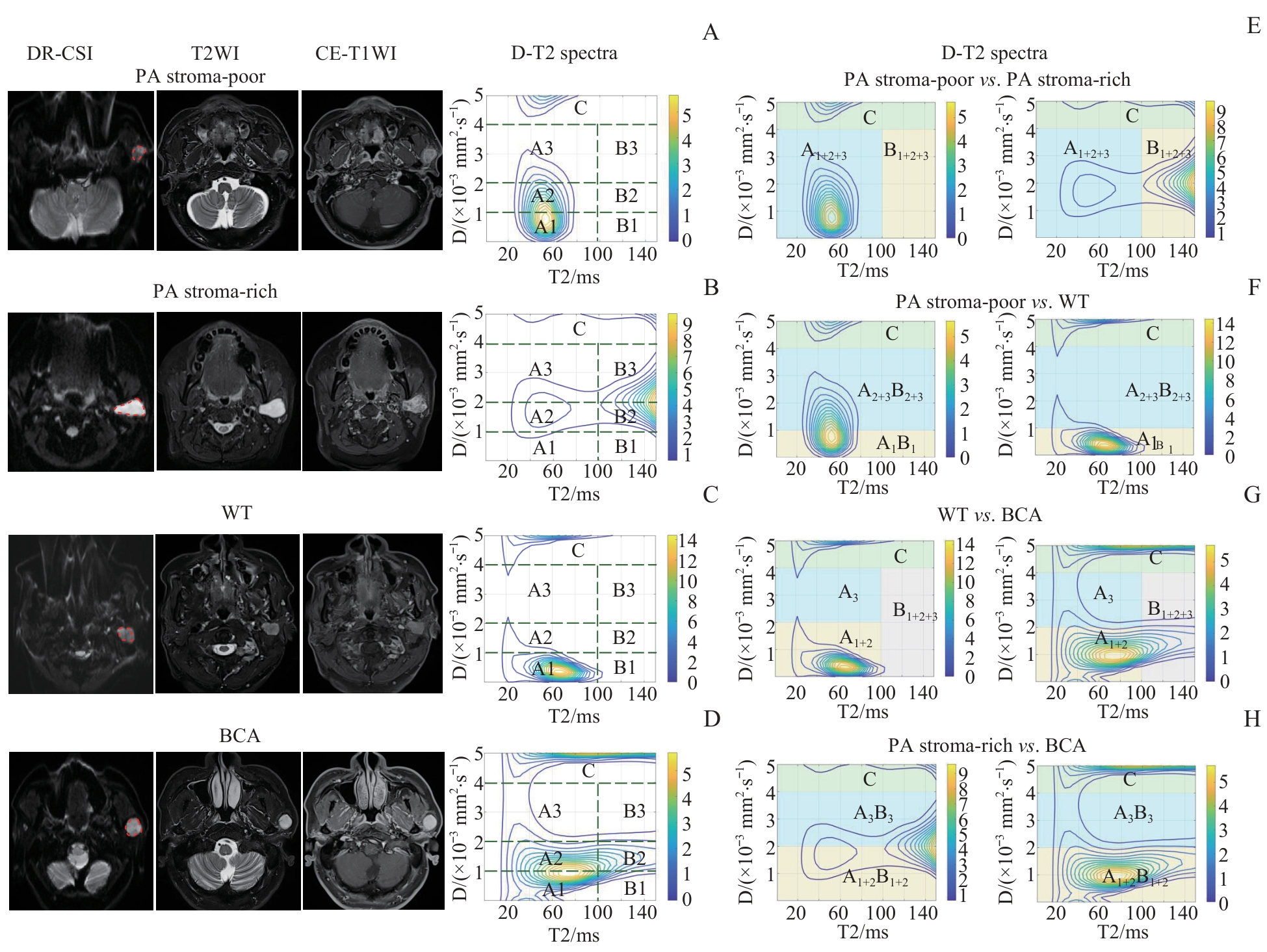
Fig 2 Representative cases of different pathological types of benign head and neck tumors
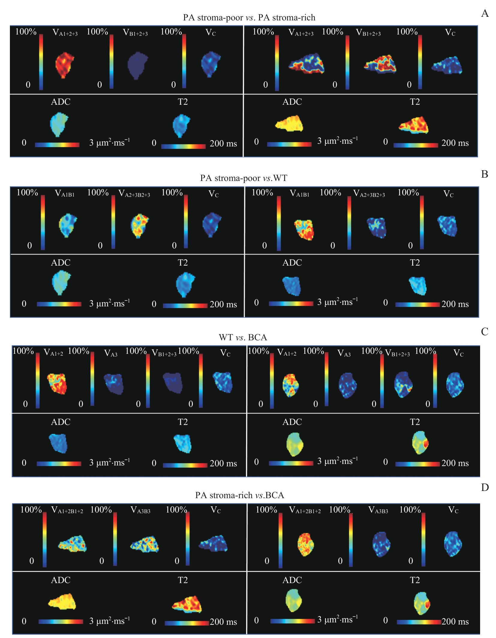
Fig 3 Representative DR-CSI volume fraction, ADC, and T2 maps of different pathological types of benign head and neck tumors
| Item | PA stroma-poor (n=9) | PA stroma-rich (n=6) | P value |
|---|---|---|---|
| ADC/(μm2·ms-1) | 1.35±0.13 | 2.04±0.10 | <0.001 |
| T2/ms | 80.32±19.89 | 155.85±23.84 | <0.001 |
| DR-CSI VA1+2+3 /% | 87.0±6.1 | 46.4±19.8 | 0.003 |
| DR-CSI VB1+2+3 /% | 3.9±3.6 | 39.9±17.6 | 0.004 |
| DR-CSI VC /% | 9.1±4.4 | 13.7±3.8 | 0.060 |
| Item | PA stroma-poor (n=9) | WT (n=9) | P value |
| ADC/(μm2·ms-1) | 1.35±0.13 | 0.87±0.21 | <0.001 |
| T2/ms | 80.32±19.89 | 67.12±5.56 | 0.086 |
| DR-CSI VA1B1 /% | 25.3±4.5 | 53.9±9.5 | <0.001 |
| DR-CSI VA2+3B2+3 /% | 67.3 (60.3, 70.5) | 16.9 (2.0, 19.2) | <0.001 |
| DR-CSI VC /% | 9.1±4.4 | 34.5±12.5 | <0.001 |
| Item | WT (n=9) | BCA (n=4) | P value |
| ADC/(μm2·ms-1) | 0.87±0.21 | 1.72±0.18 | <0.001 |
| T2/ms | 67.12±5.56 | 124.29±19.16 | 0.008 |
| DR-CSI VA1+2 /% | 53.1±9.3 | 15.4±7.0 | <0.001 |
| DR-CSI VA3 /% | 15.7 (2.0, 19.1) | 38.5 (33.0, 40.9) | 0.005 |
| DR-CSI VB1+2+3 /% | 1.1±0.9 | 29.0±11.7 | 0.017 |
| DR-CSI VC /% | 34.5±12.5 | 18.1±2.7 | 0.004 |
| Item | PA stroma-rich (n=6) | BCA (n=4) | P value |
| ADC/(μm2·ms-1) | 2.04±0.10 | 1.72±0.18 | 0.006 |
| T2/ms | 155.85±23.84 | 124.29±19.16 | 0.059 |
| DR-CSI VA1+2B1+2 /% | 40.7±3.7 | 63.0±7.0 | 0.004 |
| DR-CSI VA3B3 /% | 45.6±3.9 | 18.9±7.6 | 0.003 |
| DR-CSI VC /% | 13.7±3.8 | 18.1±2.7 | 0.080 |
Tab 3 Pairwise comparison of imaging parameters among different subtypes of benign head and neck tumors
| Item | PA stroma-poor (n=9) | PA stroma-rich (n=6) | P value |
|---|---|---|---|
| ADC/(μm2·ms-1) | 1.35±0.13 | 2.04±0.10 | <0.001 |
| T2/ms | 80.32±19.89 | 155.85±23.84 | <0.001 |
| DR-CSI VA1+2+3 /% | 87.0±6.1 | 46.4±19.8 | 0.003 |
| DR-CSI VB1+2+3 /% | 3.9±3.6 | 39.9±17.6 | 0.004 |
| DR-CSI VC /% | 9.1±4.4 | 13.7±3.8 | 0.060 |
| Item | PA stroma-poor (n=9) | WT (n=9) | P value |
| ADC/(μm2·ms-1) | 1.35±0.13 | 0.87±0.21 | <0.001 |
| T2/ms | 80.32±19.89 | 67.12±5.56 | 0.086 |
| DR-CSI VA1B1 /% | 25.3±4.5 | 53.9±9.5 | <0.001 |
| DR-CSI VA2+3B2+3 /% | 67.3 (60.3, 70.5) | 16.9 (2.0, 19.2) | <0.001 |
| DR-CSI VC /% | 9.1±4.4 | 34.5±12.5 | <0.001 |
| Item | WT (n=9) | BCA (n=4) | P value |
| ADC/(μm2·ms-1) | 0.87±0.21 | 1.72±0.18 | <0.001 |
| T2/ms | 67.12±5.56 | 124.29±19.16 | 0.008 |
| DR-CSI VA1+2 /% | 53.1±9.3 | 15.4±7.0 | <0.001 |
| DR-CSI VA3 /% | 15.7 (2.0, 19.1) | 38.5 (33.0, 40.9) | 0.005 |
| DR-CSI VB1+2+3 /% | 1.1±0.9 | 29.0±11.7 | 0.017 |
| DR-CSI VC /% | 34.5±12.5 | 18.1±2.7 | 0.004 |
| Item | PA stroma-rich (n=6) | BCA (n=4) | P value |
| ADC/(μm2·ms-1) | 2.04±0.10 | 1.72±0.18 | 0.006 |
| T2/ms | 155.85±23.84 | 124.29±19.16 | 0.059 |
| DR-CSI VA1+2B1+2 /% | 40.7±3.7 | 63.0±7.0 | 0.004 |
| DR-CSI VA3B3 /% | 45.6±3.9 | 18.9±7.6 | 0.003 |
| DR-CSI VC /% | 13.7±3.8 | 18.1±2.7 | 0.080 |
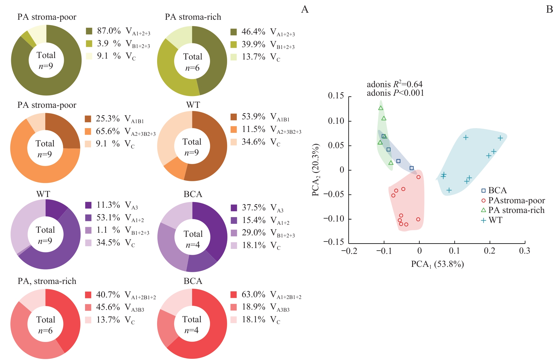
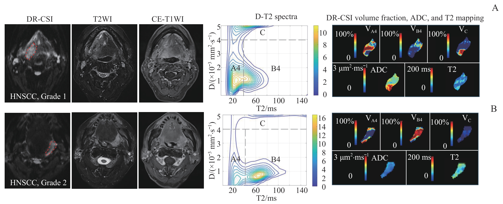
Fig 5 Representative cases of HNSCC with different pathological grades
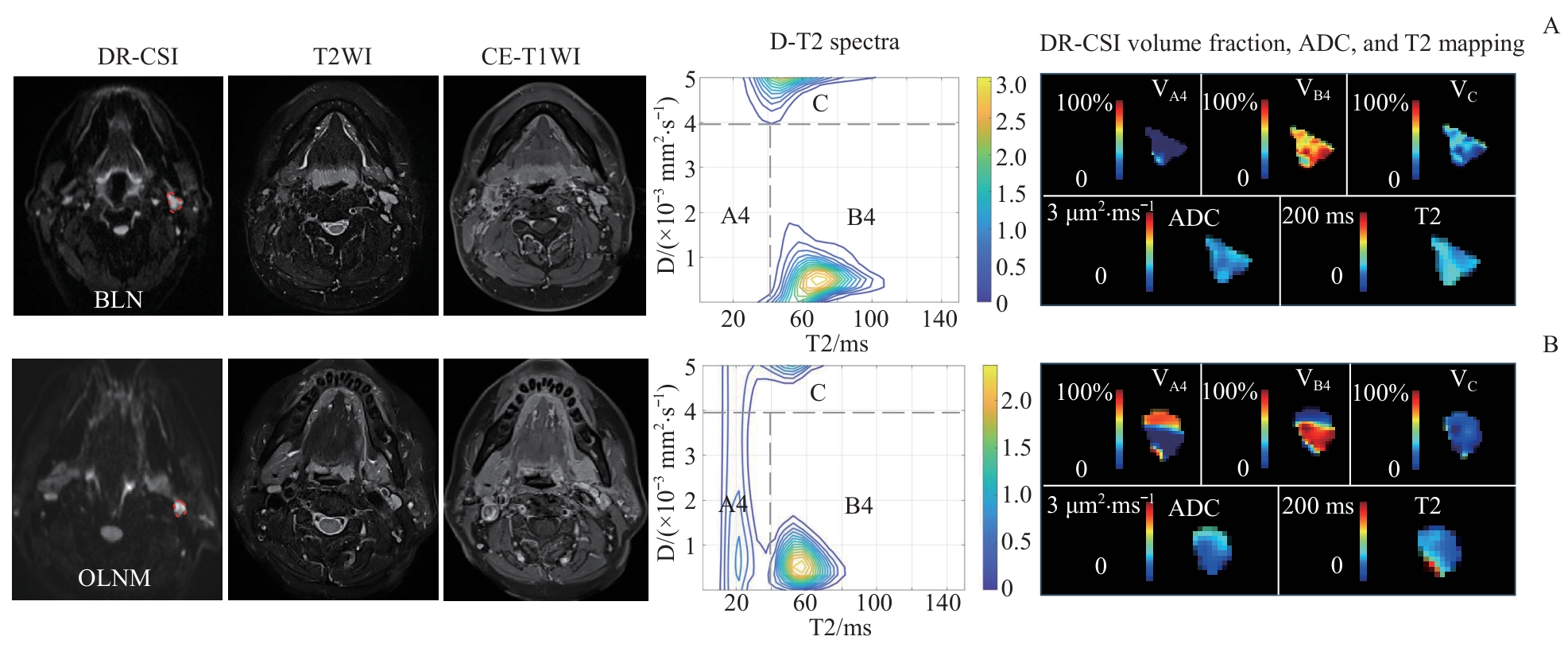
Fig 6 Representative cases of BLN and OLNM
| Item | BLN (n=20) | OLNM (n=15) | P value |
|---|---|---|---|
| SD/cm | 0.67 (0.57, 0.80) | 0.92 (0.72, 1.00) | 0.011 |
| L/S ratio | 1.46 (1.28, 1.84) | 1.51 (1.33, 1.73) | 0.805 |
| ADC/(μm2·ms-1) | 0.88±0.17 | 0.96±0.16 | 0.119 |
| T2/ms | 71.03±11.09 | 67.46±13.73 | 0.401 |
| DR-CSI VA4 /% | 16.7±9.9 | 27.3±13.0 | 0.010 |
| DR-CSI VB4 /% | 63.1±12.3 | 54.5±14.4 | 0.066 |
| DR-CSI VC /% | 20.2±8.7 | 18.2±9.6 | 0.531 |
Tab 4 Comparison of imaging parameters between BLN and OLNM groups
| Item | BLN (n=20) | OLNM (n=15) | P value |
|---|---|---|---|
| SD/cm | 0.67 (0.57, 0.80) | 0.92 (0.72, 1.00) | 0.011 |
| L/S ratio | 1.46 (1.28, 1.84) | 1.51 (1.33, 1.73) | 0.805 |
| ADC/(μm2·ms-1) | 0.88±0.17 | 0.96±0.16 | 0.119 |
| T2/ms | 71.03±11.09 | 67.46±13.73 | 0.401 |
| DR-CSI VA4 /% | 16.7±9.9 | 27.3±13.0 | 0.010 |
| DR-CSI VB4 /% | 63.1±12.3 | 54.5±14.4 | 0.066 |
| DR-CSI VC /% | 20.2±8.7 | 18.2±9.6 | 0.531 |
| Item | AUC | Sensitivity | Specificity | Cut-off |
|---|---|---|---|---|
| HNSCC malignancy assessment | ||||
| DR-CSI VA4 /% | 0.733 | 0.809 | 0.591 | 25.0 |
| DR-CSI VB4 /% | 0.730 | 0.818 | 0.660 | 49.3 |
| OLNM identification | ||||
| SD/cm | 0.753 | 0.867 | 0.700 | 0.70 |
| DR-CSI VA4 /% | 0.722 | 0.733 | 0.650 | 19.6 |
| Combined parameters | 0.843 | 0.667 | 0.900 | ‒ |
Tab 5 Diagnostic performance of imaging parameters for assessing malignancy of HNSCC and detecting OLNM
| Item | AUC | Sensitivity | Specificity | Cut-off |
|---|---|---|---|---|
| HNSCC malignancy assessment | ||||
| DR-CSI VA4 /% | 0.733 | 0.809 | 0.591 | 25.0 |
| DR-CSI VB4 /% | 0.730 | 0.818 | 0.660 | 49.3 |
| OLNM identification | ||||
| SD/cm | 0.753 | 0.867 | 0.700 | 0.70 |
| DR-CSI VA4 /% | 0.722 | 0.733 | 0.650 | 19.6 |
| Combined parameters | 0.843 | 0.667 | 0.900 | ‒ |
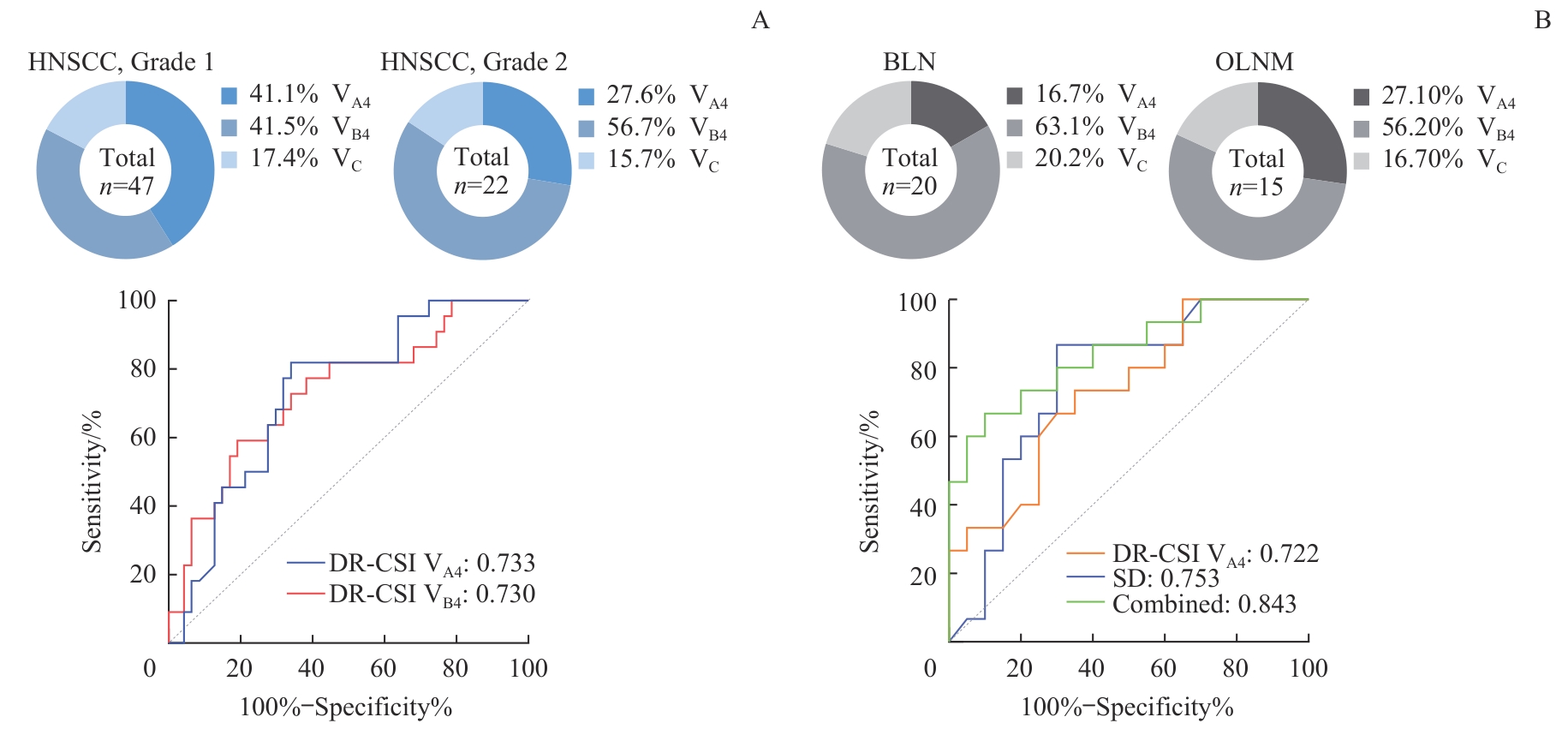
Fig 7 Diagnostic performance for imaging parameters in characterizing the malignancy of HNSCC and identifying OLNM
| [1] | BHAT G R, HYOLE R G, LI J. Head and neck cancer: current challenges and future perspectives[J]. Adv Cancer Res, 2021, 152: 67-102. |
| [2] | BRAY F, LAVERSANNE M, SUNG H, et al. Global cancer statistics 2022: GLOBOCAN estimates of incidence and mortality worldwide for 36 cancers in 185 countries[J]. CA Cancer J Clin, 2024, 74(3): 229-263. |
| [3] | CHEN F F, GE Y Q, LI S, et al. Enhanced CT-based texture analysis and radiomics score for differentiation of pleomorphic adenoma, basal cell adenoma, and Warthin tumor of the parotid gland[J]. Dentomaxillofac Radiol, 2023, 52(2): 20220009. |
| [4] | SEIFERT G, LANGROCK I, DONATH K. A pathological classification of pleomorphic adenoma of the salivary glands (author's transl)[J]. HNO, 1976, 24(12): 415-426. |
| [5] | JOHNSON D E, BURTNESS B, LEEMANS C R, et al. Head and neck squamous cell carcinoma[J]. Nat Rev Dis Primers, 2020, 6(1): 92. |
| [6] | HUANG Z G, WEN W P, MAO W. Comprehensive treatment strategies for head neck tumors[J]. Lin Chuang Er Bi Yan Hou Tou Jing Wai Ke Za Zhi, 2023, 37(9): 673-690. |
| [7] | ANTRA, PARASHAR P, HUNGYO H, et al. Unraveling molecular mechanisms of head and neck cancer[J]. Crit Rev Oncol Hematol, 2022, 178: 103778. |
| [8] | ALMANGUSH A, HEIKKINEN I, MÄKITIE A A, et al. Prognostic biomarkers for oral tongue squamous cell carcinoma: a systematic review and meta-analysis[J]. Br J Cancer, 2017, 117(6): 856-866. |
| [9] | AHN S J, CHOI S H, KIM Y J, et al. Histogram analysis of apparent diffusion coefficient map of standard and high B-value diffusion MR imaging in head and neck squamous cell carcinoma: a correlation study with histological grade[J]. Acad Radiol, 2012, 19(10): 1233-1240. |
| [10] | SHARBEL D D, ABKEMEIER M, GROVES M W, et al. Occult metastasis in laryngeal squamous cell carcinoma: a systematic review and meta-analysis[J]. Ann Otol Rhinol Laryngol, 2021, 130(1): 67-77. |
| [11] | MOURAD M A F, HIGAZI M M. MRI prognostic factors of tongue cancer: potential predictors of cervical lymph nodes metastases[J]. Radiol Oncol, 2019, 53(1): 49-56. |
| [12] | THOENY H C, DE KEYZER F, KING A D. Diffusion-weighted MR imaging in the head and neck[J]. Radiology, 2012, 263(1): 19-32. |
| [13] | ABDEL RAZEK A A K, KANDEEL A Y, SOLIMAN N, et al. Role of diffusion-weighted echo-planar MR imaging in differentiation of residual or recurrent head and neck tumors and posttreatment changes[J]. AJNR Am J Neuroradiol, 2007, 28(6): 1146-1152. |
| [14] | CHEN J, HAGIWARA M, GIVI B, et al. Assessment of metastatic lymph nodes in head and neck squamous cell carcinomas using simultaneous 18F-FDG-PET and MRI[J]. Sci Rep, 2020, 10(1): 20764. |
| [15] | DAI Y M, HU W T, WU G Y, et al. Grading clear cell renal cell carcinoma grade using diffusion relaxation correlated MR spectroscopic imaging[J]. J Magn Reson Imaging, 2024, 59(2): 699-710. |
| [16] | ZHANG Z H, WU H H, PRIESTER A, et al. Prostate microstructure in prostate cancer using 3-T MRI with diffusion-relaxation correlation spectrum imaging: validation with whole-mount digital histopathology[J]. Radiology, 2020, 296(2): 348-355. |
| [17] | TANG W Q, WANG Y, YUAN Y, et al. Assessment of tumor depth in oral tongue squamous cell carcinoma with multiparametric MRI: correlation with pathology[J]. Eur Radiol, 2022, 32(1): 254-261. |
| [18] | YANG X H, XIANG W, SUN Y, et al. Risk factors and impact of occult and skip metastasis in early-stage oral tongue squamous cell carcinoma[J]. Clin Oral Investig, 2024, 28(9): 510. |
| [19] | LIU F, HU W T, SUN Y W, et al. Exploration of interstitial fibrosis in chronic kidney disease by diffusion-relaxation correlation spectrum MR imaging: a preliminary study[J]. J Magn Reson Imaging, 2023, 58(2): 415-426. |
| [20] | STENNERT E, GUNTINAS-LICHIUS O, KLUSSMANN J P, et al. Histopathology of pleomorphic adenoma in the parotid gland: a prospective unselected series of 100 cases[J]. Laryngoscope, 2001, 111(12): 2195-2200. |
| [21] | ZHAO L H, MAO Y R, MU J, et al. The diagnostic value of Superb Microvascular Imaging in identifying benign tumors of parotid gland[J]. BMC Med Imaging, 2020, 20(1): 107. |
| [22] | YUN T J, KIM J H, KIM K H, et al. Head and neck squamous cell carcinoma: differentiation of histologic grade with standard- and high-b-value diffusion-weighted MRI[J]. Head Neck, 2013, 35(5): 626-631. |
| [23] | VIDIRI A, ASCIONE A, PILUDU F, et al. Microenvironmental factors in oral cavity squamous cell carcinoma undergoing surgery: correlation with diffusion kurtosis imaging and dynamic contrast-enhanced MRI[J]. Cancers, 2022, 15(1): 15. |
| [24] | ASTEKAR M, JOSHI A, RAMESH G, et al. Expression of vascular endothelial growth factor and microvessel density in oral tumorigenesis[J]. J Oral Maxillofac Pathol, 2012, 16(1): 22-26. |
| [25] | VIALLARD C, LARRIVÉE B. Tumor angiogenesis and vascular normalization: alternative therapeutic targets[J]. Angiogenesis, 2017, 20(4): 409-426. |
| [26] | QU J M, PAN B J, SU T, et al. T1 and T2 mapping for identifying malignant lymph nodes in head and neck squamous cell carcinoma[J]. Cancer Imaging, 2023, 23(1): 125. |
| [27] | ROFSTAD E K, STEINSLAND E, KAALHUS O, et al. Magnetic resonance imaging of human melanoma xenografts in vivo: proton spin-lattice and spin-spin relaxation times versus fractional tumour water content and fraction of necrotic tumour tissue[J]. Int J Radiat Biol, 1994, 65(3): 387-401. |
| [1] | CHEN Yinan, ZHENG Yang, ZENG Hanlin, LEI Ming. Mechanism of Fas-associated protein with death domain in promoting proliferation of head and neck squamous cell carcinoma cells [J]. Journal of Shanghai Jiao Tong University (Medical Science), 2025, 45(4): 404-414. |
| [2] | AN Junyi, CHEN Biying, CHEN Xunrui, YIN Shanshan, BIAN Zhouliang, LIU Feng. SFXN3 expression in head and neck squamous cell carcinoma and its effect on cell proliferation [J]. Journal of Shanghai Jiao Tong University (Medical Science), 2024, 44(4): 427-434. |
| [3] | ZHANG Yu-han, CHEN Xue-hua, ZHANG Hao. Down-regulation of aurora kinase A activity inhibits pro-angiogenesis, migration and invasion of Hep2 cells [J]. , 2018, 38(10): 1181-. |
| [4] | LIU Kai, MA Ling-ling, WANG Duo-ming, et al. Relationship between HPV infection and expression of HIF-1alpha and VEGF in head and neck squamous cell carcinoma and its prognostic value [J]. , 2013, 33(6): 806-. |
| Viewed | ||||||
|
Full text |
|
|||||
|
Abstract |
|
|||||