Journal of Shanghai Jiao Tong University (Medical Science) ›› 2022, Vol. 42 ›› Issue (6): 758-767.doi: 10.3969/j.issn.1674-8115.2022.06.010
• Basic research • Previous Articles Next Articles
WANG Xinpeng1( ), WANG Junying1, CAI Jiayi2, FU Wanbing1, ZHONG Hua1()
Received:2022-02-11
Accepted:2022-05-11
Online:2022-06-28
Published:2022-08-19
Contact:
ZHONG Hua
E-mail:wxpyoyo@126.com;zhh_lj@163.com
Supported by:CLC Number:
WANG Xinpeng, WANG Junying, CAI Jiayi, FU Wanbing, ZHONG Hua. Effect of low-dose decitabine on the biological behavior of bone marrow mesenchymal stem cells derived from patients with immune thrombocytopenia[J]. Journal of Shanghai Jiao Tong University (Medical Science), 2022, 42(6): 758-767.
Add to citation manager EndNote|Ris|BibTeX
URL: https://xuebao.shsmu.edu.cn/EN/10.3969/j.issn.1674-8115.2022.06.010
| ITP patient | Gender | Age/year | Platelet count/(×109 L-1) | NC subject | Gender | Age/year | Platelet count/(×109 L-1) |
|---|---|---|---|---|---|---|---|
| P1 | Female | 48 | 44 | N1 | Female | 60 | 137 |
| P2 | Female | 44 | 34 | N2 | Female | 74 | 206 |
| P3 | Female | 70 | 69 | N3 | Female | 65 | 169 |
| P4 | Female | 71 | 102 | N4 | Male | 31 | 145 |
| P5 | Male | 72 | 61 | N5 | Female | 66 | 172 |
| P6 | Female | 84 | 38 | N6 | Male | 39 | 151 |
| P7 | Male | 77 | 20 | N7 | Female | 51 | 183 |
| P8 | Female | 45 | 48 | N8 | Male | 68 | 201 |
| P9 | Female | 85 | 34 | N9 | Male | 45 | 195 |
| P10 | Male | 61 | 7 | N10 | Female | 75 | 141 |
Tab 1 Clinical information of the ITP patients and the normal controls enrolled in the study
| ITP patient | Gender | Age/year | Platelet count/(×109 L-1) | NC subject | Gender | Age/year | Platelet count/(×109 L-1) |
|---|---|---|---|---|---|---|---|
| P1 | Female | 48 | 44 | N1 | Female | 60 | 137 |
| P2 | Female | 44 | 34 | N2 | Female | 74 | 206 |
| P3 | Female | 70 | 69 | N3 | Female | 65 | 169 |
| P4 | Female | 71 | 102 | N4 | Male | 31 | 145 |
| P5 | Male | 72 | 61 | N5 | Female | 66 | 172 |
| P6 | Female | 84 | 38 | N6 | Male | 39 | 151 |
| P7 | Male | 77 | 20 | N7 | Female | 51 | 183 |
| P8 | Female | 45 | 48 | N8 | Male | 68 | 201 |
| P9 | Female | 85 | 34 | N9 | Male | 45 | 195 |
| P10 | Male | 61 | 7 | N10 | Female | 75 | 141 |
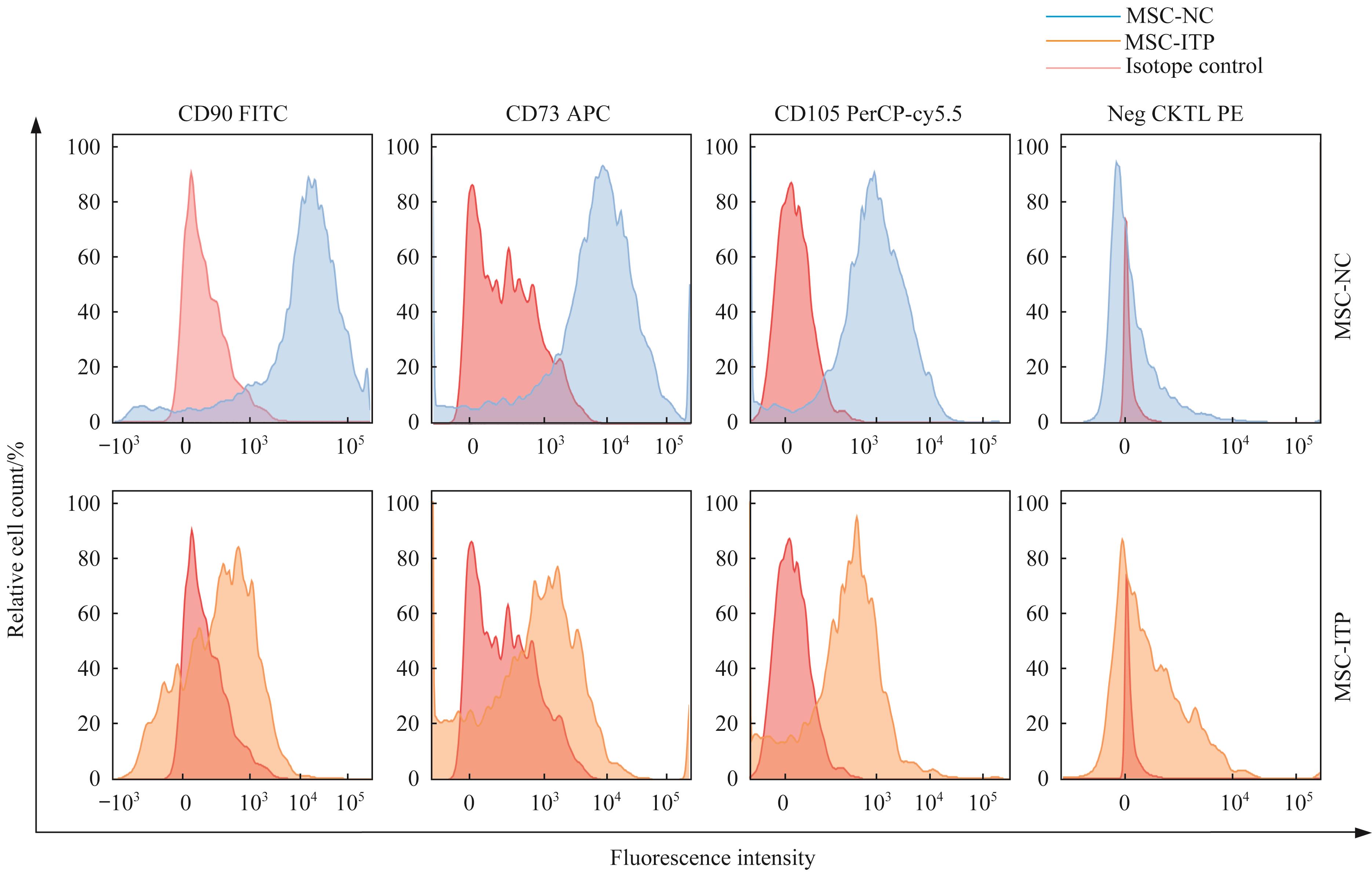
Fig 1 Phenotype of MSCs cultured in vitro identified by flow cytometry

Fig 2 Detection of proliferation and morphology of MSCs derived from ITP patients
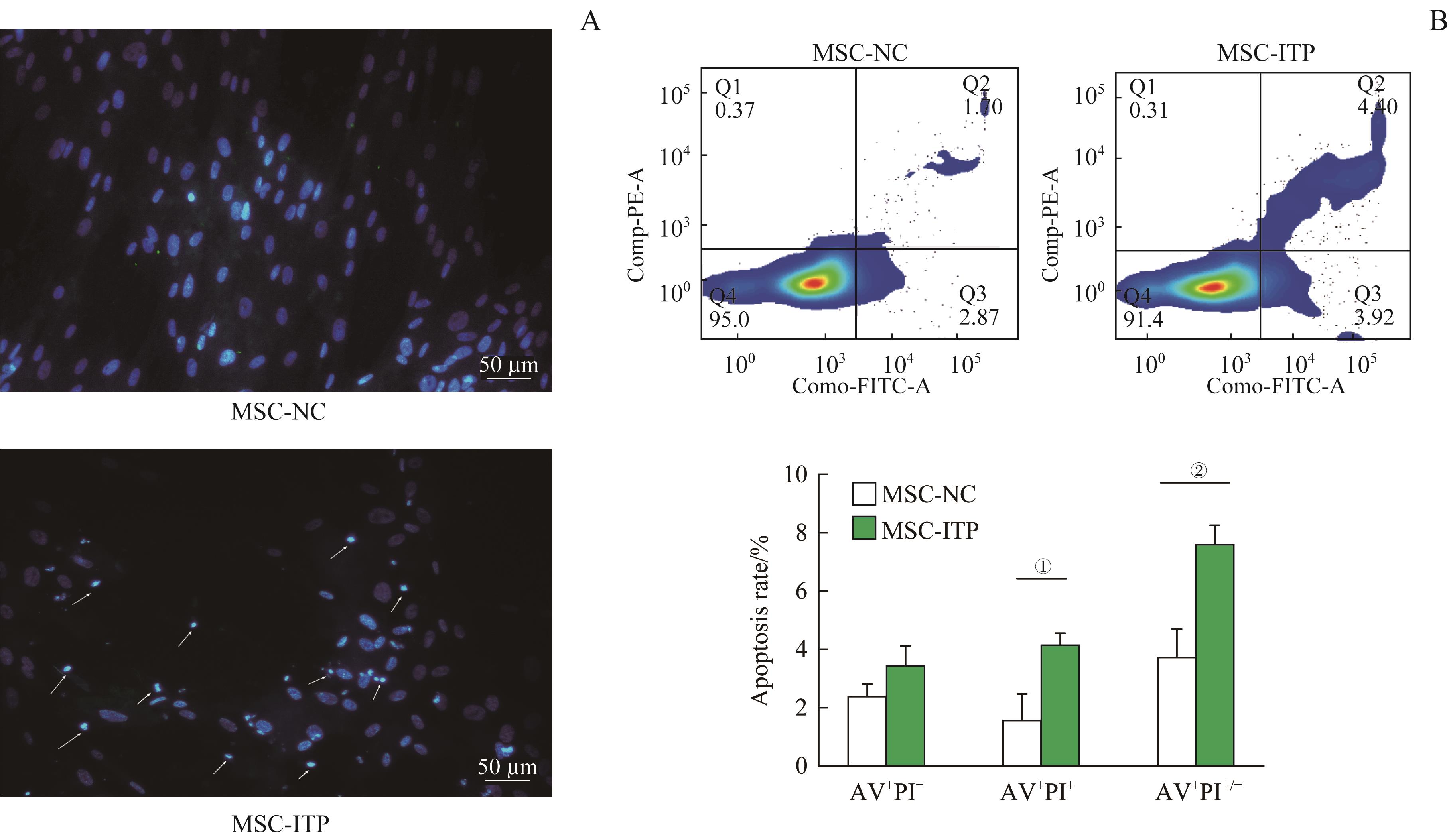
Fig 3 Detection of basal apoptosis in MSCs derived from ITP patients

Fig 4 Effect of decitabine on MSC-ITP proliferation under different doses and working hours detected by CCK-8 assay
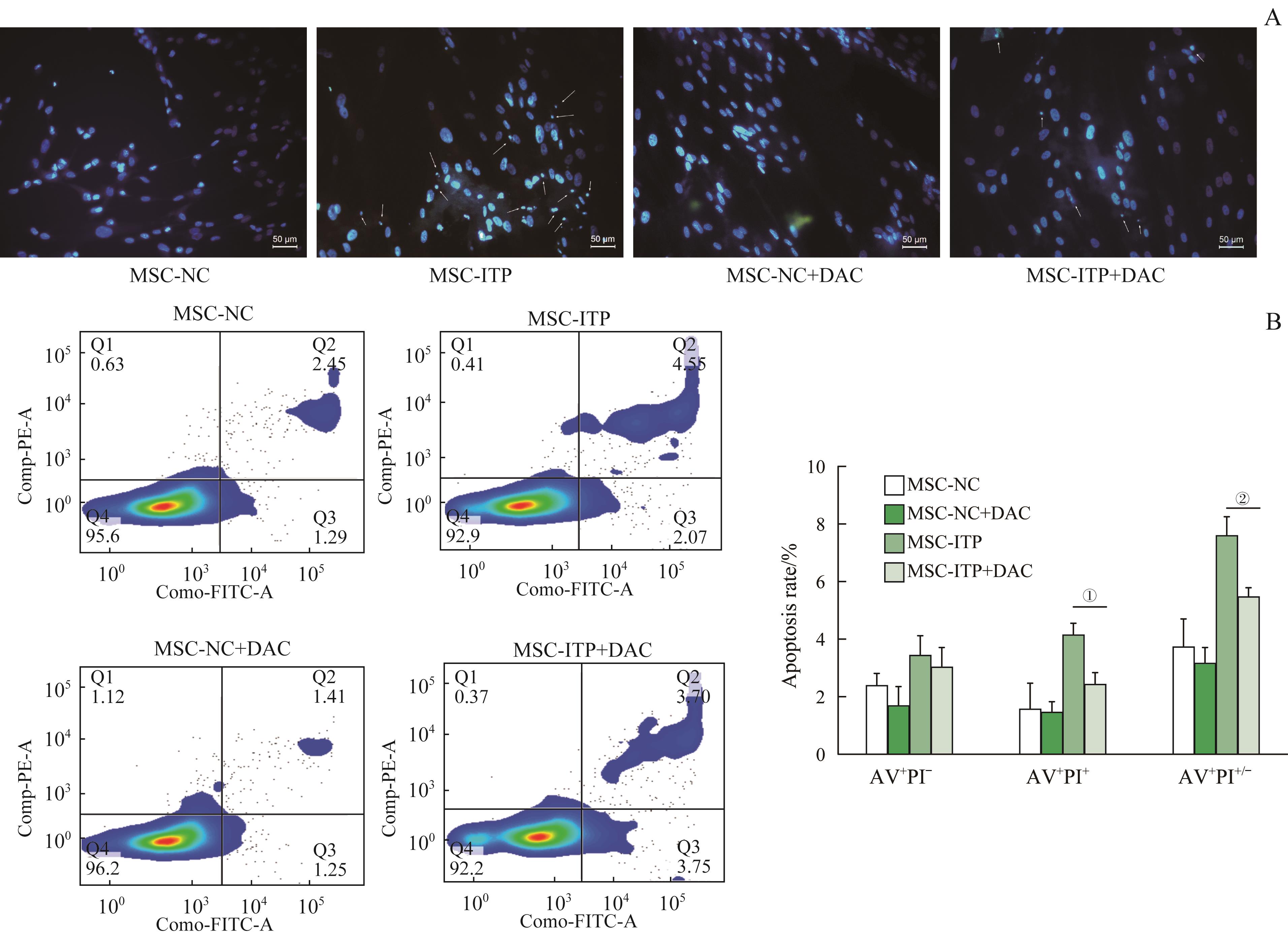
Fig 5 Apoptosis detection of MSC-ITP after 2.5 μmol/L decitabine treatment for 24 h
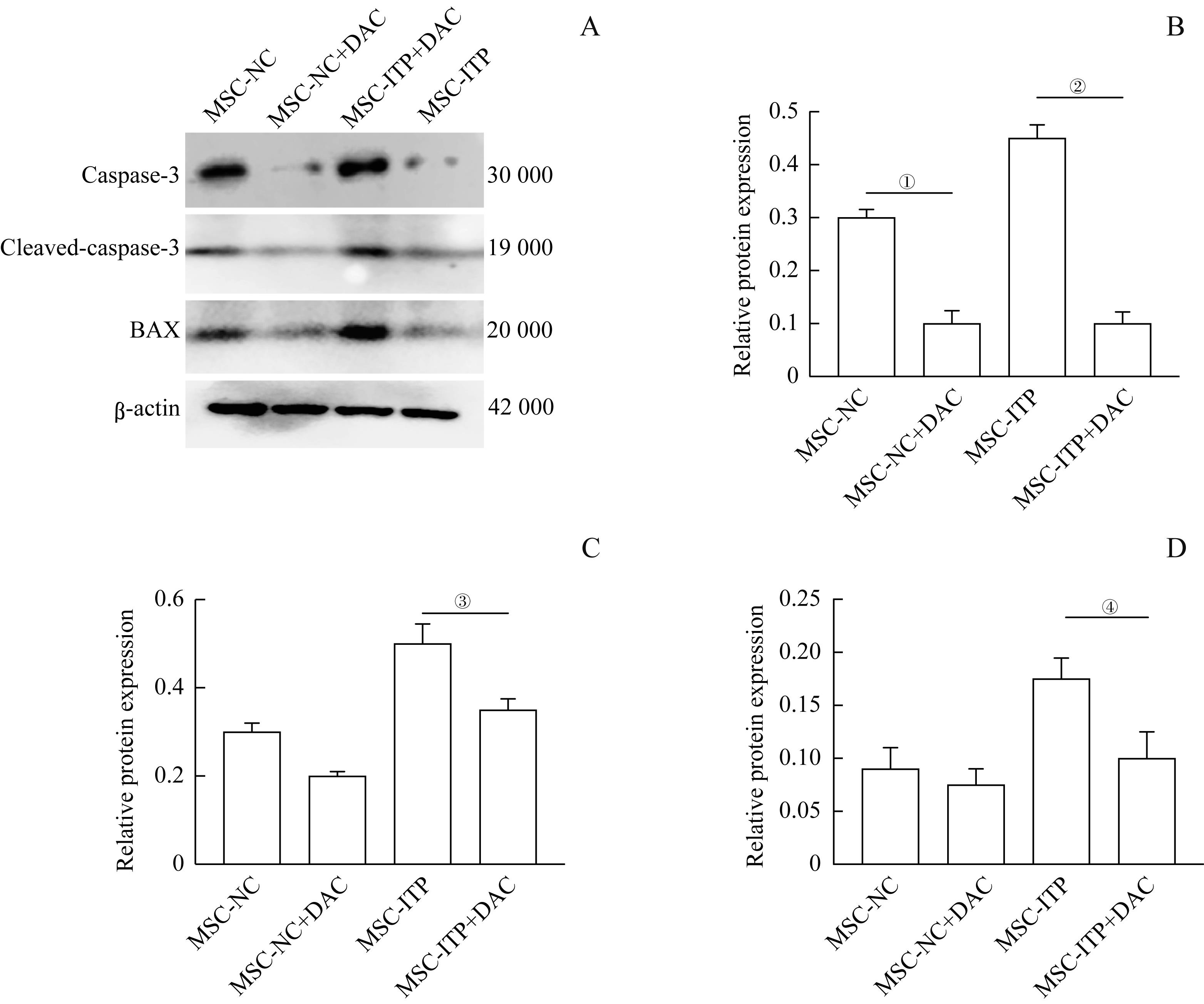
Fig 6 Expression levels of apoptosis-related proteins BAX, caspase-3 and cleaved-caspase3 detected by Western blotting
| 1 | SEMPLE J W, PROVAN D, GARVEY M B, et al. Recent progress in understanding the pathogenesis of immune thrombocytopenia[J]. Curr Opin Hematol, 2010, 17(6): 590-595. |
| 2 | REEMS J A, PINEAULT N, SUN S J. In vitro megakaryocyte production and platelet biogenesis: state of the art[J]. Transfus Med Rev, 2010, 24(1): 33-43. |
| 3 | GLENNIE S, SOEIRO I, DYSON P J, et al. Bone marrow mesenchymal stem cells induce division arrest anergy of activated T cells[J]. Blood, 2005, 105(7): 2821-2827. |
| 4 | TABERA S, PÉREZ-SIMÓN J A, DÍEZ-CAMPELO M, et al. The effect of mesenchymal stem cells on the viability, proliferation and differentiation of B-lymphocytes[J]. Haematologica, 2008, 93(9): 1301-1309. |
| 5 | TASSO R, ILENGO C, QUARTO R, et al. Mesenchymal stem cells induce functionally active T-regulatory lymphocytes in a paracrine fashion and ameliorate experimental autoimmune uveitis[J]. Invest Ophthalmol Vis Sci, 2012, 53(2): 786-793. |
| 6 | MA J, NING Y N, XU M, et al. Thalidomide corrects impaired mesenchymal stem cell function in inducing tolerogenic DCs in patients with immune thrombocytopenia[J]. Blood, 2013, 122(12): 2074-2082. |
| 7 | PÉREZ-SIMÓN J A, TABERA S, SARASQUETE M E, et al. Mesenchymal stem cells are functionally abnormal in patients with immune thrombocytopenic purpura[J]. Cytotherapy, 2009, 11(6): 698-705. |
| 8 | ZHANG D L, LI H Y, MA L, et al. The defective bone marrow-derived mesenchymal stem cells in patients with chronic immune thrombocytopenia[J]. Autoimmunity, 2014, 47(8): 519-529. |
| 9 | 王君颖, 李昕, 殷婷玉, 等. 免疫性血小板减少症患者来源的骨髓间充质细胞对巨核细胞生物学行为的影响[J]. 上海交通大学学报(医学版), 2018, 38(6): 616-623. |
| WANG J Y, LI X, YIN T Y, et al. Effects of bone marrow mesenchymal cells from immune thrombocytopenia patients on the biological behaviors of megakaryocytes[J]. J Shanghai Jiao Tong Univ (Med Sci), 2018, 38(6): 616–625. | |
| 10 | XIAO J H, ZHANG C R, ZHANG Y C, et al. Transplantation of adipose-derived mesenchymal stem cells into a murine model of passive chronic immune thrombocytopenia[J]. Transfusion, 2012, 52(12): 2551-2558. |
| 11 | ZHANG P, ZHANG G, LIU X, et al. Mesenchymal stem cells improve platelet counts in mice with immune thrombocytopenia[J]. J Cell Biochem, 2019. DOI: 10.1002/jcb.28405. |
| 12 | WANG X H, YIN X G, SUN W, et al. Intravenous infusion umbilical cord-derived mesenchymal stem cell in primary immune thrombocytopenia: a two-year follow-up[J]. Exp Ther Med, 2017, 13(5): 2255-2258. |
| 13 | LEE S, KIM H S, ROH K H, et al. DNA methyltransferase inhibition accelerates the immunomodulation and migration of human mesenchymal stem cells[J]. Sci Rep, 2015, 5: 8020. |
| 14 | LIU S Y, SHAN N N. DNA methylation plays an important role in immune thrombocytopenia[J]. Int Immunopharmacol, 2020, 83: 106390. |
| 15 | ASSIS R I F, WIENCH M, SILVÉRIO K G, et al. RG108 increases NANOG and OCT4 in bone marrow-derived mesenchymal cells through global changes in DNA modifications and epigenetic activation[J]. PLoS One, 2018, 13(12): e0207873. |
| 16 | OH Y S, JEONG S G, CHO G W. Anti-senescence effects of DNA methyltransferase inhibitor RG108 in human bone marrow mesenchymal stromal cells[J]. Biotechnol Appl Biochem, 2015, 62(5): 583-590. |
| 17 | OH Y S, KIM S H, CHO G W. Functional restoration of amyotrophic lateral sclerosis patient-derived mesenchymal stromal cells through inhibition of DNA methyltransferase[J]. Cell Mol Neurobiol, 2016, 36(4): 613-620. |
| 18 | ZHOU H, HOU Y, LIU X N, et al. Low-dose decitabine promotes megakaryocyte maturation and platelet production in healthy controls and immune thrombocytopenia[J]. Thromb Haemost, 2015, 113(5): 1021-1034. |
| 19 | RODEGHIERO F, STASI R, GERNSHEIMER T, et al. Standardization of terminology, definitions and outcome criteria in immune thrombocytopenic purpura of adults and children: report from an international working group[J]. Blood, 2009, 113(11): 2386-2393. |
| 20 | LAMBERT M P, GERNSHEIMER T B. Clinical updates in adult immune thrombocytopenia[J]. Blood, 2017, 129(21): 2829-2835. |
| 21 | SHEN Y, NILSSON S K. Bone, microenvironment and hematopoiesis[J]. Curr Opin Hematol, 2012, 19(4): 250-255. |
| 22 | SONG Y, WANG Y T, HUANG X J, et al. Abnormalities of the bone marrow immune microenvironment in patients with immune thrombocytopenia[J]. Ann Hematol, 2016, 95(6): 959-965. |
| 23 | TAO Y L, SONG D X, ZHANG F Y, et al. Transplantation of bone-marrow-derived mesenchymal stem cells into a murine model of immune thrombocytopenia[J]. Blood Coagul Fibrinolysis, 2017, 28(8): 596-601. |
| 24 | LI H Y, XUAN M, YANG R C. DNA methylation and primary immune thrombocytopenia[J]. Semin Hematol, 2013, 50(Suppl 1): S116-S126. |
| 25 | WANG J H, YI Z H, WANG S Y, et al. The effect of decitabine on megakaryocyte maturation and platelet release[J]. Thromb Haemost, 2011, 106(2): 337-343. |
| 26 | HAN P P, HOU Y, ZHAO Y J, et al. Low-dose decitabine modulates T cell homeostasis and restores immune tolerance in immune thrombocytopenia[J]. Blood, 2021, 138(8): 674-688. |
| 27 | WANG Y, WANG F X, WEN S P, et al. Artesunate-enhanced apoptosis of human high-risk myelodysplastic cells induced by the DNA methyltransferase inhibitor decitabine[J]. Oncol Lett, 2015, 9(6): 2449-2454. |
| 28 | ZHANG G, GAO X H, ZHAO X Y, et al. Decitabine inhibits the proliferation of human T-cell acute lymphoblastic leukemia molt4 cells and promotes apoptosis partly by regulating the PI3K/AKT/mTOR pathway[J]. Oncol Lett, 2021, 21(5): 340. |
| 29 | WANG L, AMOOZGAR Z, HUANG J, et al. Decitabine enhances lymphocyte migration and function and synergizes with CTLA-4 blockade in a murine ovarian cancer model[J]. Cancer Immunol Res, 2015, 3(9): 1030-1041. |
| 30 | RÜGER B M, BUCHACHER T, GIUREA A, et al. Vascular morphogenesis in the context of inflammation: self-organization in a fibrin-based 3D culture system[J]. Front Physiol, 2018, 9: 679. |
| 31 | ELMORE S. Apoptosis: a review of programmed cell death[J]. Toxicol Pathol, 2007, 35(4): 495-516. |
| 32 | FERRI K F, KROEMER G. Mitochondria: the suicide organelles[J]. BioEssays, 2001, 23(2): 111-115. |
| [1] | LIU Yuanqi, SUN Siyuan, DAI Qinggang, JIANG Lingyong, SHEN Guofang. Dual-directional effect of all-trans retinoic acid on osteogenic differentiation of jaw bone marrow mesenchymal stem cells in vitro [J]. Journal of Shanghai Jiao Tong University (Medical Science), 2024, 44(9): 1083-1093. |
| [2] | SHI Lingling, CHENG Yanyong, ZHANG Lei. Effects of sevoflurane exposure on proliferation and differentiation of primary oligodendrocytes [J]. Journal of Shanghai Jiao Tong University (Medical Science), 2024, 44(9): 1115-1123. |
| [3] | ZHANG Yong, LI Weihong, CHENG Zhipeng, WANG bin, WANG Siheng, WANG Yubin. Research status of receptor-interacting protein kinase 1 in regulating cancer progression and immune response [J]. Journal of Shanghai Jiao Tong University (Medical Science), 2024, 44(6): 788-794. |
| [4] | YANG Jingxiao, JIA Ziyao, WU Wenguang, WU Xiangsong, ZHANG Fei, LI Huaifeng, ZHU Yidi, LI Maolan. Effect of BRCA1 R1325K mutation on proliferation and apoptosis of gallbladder cancer cells [J]. Journal of Shanghai Jiao Tong University (Medical Science), 2023, 43(9): 1071-1079. |
| [5] | LI Xuran, TAO Shicong, GUO Shangchun. Ameliorative effects on osteoporosis of small extracellular vesicles derived from bone marrow mesenchymal stem cells [J]. Journal of Shanghai Jiao Tong University (Medical Science), 2023, 43(4): 406-416. |
| [6] | WANG Xuemin, WANG Yanan, NIU Aiqin, YE Ying, LI Fei. MicroRNA-30b-5p inhibits autophagy in ovarian granulosa cells in polycystic ovary syndrome rats by targeting Atg5 [J]. Journal of Shanghai Jiao Tong University (Medical Science), 2023, 43(1): 20-28. |
| [7] | LIU Hongqiang, LU Yanqing, GAO Yuxuan, WANG Yiyun, WANG Chuandong, ZHANG Xiaoling. Construction of OPEI vector for silencing TRAF6 to promote cartilage regeneration in inflammatory environment [J]. Journal of Shanghai Jiao Tong University (Medical Science), 2022, 42(7): 846-857. |
| [8] | LI Xinyu, ZUO Bin, WANG Wen, NIU Xiaoyin, WENG Zhen, HE Yang. Levels of adiponectin in the peripheral blood of patients with immune thrombocytopenia and its effect on the differentiation of megakaryocyte cell line [J]. Journal of Shanghai Jiao Tong University (Medical Science), 2022, 42(7): 866-874. |
| [9] | HU Zhexuan, ZHANG Xin, WO Lulu, LI Jingchi, WANG Jiao, ZHOU Cixiang, ZHAO Qian. Study on the function of TRMT61A in liver cancer cell and its mechanism [J]. Journal of Shanghai Jiao Tong University (Medical Science), 2022, 42(6): 742-750. |
| [10] | DU Lin, HE Ping. Effect of Leptospira interrogans 56606v on apoptosis of murine neutrophil [J]. Journal of Shanghai Jiao Tong University (Medical Science), 2022, 42(11): 1517-1523. |
| [11] | Jian ZHANG, Fei SONG, Xiqiao WANG. Role of autosis of fibroblasts in hypertrophic scar regression [J]. JOURNAL OF SHANGHAI JIAOTONG UNIVERSITY (MEDICAL SCIENCE), 2022, 42(1): 44-50. |
| [12] | Jing-jing LIU, Hui-jie HU, Zi-kai LIU, Ming-ke SONG. Suppressing effect of the Na+/Ca2+ exchanger blocker CB-DMB on the growth of human glioblastoma cells [J]. JOURNAL OF SHANGHAI JIAOTONG UNIVERSITY (MEDICAL SCIENCE), 2021, 41(6): 710-716. |
| [13] | Meng-di LIU, Yi-lian PAN, Xiao-na ZHU, Shuo YANG, Qian YANG, Yun YU. Molecular mechanism of apoptosis-inducing factor regulated by FBXO22 and its role in cancer cell apoptosis [J]. JOURNAL OF SHANGHAI JIAOTONG UNIVERSITY (MEDICAL SCIENCE), 2021, 41(4): 427-433. |
| [14] | Tong TONG, Xing QIN, Fei XIE, Ying-ying JIANG, Jian-bo SHI, Jian-jun ZHANG. USP47 induces cisplatin resistance in head and neck squamous cell carcinoma by up-regulation of anti-apoptotic proteins [J]. JOURNAL OF SHANGHAI JIAOTONG UNIVERSITY (MEDICAL SCIENCE), 2021, 41(4): 434-441. |
| [15] | Run-ze YANG, Wen-ning XU, Huo-liang ZHENG, Sheng-dan JIANG. Effects of exosomes derived from human umbilical vein endothelial cells on apoptosis of pre-chondrogenic cells stimulated by inflammatory factors [J]. JOURNAL OF SHANGHAI JIAOTONG UNIVERSITY (MEDICAL SCIENCE), 2021, 41(2): 147-153. |
| Viewed | ||||||
|
Full text |
|
|||||
|
Abstract |
|
|||||