Journal of Shanghai Jiao Tong University (Medical Science) ›› 2025, Vol. 45 ›› Issue (7): 900-909.doi: 10.3969/j.issn.1674-8115.2025.07.012
• Clinical research • Previous Articles Next Articles
WANG Rui, YUAN Ying, TAO Xiaofeng( )
)
Received:2024-12-30
Accepted:2025-04-08
Online:2025-07-28
Published:2025-07-28
Contact:
TAO Xiaofeng
E-mail:cjr.taoxiaofeng@vip.163.com
CLC Number:
WANG Rui, YUAN Ying, TAO Xiaofeng. Application value of synthetic magnetic resonance imaging in predicting cervical lymph node metastasis of oral cancer[J]. Journal of Shanghai Jiao Tong University (Medical Science), 2025, 45(7): 900-909.
Add to citation manager EndNote|Ris|BibTeX
URL: https://xuebao.shsmu.edu.cn/EN/10.3969/j.issn.1674-8115.2025.07.012
| Indicator | Non-metastasis (n=42) | Metastasis (n=19) | P value |
|---|---|---|---|
| Age/year | 56.3±16.2 | 54.9±12.5 | 0.486 |
| Female/n (%) | 15 (35.7) | 2 (10.5) | 0.066 |
| Anatomical site/n (%) | 0.306 | ||
| Tongue | 32 (76.2) | 18 (94.7) | |
| Buccal mucosa | 10 (23.8) | 1 (5.3) | |
| T stage/n (%) | 0.094 | ||
| T1/T2 | 22 (52.4) | 5 (26.3) | |
| T3/T4 | 20 (47.6) | 14 (73.7) | |
| DOI/n (%) | 0.094 | ||
| ≤10 mm | 22 (52.4) | 5 (26.3) | |
| >10 mm | 20 (47.6) | 14 (73.7) | |
| MRI diagnostic report/n (%) | |||
| Positive | 9 (21.4) | 14 (73.7) | <0.001 |
| Negative | 33 (78.6) | 5 (26.3) |
Tab 1 Clinical characteristics of patients in the two groups
| Indicator | Non-metastasis (n=42) | Metastasis (n=19) | P value |
|---|---|---|---|
| Age/year | 56.3±16.2 | 54.9±12.5 | 0.486 |
| Female/n (%) | 15 (35.7) | 2 (10.5) | 0.066 |
| Anatomical site/n (%) | 0.306 | ||
| Tongue | 32 (76.2) | 18 (94.7) | |
| Buccal mucosa | 10 (23.8) | 1 (5.3) | |
| T stage/n (%) | 0.094 | ||
| T1/T2 | 22 (52.4) | 5 (26.3) | |
| T3/T4 | 20 (47.6) | 14 (73.7) | |
| DOI/n (%) | 0.094 | ||
| ≤10 mm | 22 (52.4) | 5 (26.3) | |
| >10 mm | 20 (47.6) | 14 (73.7) | |
| MRI diagnostic report/n (%) | |||
| Positive | 9 (21.4) | 14 (73.7) | <0.001 |
| Negative | 33 (78.6) | 5 (26.3) |
| Variable | Metastasis | Non- metastasis | P value | AUC (95%CI) | Sensitivity/% | Specificity/% | Accuracy/% | ||
|---|---|---|---|---|---|---|---|---|---|
| Univariate | Multivariate | ||||||||
| Tumor in situ | 0.798 (0.673‒0.924) | 78.95 | 83.33 | 73.8 | |||||
| T1 | P10/ms | 906.88±146.33 | 768.07±156.07 | 0.001 | 0.007 | ||||
| Total energy/(×1010 ms-1) | 2.01±4.53 | 5.25±6.83 | 0.004 | 0.077 | |||||
| PD | Total energy/(×108 ms-1) | 1.68±2.01 | 0.65±0.13 | 0.003 | 0.048 | ||||
| 5 mm | 0.812 (0.689‒0.935) | 78.95 | 80.95 | 72.1 | |||||
| T1 | Mean/ms | 1 149.42±143.60 | 996.79±160.04 | 0.001 | 0.701 | ||||
| Median/ms | 1 149±143.6 | 996.8±160.0 | <0.001 | 0.131 | |||||
| Total energy/(×1010 ms-1) | 7.53±7.18 | 3.61±6.14 | 0.001 | 0.049 | |||||
| PD | Total energy/(×108 ms-1) | 2.69±2.28 | 1.42±1.71 | 0.004 | 0.049 | ||||
| 7 mm | 0.822 (0.701‒0.944) | 84.21 | 78.57 | 73.8 | |||||
| T1 | Mean/ms | 1 208.57±158.09 | 1064.47±184.70 | 0.001 | 0.916 | ||||
| Median/ms | 1 114±132.8 | 961.1±147.1 | 0.001 | 0.041 | |||||
| Total energy/(×1010 ms-1) | 8.58±7.89 | 4.34±6.89 | 0.001 | 0.023 | |||||
| PD | Total energy/(×108 ms-1) | 3.21±2.71 | 1.80±1.96 | 0.005 | 0.026 | ||||
| 10 mm | 0.891 (0.790‒0.992) | 78.95 | 92.86 | 86.9 | |||||
| T1 | Mean/ms | 1 175±152.8 | 1040±178.4 | 0.006 | 0.780 | ||||
| Median/ms | 1 079.24±127.72 | 931.57±156.00 | 0.013 | 0.019 | |||||
| Total energy/(×1010 ms-1) | 1.02±7.64 | 5.64±7.66 | 0.005 | 0.007 | |||||
| PD | Total energy/(×108 ms-1) | 4.00±2.51 | 2.48±2.20 | 0.002 | 0.008 | ||||
| 12 mm | 0.897 (0.800‒0.995) | 78.95 | 92.86 | 85.2 | |||||
| T1 | Mean/ms | 1 164±164.5 | 1033±177.1 | 0.007 | 0.733 | ||||
| Median/ms | 1 068.05±121.88 | 922.69±155.50 | 0.001 | 0.015 | |||||
| Total energy/(×1010 ms-1) | 10.88±7.80 | 6.16±7.89 | 0.001 | 0.005 | |||||
| PD | Total energy/(×108 ms-1) | 4.34±2.60 | 2.76±2.27 | 0.003 | 0.005 | ||||
| 15 mm | 0.746 (0.612‒0.870) | 84.21 | 57.14 | 73.8 | |||||
| T1 | Median/ms | 982.02±245.12 | 886.61±203.35 | 0.008 | 0.657 | ||||
| Total energy/(×1010 ms-1) | 13.0±10.53 | 7.60±8.74 | 0.004 | 0.023 | |||||
| PD | Total energy/(×1010 ms-1) | 239.61±149.49 | 60.69±26.65 | 0.006 | 0.037 | ||||
Tab 2 Histogram characteristics of intratumoral and peritumoral extensions of different ranges
| Variable | Metastasis | Non- metastasis | P value | AUC (95%CI) | Sensitivity/% | Specificity/% | Accuracy/% | ||
|---|---|---|---|---|---|---|---|---|---|
| Univariate | Multivariate | ||||||||
| Tumor in situ | 0.798 (0.673‒0.924) | 78.95 | 83.33 | 73.8 | |||||
| T1 | P10/ms | 906.88±146.33 | 768.07±156.07 | 0.001 | 0.007 | ||||
| Total energy/(×1010 ms-1) | 2.01±4.53 | 5.25±6.83 | 0.004 | 0.077 | |||||
| PD | Total energy/(×108 ms-1) | 1.68±2.01 | 0.65±0.13 | 0.003 | 0.048 | ||||
| 5 mm | 0.812 (0.689‒0.935) | 78.95 | 80.95 | 72.1 | |||||
| T1 | Mean/ms | 1 149.42±143.60 | 996.79±160.04 | 0.001 | 0.701 | ||||
| Median/ms | 1 149±143.6 | 996.8±160.0 | <0.001 | 0.131 | |||||
| Total energy/(×1010 ms-1) | 7.53±7.18 | 3.61±6.14 | 0.001 | 0.049 | |||||
| PD | Total energy/(×108 ms-1) | 2.69±2.28 | 1.42±1.71 | 0.004 | 0.049 | ||||
| 7 mm | 0.822 (0.701‒0.944) | 84.21 | 78.57 | 73.8 | |||||
| T1 | Mean/ms | 1 208.57±158.09 | 1064.47±184.70 | 0.001 | 0.916 | ||||
| Median/ms | 1 114±132.8 | 961.1±147.1 | 0.001 | 0.041 | |||||
| Total energy/(×1010 ms-1) | 8.58±7.89 | 4.34±6.89 | 0.001 | 0.023 | |||||
| PD | Total energy/(×108 ms-1) | 3.21±2.71 | 1.80±1.96 | 0.005 | 0.026 | ||||
| 10 mm | 0.891 (0.790‒0.992) | 78.95 | 92.86 | 86.9 | |||||
| T1 | Mean/ms | 1 175±152.8 | 1040±178.4 | 0.006 | 0.780 | ||||
| Median/ms | 1 079.24±127.72 | 931.57±156.00 | 0.013 | 0.019 | |||||
| Total energy/(×1010 ms-1) | 1.02±7.64 | 5.64±7.66 | 0.005 | 0.007 | |||||
| PD | Total energy/(×108 ms-1) | 4.00±2.51 | 2.48±2.20 | 0.002 | 0.008 | ||||
| 12 mm | 0.897 (0.800‒0.995) | 78.95 | 92.86 | 85.2 | |||||
| T1 | Mean/ms | 1 164±164.5 | 1033±177.1 | 0.007 | 0.733 | ||||
| Median/ms | 1 068.05±121.88 | 922.69±155.50 | 0.001 | 0.015 | |||||
| Total energy/(×1010 ms-1) | 10.88±7.80 | 6.16±7.89 | 0.001 | 0.005 | |||||
| PD | Total energy/(×108 ms-1) | 4.34±2.60 | 2.76±2.27 | 0.003 | 0.005 | ||||
| 15 mm | 0.746 (0.612‒0.870) | 84.21 | 57.14 | 73.8 | |||||
| T1 | Median/ms | 982.02±245.12 | 886.61±203.35 | 0.008 | 0.657 | ||||
| Total energy/(×1010 ms-1) | 13.0±10.53 | 7.60±8.74 | 0.004 | 0.023 | |||||
| PD | Total energy/(×1010 ms-1) | 239.61±149.49 | 60.69±26.65 | 0.006 | 0.037 | ||||
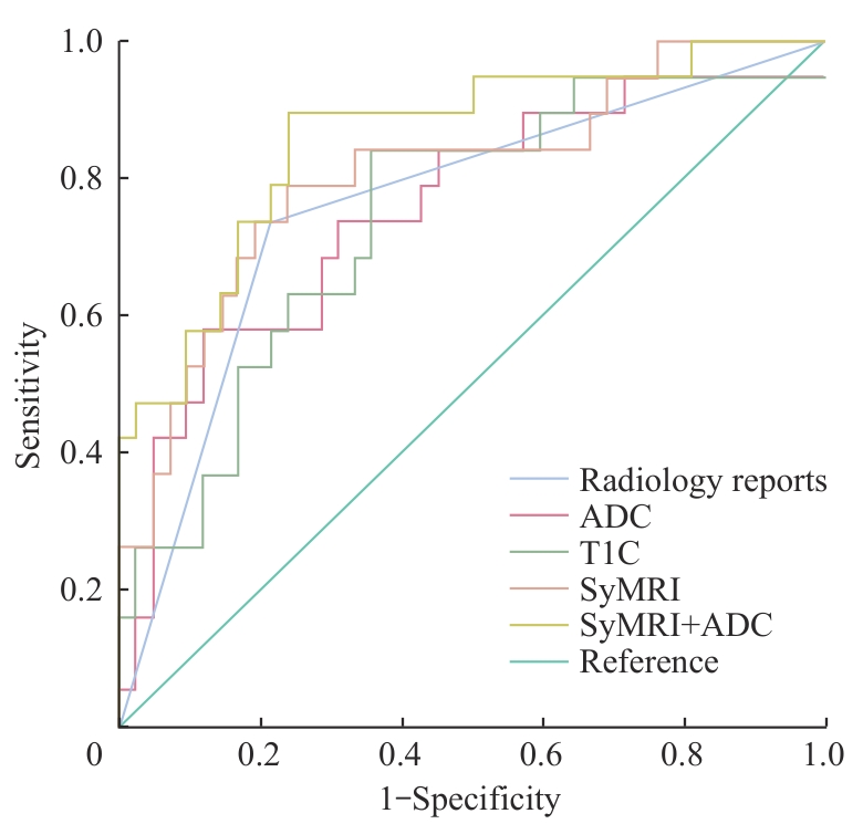
Fig 1 ROC curves of intra-tumoral histogram characteristics
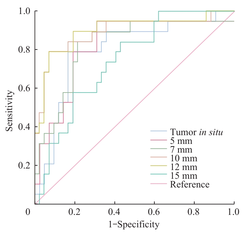
Fig 2 Predictive performance of histogram features of different peritumoral extension ranges
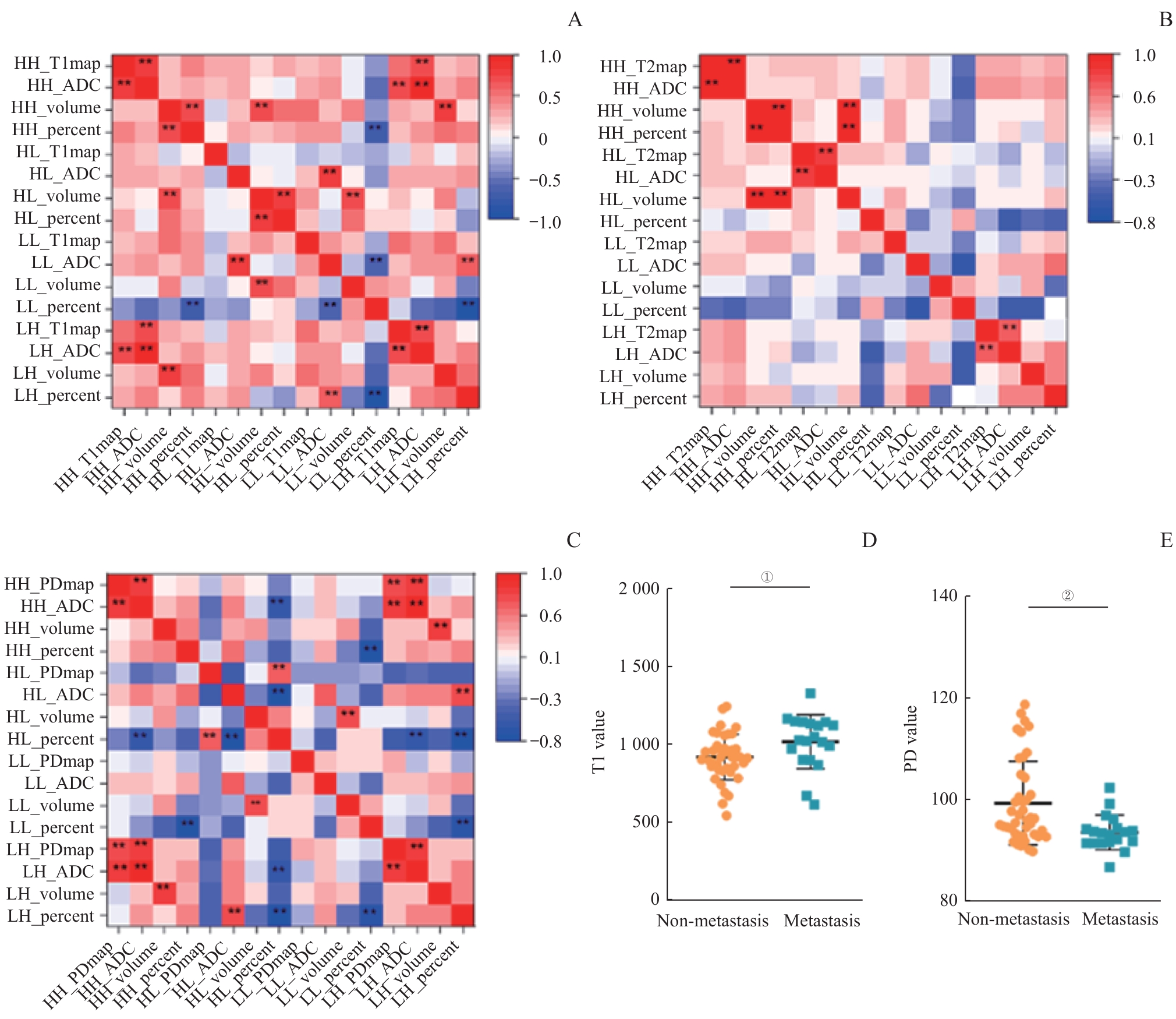
Fig 3 Heatmap of correlation analysis among habitat characteristics in different sub-regions and analysis of characteristic differences
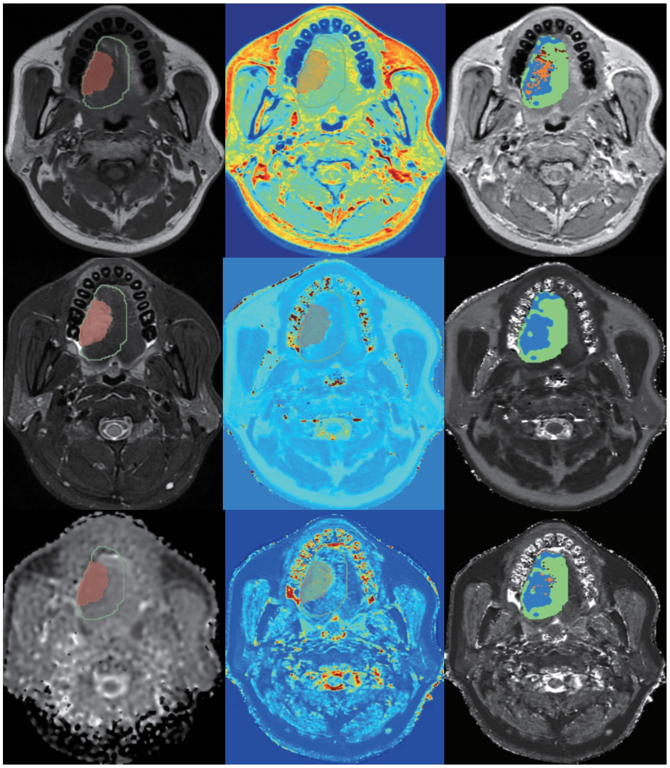
Fig 4 Results of habitat analysis based on 12 mm peritumoral extension
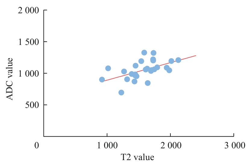
Fig 5 Correlation between T2 and ADC values
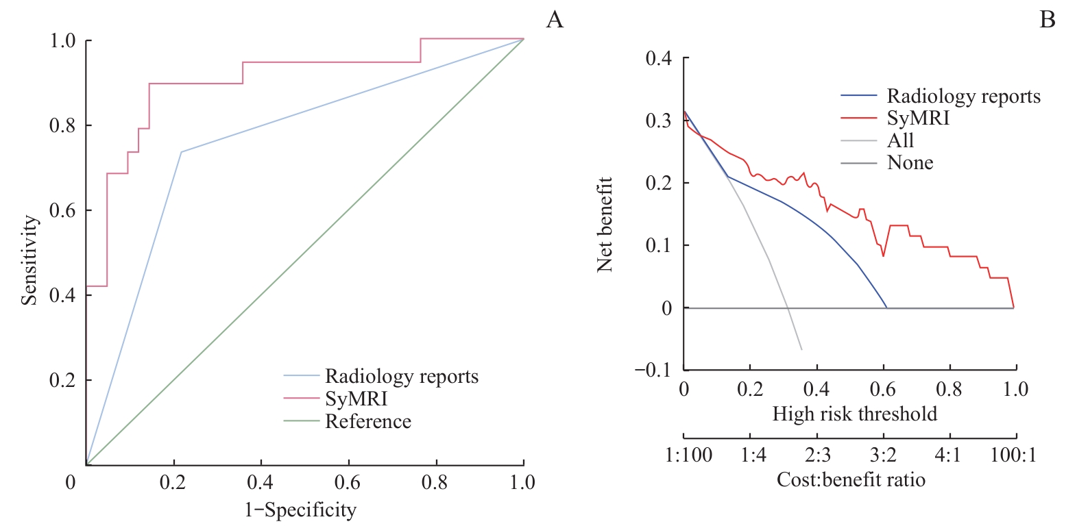
Fig 6 ROC and decision curves of the final established model
| [1] | BRAV F, LAVERSANNE M, SUNG H, et al. Global cancer statistics 2022: GLOBOCAN estimates of incidence and mortality worldwide for 36 cancers in 185 countries[J]. CA Cancer J Clin, 2024, 74: 229-263. |
| [2] | RADBRUCH A. Gadolinium deposition in the brain: we need to differentiate between chelated and dechelated gadolinium[J]. Radiology, 2018, 288(2): 434-435. |
| [3] | 崔亚东, 李春媚, 陈敏. 合成MRI技术临床应用进展[J]. 中华放射学杂志, 2021, 55(6): 677-681. |
| CUI Y D, LI C M, CHEN M. Progress in clinical application of synthetic MRI technology [J]. Chinese Journal of Radiology, 2021, 55(6): 677-681. | |
| [4] | MAI J, ABUBRIG M, LEHMANN T, et al. T2 mapping in prostate cancer[J]. Invest Radiol, 2019, 54(3): 146-152. |
| [5] | WANG F, YANG Q, ZHANG Y P, et al. 3D variable flip angle T1 mapping for differentiating benign and malignant liver lesions at 3T: comparison with diffusion weighted imaging[J]. BMC Med Imaging, 2022, 22(1): 146. |
| [6] | GE Y X, HU S D, WANG Z, et al. Feasibility and reproducibility of T2 mapping and DWI for identifying malignant lymph nodes in rectal cancer[J]. Eur Radiol, 2021, 31(5): 3347-3354. |
| [7] | WANG P, HU S D, WANG X Y, et al. Synthetic MRI in differentiating benign from metastatic retropharyngeal lymph node: combination with diffusion-weighted imaging[J]. Eur Radiol, 2023, 33(1): 152-161. |
| [8] | ZHAO L, LIANG M, SHI Z, et al. Preoperative volumetric synthetic magnetic resonance imaging of the primary tumor for a more accurate prediction of lymph node metastasis in rectal cancer[J]. Quant Imaging Med Surg, 2021, 11(5): 1805-1816. |
| [9] | JARDIM J F, GONDAK R, GALVIS M M, et al. A decreased peritumoral CD1a+ cell number predicts a worse prognosis in oral squamous cell carcinoma[J]. Histopathology, 2018, 72(6): 905-913. |
| [10] | 赵楠楠, 朱芸, 汤晓敏, 等. 基于瘤内及瘤周MRI影像组学列线图预测乳腺癌腋窝淋巴结转移[J]. 磁共振成像, 2023, 14(3): 81-87, 94. |
| ZHAO N N, ZHU Y, TANG X M, et al. Prediction of axillary lymph node metastasis in breast cancer based on intratumoral and peritumoral MRI radiomics nomogram[J]. Chinese Journal of Magnetic Resonance Imaging, 2023, 14(3): 81-87, 94. | |
| [11] | CUI Y, THA K K, TERASAKA S, et al. Prognostic imaging biomarkers in glioblastoma: development and independent validation on the basis of multiregion and quantitative analysis of MR images[J]. Radiology, 2016, 278(2): 546-553. |
| [12] | PARK J E, KIM H S, KIM N, et al. Spatiotemporal heterogeneity in multiparametric physiologic MRI is associated with patient outcomes in IDH-wildtype glioblastoma[J]. Clin Cancer Res, 2021, 27(1): 237-245. |
| [13] | 傅旖, 马辰莺, 张露, 等. 生境分析在恶性肿瘤影像组学中的研究进展[J]. 国际肿瘤学杂志, 2024, (5): 292-297. |
| FU Y, MA C Y, ZHANG L, et al. Research progress of habitat analysis in radiomics of malignant tumors[J]. Journal of International Oncology, 2024, (5): 292-297. | |
| [14] | GOURTSOYIANNI S, DOUMOU G, PREZZI D, et al. Primary rectal cancer: repeatability of global and local-regional MR imaging texture features[J]. Radiology, 2017, 284(2): 552-561. |
| [15] | CUI Y F, YANG X T, DU X S, et al. Whole-tumour diffusion kurtosis MR imaging histogram analysis of rectal adenocarcinoma: correlation with clinical pathologic prognostic factors[J]. Eur Radiol, 2018, 28(4): 1485-1494. |
| [16] | YANG L Q, LIU D, FANG X, et al. Rectal cancer: can T2WI histogram of the primary tumor help predict the existence of lymph node metastasis?[J]. Eur Radiol, 2019, 29(12): 6469-6476. |
| [17] | REN J L, YUAN Y, TAO X F. Histogram analysis of diffusion-weighted imaging and dynamic contrast-enhanced MRI for predicting occult lymph node metastasis in early-stage oral tongue squamous cell carcinoma[J]. Eur Radiol, 2022, 32(4): 2739-2747. |
| [18] | REN J, LI Y, LIU X Y, et al. Diagnostic performance of ADC values and MRI-based radiomics analysis for detecting lymph node metastasis in patients with cervical cancer: a systematic review and meta-analysis[J]. Eur J Radiol, 2022, 156: 110504. |
| [19] | YANG X, LU Z, TAN X Y, et al. Evaluating the added value of synthetic magnetic resonance imaging in predicting sentinel lymph node status in breast cancer[J]. Quant Imaging Med Surg, 2024, 14(6): 3789-3802. |
| [20] | ROONEY W D, JOHNSON G, LI X, et al. Magnetic field and tissue dependencies of human brain longitudinal 1H2O relaxation in vivo[J]. Magn Reson Med, 2007, 57(2): 308-318. |
| [21] | IORDANISHVILI E, SCHALL M, LOUÇÃO R, et al. Quantitative MRI of cerebral white matter hyperintensities: a new approach towards understanding the underlying pathology[J]. Neuroimage, 2019, 202: 116077. |
| [22] | DENG S Z, WANG S M, SHI X K, et al. Microenvironment in oral potentially malignant disorders: multi-dimensional characteristics and mechanisms of carcinogenesis[J]. Int J Mol Sci, 2022, 23(16): 8940. |
| [23] | 谢佳培, 张卫东, 朱婧怡, 等. 磁共振T1、T2值在脑胶质瘤分级及细胞增殖活性预测中的临床价值[J]. 磁共振成像, 2021, 12(1):15-20. |
| XIE J P, ZHANG W D, ZHU J Y, et al. The clinical value of MRI T1 and T2 values in predicting glioma grading and cell proliferation activity [J]. Chinese Journal of Magnetic Resonance Imaging, 2021, 12(1): 15-20. | |
| [24] | JOHNSON D E, BURTNESS B, LEEMANS C R, et al. Head and neck squamous cell carcinoma[J]. Nat Rev Dis Primers, 2020, 6(1): 92. |
| [25] | 章梓郁, 杜楠楠, 李源茂, 等. 肿瘤淋巴结转移: 肿瘤细胞与免疫系统的博弈[J]. 中华转移性肿瘤杂志, 2022, 5(4): 357-361. |
| ZHANG Z Y, DU N N, LI Y M, et al. Lymph node metastasis of tumor: the battle between tumor cells and immune system[J]. Chinese Journal of Metastatic Cancer, 2022, 5(4): 357-361. | |
| [26] | GRACIEN R M, REITZ S C, HOF S M, et al. Changes and variability of proton density and T1 relaxation times in early multiple sclerosis: MRI markers of neuronal damage in the cerebral cortex[J]. Eur Radiol, 2016, 26(8): 2578-2586. |
| [27] | CHO Y A, YOON H J, LEE J I, et al. Relationship between the expressions of PD-L1 and tumor-infiltrating lymphocytes in oral squamous cell carcinoma[J]. Oral Oncol, 2011, 47(12): 1148-1153. |
| [28] | MERMOD M, BONGIOVANNI M, PETROVA T V, et al. Prediction of occult lymph node metastasis in squamous cell carcinoma of the oral cavity and the oropharynx using peritumoral Prospero homeobox protein 1 lymphatic nuclear quantification[J]. Head Neck, 2016, 38(9): 1407-1415. |
| [29] | WU Q X, WANG S, CHEN X, et al. Radiomics analysis of magnetic resonance imaging improves diagnostic performance of lymph node metastasis in patients with cervical cancer[J]. Radiother Oncol, 2019, 138: 141-148. |
| [30] | WANG Y X, SHANG Y Y, GUO Y X, et al. Clinical study on the prediction of ALN metastasis based on intratumoral and peritumoral DCE-MRI radiomics and clinico-radiological characteristics in breast cancer[J]. Front Oncol, 2024, 14: 1357145. |
| [31] | BAI H L, XIA W, JI X F, et al. Multiparametric magnetic resonance imaging-based peritumoral radiomics for preoperative prediction of the presence of extracapsular extension with prostate cancer[J]. J Magn Reson Imaging, 2021, 54(4): 1222-1230. |
| [32] | WANG F, TAN R K, FENG K, et al. Magnetic resonance imaging-based radiomics features associated with depth of invasion predicted lymph node metastasis and prognosis in tongue cancer[J]. J Magn Reson Imaging, 2022, 56(1): 196-209. |
| [33] | WANG S X, LIU X W, WU Y, et al. Habitat-based radiomics enhances the ability to predict lymphovascular space invasion in cervical cancer: a multi-center study[J]. Front Oncol, 2023, 13: 1252074. |
| [34] | HUANG H Z, CHEN H, ZHENG D Z, et al. Habitat-based radiomics analysis for evaluating immediate response in colorectal cancer lung metastases treated by radiofrequency ablation[J]. Cancer Imaging, 2024, 24(1): 44. |
| Viewed | ||||||
|
Full text |
|
|||||
|
Abstract |
|
|||||