JOURNAL OF SHANGHAI JIAOTONG UNIVERSITY (MEDICAL SCIENCE) ›› 2021, Vol. 41 ›› Issue (5): 695-700.doi: 10.3969/j.issn.1674-8115.2021.05.023
• Techniques and methods • Previous Articles
Hao GU1( ), Xi YANG1(), Zi-min ZHANG2, Yun-bo JIN1, Li HU1, Hui CHEN1(), Xiao-xi LIN1()
Online:2021-05-28
Published:2021-05-27
Contact:
Hui CHEN,Xiao-xi LIN
E-mail:drguhao9prs@gmail.com;chenhui9801640@163.com;linxiaoxi@126.com
Supported by:CLC Number:
Hao GU, Xi YANG, Zi-min ZHANG, Yun-bo JIN, Li HU, Hui CHEN, Xiao-xi LIN. Image-fusion-based surgical navigation in sclerotherapy for low-flow vascular malformations in the risky regions of head and neck[J]. JOURNAL OF SHANGHAI JIAOTONG UNIVERSITY (MEDICAL SCIENCE), 2021, 41(5): 695-700.
Add to citation manager EndNote|Ris|BibTeX
URL: https://xuebao.shsmu.edu.cn/EN/10.3969/j.issn.1674-8115.2021.05.023
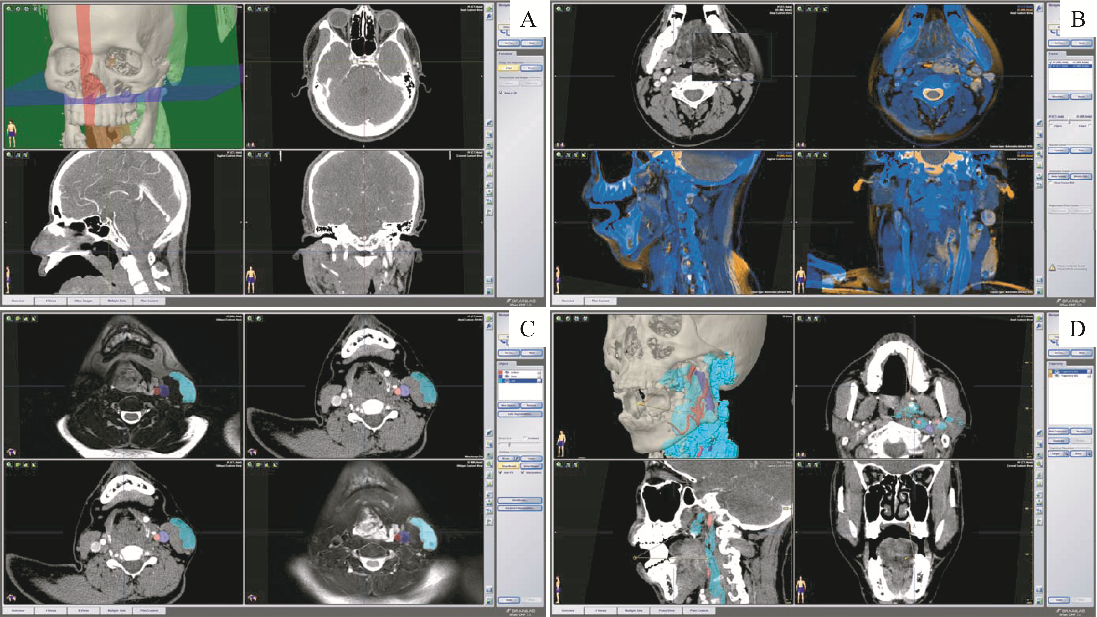
Fig 1 Preoperative planning with the software of surgical navigation system
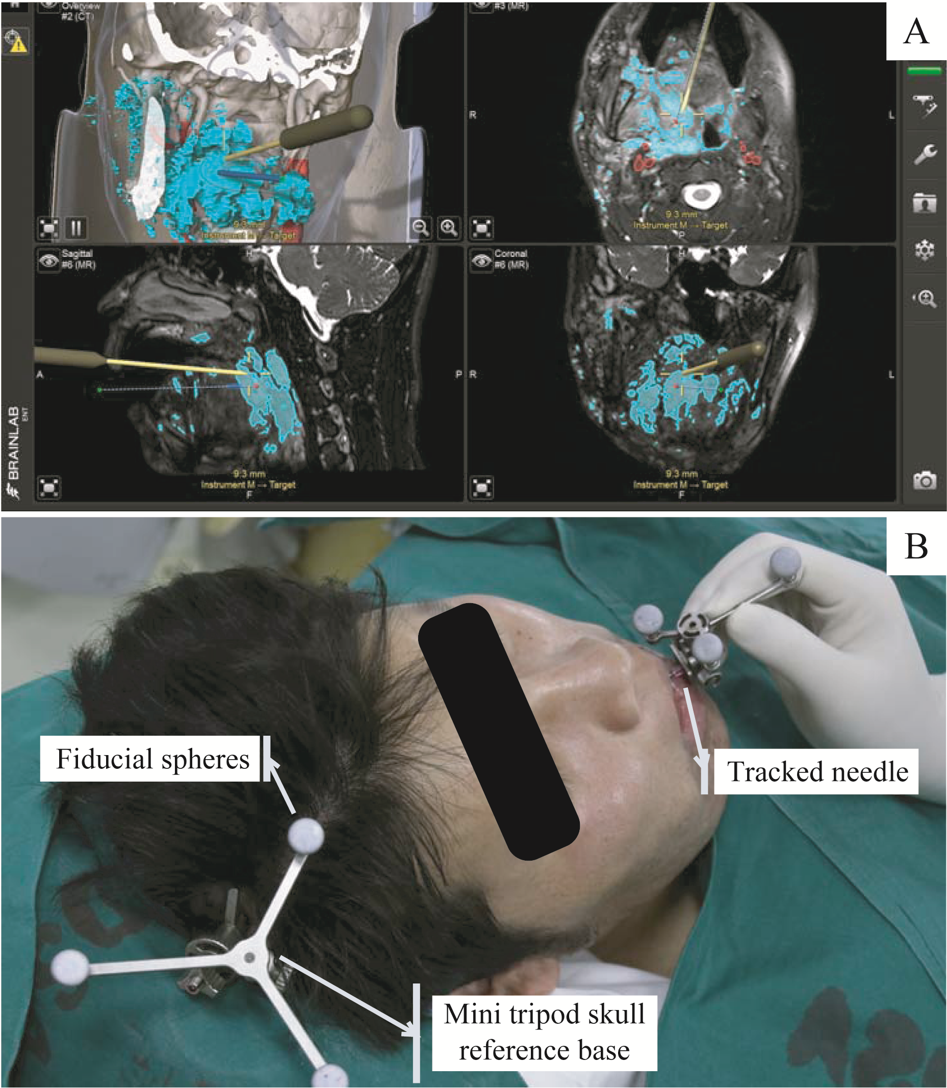
Fig 2 Navigation-guided needle puncture during sclerotherapy
| Patient No. | Sex | Age/year | Site | Diagnosis | Previous treatment | Local symptom |
|---|---|---|---|---|---|---|
| 1 | Female | 47 | Left orbit | VM | Percutaneous sclerotherapy | Argamblyopia caused by blepharoptosis and swelling |
| 2 | Female | 3 | Left orbit | LM | Percutaneous sclerotherapy | |
| 3 | Female | 19 | Left orbit | VM | Percutaneous sclerotherapy and laser | |
| 4 | Male | 6 | Right orbit | LM | Percutaneous sclerotherapy | |
| 5 | Female | 8 | Left orbit | LM | Percutaneous sclerotherapy | |
| 6 | Male | 16 | Bilateral orbit | VM | Percutaneous sclerotherapy | Swelling |
| 7 | Male | 27 | Left face and neck | VM | Percutaneous sclerotherapy and laser | Dyspnoea (before tracheotomy) |
| 8 | Male | 29 | Right face and neck | VM | Percutaneous sclerotherapy, laser, and orthodontic orthognathic treatment | |
| 9 | Male | 33 | Bilateral face and neck | VM | Laryngoscope-guided sclerotherapy, resection of phlebolith, and percutaneous sclerotherapy | |
| 10 | Male | 21 | Bilateral face and neck | VM | Percutaneous sclerotherapy |
Tab 1 General information of the subjects
| Patient No. | Sex | Age/year | Site | Diagnosis | Previous treatment | Local symptom |
|---|---|---|---|---|---|---|
| 1 | Female | 47 | Left orbit | VM | Percutaneous sclerotherapy | Argamblyopia caused by blepharoptosis and swelling |
| 2 | Female | 3 | Left orbit | LM | Percutaneous sclerotherapy | |
| 3 | Female | 19 | Left orbit | VM | Percutaneous sclerotherapy and laser | |
| 4 | Male | 6 | Right orbit | LM | Percutaneous sclerotherapy | |
| 5 | Female | 8 | Left orbit | LM | Percutaneous sclerotherapy | |
| 6 | Male | 16 | Bilateral orbit | VM | Percutaneous sclerotherapy | Swelling |
| 7 | Male | 27 | Left face and neck | VM | Percutaneous sclerotherapy and laser | Dyspnoea (before tracheotomy) |
| 8 | Male | 29 | Right face and neck | VM | Percutaneous sclerotherapy, laser, and orthodontic orthognathic treatment | |
| 9 | Male | 33 | Bilateral face and neck | VM | Laryngoscope-guided sclerotherapy, resection of phlebolith, and percutaneous sclerotherapy | |
| 10 | Male | 21 | Bilateral face and neck | VM | Percutaneous sclerotherapy |
| Patient No. | Region | Approach | Result of puncture | Sclerosant | Complication | Follow-up/month |
|---|---|---|---|---|---|---|
| 1 | Left retrobulbar | Percutaneously | Success | B | None | 16 |
| 2 | Left retrobulbar | Percutaneously | Success | B | None | 9 |
| 3 | Left nasal intraorbital | Percutaneously | Success | BPF | None | 3 |
| Left inferior intraorbital | Percutaneously | Success | BPF | None | ||
| 4 | Right superior nasal intraorbital | Percutaneously | Success | B | None | 4 |
| Right inferior temporal intraorbital | Percutaneously | Success | B | None | ||
| 5 | Left superior nasal intraorbital | Percutaneously | Failure | No delivery | None | 6 |
| 6 | Right nasal intraorbital | Percutaneously | Success | BPF | None | 4 |
| Left retrobulbar | Percutaneously | Success | BPF | None | ||
| 7 | ||||||
| 1st procedure | Left nasopharynx | Transorally | Success | No delivery① | None | 2.5 |
| Lingual radix | Transorally | Success | BPF | None | ||
| 2nd procedure | Left lingual radix | Transorally | Success | BPF | None | 7.5 |
| Left nasopharynx | Transorally | Failure | No delivery | None | ||
| 3rd procedure | Left nasopharynx | Transorally | Failure | No delivery | None | 3 |
| Posterior wall of left oropharynx | Percutaneously through mouth floor | Success | BPF | None | ||
| 4th procedure | Left nasopharynx | Transorally | Success | BPF | None | 5 |
| Posterior wall of left oropharynx | Percutaneously through mouth floor | Failure | No delivery | None | ||
| 8 | ||||||
| 1st procedure | Posterior wall of right nasopharynx and oropharynx | Transorally | Success | BPF | None | 3.5 |
| 2nd procedure | Right lingual radix | Transorally | Success | BPF | None | 8.5 |
| Posterior wall of right nasopharynx and oropharynx | Transorally | Success | A & BPF | None | ||
| 3rd procedure | Right lingual radix | Percutaneously through mouth floor | Success | BPF | None | 8 |
| 9 | ||||||
| 1st procedure | Left supraglottic | Transorally | Success | BPF | None | 4 |
| Right nasopharynx | Transorally | Failure | No delivery | None | ||
| Glottis | Percutaneously | Success | A | Pneumonia caused by glottic ulcer | ||
| 2nd procedure | Left laryngopharynx | Percutaneously through mouth floor | Success | BPF | None | 1.5 |
| Right nasopharynx | Transorally | Failure | No delivery | None | ||
| 3rd procedure | Left supraglottic | Percutaneously through mouth floor | Success | BPF | None | 11 |
| 10 | ||||||
| 1st procedure | Right nasopharynx | Transorally | Success | BPF | None | 6 |
| 2nd procedure | Right oropharynx | Transorally | Success | A & BPF | None | 5 |
| 3rd procedure | Pre-epiglottic space | Percutaneously through mouth floor | Success | BPF | None | 3 |
Tab 2 Puncture outcomes and details of navigation-assisted procedures
| Patient No. | Region | Approach | Result of puncture | Sclerosant | Complication | Follow-up/month |
|---|---|---|---|---|---|---|
| 1 | Left retrobulbar | Percutaneously | Success | B | None | 16 |
| 2 | Left retrobulbar | Percutaneously | Success | B | None | 9 |
| 3 | Left nasal intraorbital | Percutaneously | Success | BPF | None | 3 |
| Left inferior intraorbital | Percutaneously | Success | BPF | None | ||
| 4 | Right superior nasal intraorbital | Percutaneously | Success | B | None | 4 |
| Right inferior temporal intraorbital | Percutaneously | Success | B | None | ||
| 5 | Left superior nasal intraorbital | Percutaneously | Failure | No delivery | None | 6 |
| 6 | Right nasal intraorbital | Percutaneously | Success | BPF | None | 4 |
| Left retrobulbar | Percutaneously | Success | BPF | None | ||
| 7 | ||||||
| 1st procedure | Left nasopharynx | Transorally | Success | No delivery① | None | 2.5 |
| Lingual radix | Transorally | Success | BPF | None | ||
| 2nd procedure | Left lingual radix | Transorally | Success | BPF | None | 7.5 |
| Left nasopharynx | Transorally | Failure | No delivery | None | ||
| 3rd procedure | Left nasopharynx | Transorally | Failure | No delivery | None | 3 |
| Posterior wall of left oropharynx | Percutaneously through mouth floor | Success | BPF | None | ||
| 4th procedure | Left nasopharynx | Transorally | Success | BPF | None | 5 |
| Posterior wall of left oropharynx | Percutaneously through mouth floor | Failure | No delivery | None | ||
| 8 | ||||||
| 1st procedure | Posterior wall of right nasopharynx and oropharynx | Transorally | Success | BPF | None | 3.5 |
| 2nd procedure | Right lingual radix | Transorally | Success | BPF | None | 8.5 |
| Posterior wall of right nasopharynx and oropharynx | Transorally | Success | A & BPF | None | ||
| 3rd procedure | Right lingual radix | Percutaneously through mouth floor | Success | BPF | None | 8 |
| 9 | ||||||
| 1st procedure | Left supraglottic | Transorally | Success | BPF | None | 4 |
| Right nasopharynx | Transorally | Failure | No delivery | None | ||
| Glottis | Percutaneously | Success | A | Pneumonia caused by glottic ulcer | ||
| 2nd procedure | Left laryngopharynx | Percutaneously through mouth floor | Success | BPF | None | 1.5 |
| Right nasopharynx | Transorally | Failure | No delivery | None | ||
| 3rd procedure | Left supraglottic | Percutaneously through mouth floor | Success | BPF | None | 11 |
| 10 | ||||||
| 1st procedure | Right nasopharynx | Transorally | Success | BPF | None | 6 |
| 2nd procedure | Right oropharynx | Transorally | Success | A & BPF | None | 5 |
| 3rd procedure | Pre-epiglottic space | Percutaneously through mouth floor | Success | BPF | None | 3 |
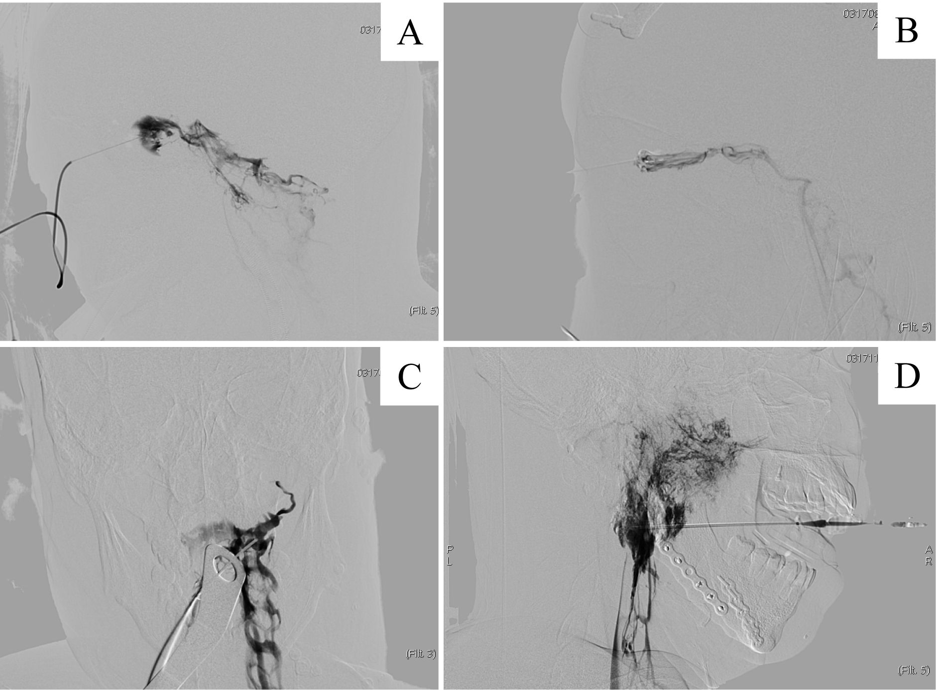
Fig 3 Intraoperative DSA images
| 1 | Mulliken JB, Glowacki J. Hemangiomas and vascular malformations in infants and children: a classification based on endothelial characteristics[J]. Plast Reconstr Surg, 1982, 69(3): 412-422. |
| 2 | Mulliken JB, Fishman SJ, Burrows PE. Vascular anomalies[J]. Curr Probl Surg, 2000, 37(8): 517-584. |
| 3 | 中华医学会整形外科分会血管瘤和脉管畸形学组. 血管瘤和脉管畸形的诊断及治疗指南(2019版)[J]. 组织工程与重建外科杂志, 2019, 15(5): 277-317. |
| 4 | Heit JJ, Do HM, Prestigiacomo CJ, et al. Guidelines and parameters: percutaneous sclerotherapy for the treatment of head and neck venous and lymphatic malformations[J]. J Neurointerv Surg, 2017, 9(6): 611-617. |
| 5 | Leonard S, Sinha A, Reiter A, et al. Evaluation and stability analysis of video-based navigation system for functional endoscopic sinus surgery on in vivo clinical data[J]. IEEE Trans Med Imaging, 2018, 37(10): 2185-2195. |
| 6 | Nesbit GM, Nesbit EG, Hamilton BE. Integrated cone-beam CT and fluoroscopic navigation in treatment of head and neck vascular malformations and tumors[J]. J Neurointerv Surg, 2011, 3(2): 186-190. |
| 7 | Partovi S, Lu ZA, Vidal L, et al. Real-time MRI-guided percutaneous sclerotherapy treatment of venous low-flow malformations in the head and neck[J]. Phlebology, 2018, 33(5): 344-352. |
| 8 | Unsgaard G, Ommedal S, Rygh OM, et al. Operation of arteriovenous malformations assisted by stereoscopic navigation-controlled display of preoperative magnetic resonance angiography and intraoperative ultrasound angiography[J]. Neurosurgery, 2005, 56(2): ons281-ons290. |
| 9 | Gonzalez LF, Albuquerque FC, Boom S, et al. Image-guided resection of embolized cerebral arteriovenous malformations based on catheter-based angiography[J]. Neurosurgery, 2010, 67(2): 471-475. |
| 10 | Raza SM, Papadimitriou K, Gandhi D, et al. Intra-arterial intraoperative computed tomography angiography guided navigation: a new technique for localization of vascular pathology[J]. Neurosurgery, 2012, 71(2 ): ons240-ons252. |
| 11 | Ernemann U, Westendorff C, Troitzsch D, et al. Navigation-assisted sclerotherapy of orbital venolymphatic malformation: a new guidance technique for percutaneous treatment of low-flow vascular malformations[J]. AJNR Am J Neuroradiol, 2004, 25(10): 1792-1795. |
| 12 | Mamlouk MD, Nicholson AD, Cooke DL, et al. Tips and tricks to optimize MRI protocols for cutaneous vascular anomalies[J]. Clin Imaging, 2017, 45: 71-80. |
| 13 | Compter I, Peerlings J, Eekers DB, et al. Technical feasibility of integrating 7 T anatomical MRI in image-guided radiotherapy of glioblastoma: a preparatory study[J]. MAGMA, 2016, 29(3): 591-603. |
| 14 | Chhabra A, Faridian-Aragh N, Chalian M, et al. High-resolution 3-T MR neurography of peroneal neuropathy[J]. Skeletal Radiol, 2012, 41(3): 257-271. |
| 15 | Hong GX, Yang ZY, Chu JP, et al. Three-dimensional MRI with contrast diagnosis of diseases involving peripheral oculomotor nerve[J]. Clin Imaging, 2012, 36(6): 674-679. |
| 16 | Algin O, Turkbey B. Evaluation of aqueductal stenosis by 3D sampling perfection with application-optimized contrasts using different flip angle evolutions sequence: preliminary results with 3T MR imaging[J]. AJNR Am J Neuroradiol, 2012, 33(4): 740-746. |
| 17 | Lu SS, Ge S, Su CQ, et al. MRI of plaque characteristics and relationship with downstream perfusion and cerebral infarction in patients with symptomatic middle cerebral artery stenosis[J]. J Magn Reson Imaging, 2018, 48(1): 66-73. |
| [1] | Yin Wenyu, Wang Ruijie, Ma Hailong, Jin Shufang. Study on how histone lysine lactylation regulates zinc finger protein 668 to facilitate proliferation of head and neck squamous cell carcinoma cells [J]. Journal of Shanghai Jiao Tong University (Medical Science), 2026, 46(2): 151-162. |
| [2] | LI Siyu, CHEN Ya, HU Wentao, DAI Yongming, WU Yingwei. Using diffusion-relaxation correlation spectroscopic imaging to assess the heterogeneity of head and neck tumors and identify occult lymph node metastasis [J]. Journal of Shanghai Jiao Tong University (Medical Science), 2025, 45(9): 1202-1213. |
| [3] | CHEN Yinan, ZHENG Yang, ZENG Hanlin, LEI Ming. Mechanism of Fas-associated protein with death domain in promoting proliferation of head and neck squamous cell carcinoma cells [J]. Journal of Shanghai Jiao Tong University (Medical Science), 2025, 45(4): 404-414. |
| [4] | AN Junyi, CHEN Biying, CHEN Xunrui, YIN Shanshan, BIAN Zhouliang, LIU Feng. SFXN3 expression in head and neck squamous cell carcinoma and its effect on cell proliferation [J]. Journal of Shanghai Jiao Tong University (Medical Science), 2024, 44(4): 427-434. |
| [5] | GU Fen, WANG Yueping, YANG Wenyu, ZHAO Xiaomei, TANG Yan, SHEN Shukun, MAO Yan, ZHANG Jinfeng, WU Yifan, ZHANG Yuanyuan, YANG Yue, ZHANG Jianchun, YU Hong, WANG Lan, HAO Guihua, HOU Lili. Expert consensus on postoperative rehabilitation nursing of patients with head and neck cancer [J]. Journal of Shanghai Jiao Tong University (Medical Science), 2023, 43(10): 1289-1296. |
| [6] | CHEN Weihong, HOU Lili, YANG Ling, MAO Yan, ZHANG Jinfeng. Efficacy of preventive intervention of cryotherapy on radiation-induced oral mucositis in head and neck cancer: a meta-analysis [J]. Journal of Shanghai Jiao Tong University (Medical Science), 2022, 42(5): 635-645. |
| [7] | Yu-sheng LU, Wen-yi YANG, Hai-long MA, Jing-zhou HU. Impact of Ras association domain family 5 on cell migration and invasion of head and neck squamous cell carcinoma [J]. JOURNAL OF SHANGHAI JIAOTONG UNIVERSITY (MEDICAL SCIENCE), 2021, 41(6): 717-723. |
| [8] | Tong TONG, Xing QIN, Fei XIE, Ying-ying JIANG, Jian-bo SHI, Jian-jun ZHANG. USP47 induces cisplatin resistance in head and neck squamous cell carcinoma by up-regulation of anti-apoptotic proteins [J]. JOURNAL OF SHANGHAI JIAOTONG UNIVERSITY (MEDICAL SCIENCE), 2021, 41(4): 434-441. |
| [9] | Yong-chuan LI, Xin-wu LU, Zhi-hua ZHANG, Jin-bao QIN, Xing ZHANG. Analysis of the efficacy of endovascular thermal ablation (980 nm laser) combined with foam sclerotherapy in day surgery for lower extremity varicose vein [J]. JOURNAL OF SHANGHAI JIAOTONG UNIVERSITY (MEDICAL SCIENCE), 2021, 41(10): 1308-1312. |
| [10] | XIA Zhi-peng1, YUAN Ying2, YANG Xi2, GU Hao2, LIN Xiao-xi 2#, TAO Xiao-feng1#. Value of dynamic contrast-enhanced MRI in selecting sclerosants for endovascular sclerosis of venous malformation [J]. JOURNAL OF SHANGHAI JIAOTONG UNIVERSITY (MEDICAL SCIENCE), 2020, 40(7): 873-880. |
| [11] | ZHANG Yu-han, CHEN Xue-hua, ZHANG Hao. Down-regulation of aurora kinase A activity inhibits pro-angiogenesis, migration and invasion of Hep2 cells [J]. , 2018, 38(10): 1181-. |
| [12] | QU Yang, AI Song-tao, YANG Fei, ZHANG Heng-hui, WANG Yi-ping, TAO Xiao-feng, HAO Yong-qiang, DAI Ke-rong. Application of CT/MRI image registration and fusion combined with 3D printing technique in pre-surgical planning of refractory pelvic tumors#br# [J]. , 2017, 37(9): 1239-. |
| [13] | DING Yu, WANG Xin, WANG Bing, ZHAO Hai-guang. Analysis of short to medium-term efficiency of 1 470 nm laser with foam sclerotherapy to treat varicose veins of lower extremity [J]. , 2017, 37(8): 1138-. |
| [14] | WANG Shu-ze, WANG Xu-dong . Advances in the application of intraoperative computed tomography to surgeries [J]. , 2017, 37(6): 876-. |
| [15] | LIU Chun-jie, LI Ting-ting, LI Fen, XU Rang . Analysis of clinical characteristics and death risk factors of patients with heterotaxy syndrome [J]. , 2016, 36(12): 1772-. |
| Viewed | ||||||
|
Full text |
|
|||||
|
Abstract |
|
|||||