Journal of Shanghai Jiao Tong University (Medical Science) ›› 2022, Vol. 42 ›› Issue (5): 617-623.doi: 10.3969/j.issn.1674-8115.2022.05.009
• Clinical research • Previous Articles Next Articles
ZHU Tianyu1,2( ), ZONG Chunyan1,2, XU Shiqiong1,2, GE Shengfang1,2, FAN Xianqun1,2, JIA Renbing1,2()
Received:2022-03-19
Accepted:2022-05-25
Online:2022-05-28
Published:2022-05-28
Contact:
JIA Renbing
E-mail:zzzty555@sjtu.edu.cn;renbingjia@sjtu.edu.cn
Supported by:CLC Number:
ZHU Tianyu, ZONG Chunyan, XU Shiqiong, GE Shengfang, FAN Xianqun, JIA Renbing. Relationship between histopathological features, Ki-67 expression and prognosis of conjunctival melanoma[J]. Journal of Shanghai Jiao Tong University (Medical Science), 2022, 42(5): 617-623.
Add to citation manager EndNote|Ris|BibTeX
URL: https://xuebao.shsmu.edu.cn/EN/10.3969/j.issn.1674-8115.2022.05.009
| Variable | n(%) | Variable | n(%) |
|---|---|---|---|
| Laterality | Ulceration | ||
| Left | 18 (50.0) | Absent | 23 (63.9) |
| Right | 18 (50.0) | Present | 13 (36.1) |
| Lacrimal caruncle involvement | Regression | ||
| Yes | 11 (30.6) | Absent | 29 (80.6) |
| No | 25 (69.4) | Present | 7 (19.4) |
| Fornix involvement | Perineural invasion | ||
| Yes | 8 (22.2) | Absent | 33 (91.7) |
| No | 28 (77.8) | Present | 3 (8.3) |
| Palpebral conjunctiva involvement | Vascular invasion | ||
| Yes | 15 (41.7) | Absent | 28 (77.8) |
| No | 21 (58.3) | Present | 8 (22.2) |
| Local recurrence (during follow-up) | Ki-67 index | ||
| Yes | 16 (44.4) | ≤20% | 22 (61.1) |
| No | 20 (55.6) | >20% | 14 (38.9) |
| AJCC cT category | Histological type | ||
| T1 | 1 (2.8) | Superficial spreading | 18 (50.0) |
| T2 | 12 (33.3) | Nodular | 12 (33.3) |
| T3 | 23 (63.9) | Lentiginous | 4 (11.1) |
| T4 | 0 (0) | Unknown | 2 (5.6) |
| Distant metastasis (during follow-up) | Disease-specific death (during follow-up) | ||
| Yes | 17 (47.2) | Yes | 16 (44.4) |
| No | 19 (52.8) | No | 20 (55.6) |
| Breslow thickness | TILs | ||
| ≤2 mm | 7 (19.4) | Absent | 6 (16.7) |
| >2 mm and ≤4 mm | 11 (30.6) | Nonbrisk | 26 (72.2) |
| >4 mm | 18 (50.0) | Brisk | 4 (11.1) |
Tab 1 Characteristics and histopathological features of CoM patients
| Variable | n(%) | Variable | n(%) |
|---|---|---|---|
| Laterality | Ulceration | ||
| Left | 18 (50.0) | Absent | 23 (63.9) |
| Right | 18 (50.0) | Present | 13 (36.1) |
| Lacrimal caruncle involvement | Regression | ||
| Yes | 11 (30.6) | Absent | 29 (80.6) |
| No | 25 (69.4) | Present | 7 (19.4) |
| Fornix involvement | Perineural invasion | ||
| Yes | 8 (22.2) | Absent | 33 (91.7) |
| No | 28 (77.8) | Present | 3 (8.3) |
| Palpebral conjunctiva involvement | Vascular invasion | ||
| Yes | 15 (41.7) | Absent | 28 (77.8) |
| No | 21 (58.3) | Present | 8 (22.2) |
| Local recurrence (during follow-up) | Ki-67 index | ||
| Yes | 16 (44.4) | ≤20% | 22 (61.1) |
| No | 20 (55.6) | >20% | 14 (38.9) |
| AJCC cT category | Histological type | ||
| T1 | 1 (2.8) | Superficial spreading | 18 (50.0) |
| T2 | 12 (33.3) | Nodular | 12 (33.3) |
| T3 | 23 (63.9) | Lentiginous | 4 (11.1) |
| T4 | 0 (0) | Unknown | 2 (5.6) |
| Distant metastasis (during follow-up) | Disease-specific death (during follow-up) | ||
| Yes | 17 (47.2) | Yes | 16 (44.4) |
| No | 19 (52.8) | No | 20 (55.6) |
| Breslow thickness | TILs | ||
| ≤2 mm | 7 (19.4) | Absent | 6 (16.7) |
| >2 mm and ≤4 mm | 11 (30.6) | Nonbrisk | 26 (72.2) |
| >4 mm | 18 (50.0) | Brisk | 4 (11.1) |
| Variable | Ref | Local recurrence | Distant metastasis | Disease-specific death | |||
|---|---|---|---|---|---|---|---|
| OR(95%CI) | P | OR(95%CI) | P | OR(95%CI) | P | ||
| Breslow thickness | ‒ | 1.36 (1.06‒1.75) | 0.017 | 1.04 (0.89‒1.23) | 0.606 | 1.06 (0.89‒1.25) | 0.525 |
| Breslow thickness (>4 mm) | ≤4 mm | 4.09 (1.01‒16.58) | 0.049 | 3.14 (0.80‒12.28) | 0.100 | 4.09 (1.01‒16.58) | 0.049 |
| Ulceration | Absent | 5.14 (1.18‒22.48) | 0.030 | 2.49 (0.62‒10.06) | 0.201 | 3.00 (0.73‒12.27) | 0.126 |
| TILs (nonbrisk) | Absent | 0.73 (0.12‒4.35) | 0.733 | 5.00 (0.51‒48.91) | 0.167 | 4.29 (0.44‒41.95) | 0.211 |
| TILs (brisk) | Absent | 1.00 (0.08‒12.56) | 1.000 | 15.00 (0.66‒339.55) | 0.089 | 15.00 (0.66‒339.55) | 0.089 |
| Mitotic rate | ‒ | 1.05 (0.97‒1.15) | 0.208 | 1.03 (0.95‒1.12) | 0.485 | 1.03 (0.95‒1.11) | 0.504 |
| Regression | Absent | 0.92 (0.17‒4.89) | 0.925 | 9.82 (1.04‒92.78) | 0.046 | 4.10 (0.67‒24.83) | 0.126 |
| Perineural invasion | Absent | 2.71 (0.22‒32.99) | 0.433 | 2.40 (0.20‒29.13) | 0.492 | 2.71 (0.22‒32.99) | 0.433 |
| Vascular invasion | Absent | 2.58 (0.51‒13.01) | 0.252 | 2.22 (0.44‒11.18) | 0.333 | 2.58 (0.51‒13.01) | 0.252 |
Tab 2 Binary Logistic regression analysis of the relationship between histopathological factors and prognosis
| Variable | Ref | Local recurrence | Distant metastasis | Disease-specific death | |||
|---|---|---|---|---|---|---|---|
| OR(95%CI) | P | OR(95%CI) | P | OR(95%CI) | P | ||
| Breslow thickness | ‒ | 1.36 (1.06‒1.75) | 0.017 | 1.04 (0.89‒1.23) | 0.606 | 1.06 (0.89‒1.25) | 0.525 |
| Breslow thickness (>4 mm) | ≤4 mm | 4.09 (1.01‒16.58) | 0.049 | 3.14 (0.80‒12.28) | 0.100 | 4.09 (1.01‒16.58) | 0.049 |
| Ulceration | Absent | 5.14 (1.18‒22.48) | 0.030 | 2.49 (0.62‒10.06) | 0.201 | 3.00 (0.73‒12.27) | 0.126 |
| TILs (nonbrisk) | Absent | 0.73 (0.12‒4.35) | 0.733 | 5.00 (0.51‒48.91) | 0.167 | 4.29 (0.44‒41.95) | 0.211 |
| TILs (brisk) | Absent | 1.00 (0.08‒12.56) | 1.000 | 15.00 (0.66‒339.55) | 0.089 | 15.00 (0.66‒339.55) | 0.089 |
| Mitotic rate | ‒ | 1.05 (0.97‒1.15) | 0.208 | 1.03 (0.95‒1.12) | 0.485 | 1.03 (0.95‒1.11) | 0.504 |
| Regression | Absent | 0.92 (0.17‒4.89) | 0.925 | 9.82 (1.04‒92.78) | 0.046 | 4.10 (0.67‒24.83) | 0.126 |
| Perineural invasion | Absent | 2.71 (0.22‒32.99) | 0.433 | 2.40 (0.20‒29.13) | 0.492 | 2.71 (0.22‒32.99) | 0.433 |
| Vascular invasion | Absent | 2.58 (0.51‒13.01) | 0.252 | 2.22 (0.44‒11.18) | 0.333 | 2.58 (0.51‒13.01) | 0.252 |
| Variable | Ki-67 index | χ2 | P | |
|---|---|---|---|---|
| ≤20% | >20% | |||
| Breslow thickness | 1.870 | 0.305 | ||
| ≤4 mm | 13 (72.2) | 5 (27.8) | ||
| >4 mm | 9 (50.0) | 9 (50.0) | ||
| Ulceration | 4.392 | 0.073 | ||
| Absent | 17 (73.9) | 6 (26.1) | ||
| Present | 5 (38.5) | 8 (61.5) | ||
| TILs | 2.338 | 0.181 | ||
| Absent | 2 (33.3) | 4 (66.7) | ||
| Present | 20 (66.7) | 10 (33.3) | ||
| Regression | 0.389 | 0.681 | ||
| Absent | 17 (58.6) | 12 (41.4) | ||
| Present | 5 (71.4) | 2 (28.6) | ||
| Perineural invasion | 0.043 | 1.000 | ||
| Absent | 20 (60.6) | 13 (39.4) | ||
| Present | 2 (66.7) | 1 (33.3) | ||
| Vascular invasion | 0.008 | 1.000 | ||
| Absent | 17 (60.7) | 11 (39.3) | ||
| Present | 5 (62.5) | 3 (37.5) | ||
| Local recurrence | 6.756 | 0.016 | ||
| Absent | 16 (80.0) | 4 (20.0) | ||
| Present | 6 (37.5) | 10 (62.5) | ||
| Nodal metastasis | 0.719 | 0.462 | ||
| Absent | 17 (65.4) | 9 (34.6) | ||
| Present | 5 (50.0) | 5 (50.0) | ||
| Distant metastasis | 2.676 | 0.171 | ||
| Absent | 14 (73.7) | 5 (26.3) | ||
| Present | 8 (47.1) | 9 (52.9) | ||
| Disease-specific death | 3.653 | 0.087 | ||
| Absent | 15 (75.0) | 5 (25.0) | ||
| Present | 7 (43.8) | 9 (56.3) | ||
Tab 3 Correlation of Ki-67 index with histopathological factors and prognosis
| Variable | Ki-67 index | χ2 | P | |
|---|---|---|---|---|
| ≤20% | >20% | |||
| Breslow thickness | 1.870 | 0.305 | ||
| ≤4 mm | 13 (72.2) | 5 (27.8) | ||
| >4 mm | 9 (50.0) | 9 (50.0) | ||
| Ulceration | 4.392 | 0.073 | ||
| Absent | 17 (73.9) | 6 (26.1) | ||
| Present | 5 (38.5) | 8 (61.5) | ||
| TILs | 2.338 | 0.181 | ||
| Absent | 2 (33.3) | 4 (66.7) | ||
| Present | 20 (66.7) | 10 (33.3) | ||
| Regression | 0.389 | 0.681 | ||
| Absent | 17 (58.6) | 12 (41.4) | ||
| Present | 5 (71.4) | 2 (28.6) | ||
| Perineural invasion | 0.043 | 1.000 | ||
| Absent | 20 (60.6) | 13 (39.4) | ||
| Present | 2 (66.7) | 1 (33.3) | ||
| Vascular invasion | 0.008 | 1.000 | ||
| Absent | 17 (60.7) | 11 (39.3) | ||
| Present | 5 (62.5) | 3 (37.5) | ||
| Local recurrence | 6.756 | 0.016 | ||
| Absent | 16 (80.0) | 4 (20.0) | ||
| Present | 6 (37.5) | 10 (62.5) | ||
| Nodal metastasis | 0.719 | 0.462 | ||
| Absent | 17 (65.4) | 9 (34.6) | ||
| Present | 5 (50.0) | 5 (50.0) | ||
| Distant metastasis | 2.676 | 0.171 | ||
| Absent | 14 (73.7) | 5 (26.3) | ||
| Present | 8 (47.1) | 9 (52.9) | ||
| Disease-specific death | 3.653 | 0.087 | ||
| Absent | 15 (75.0) | 5 (25.0) | ||
| Present | 7 (43.8) | 9 (56.3) | ||
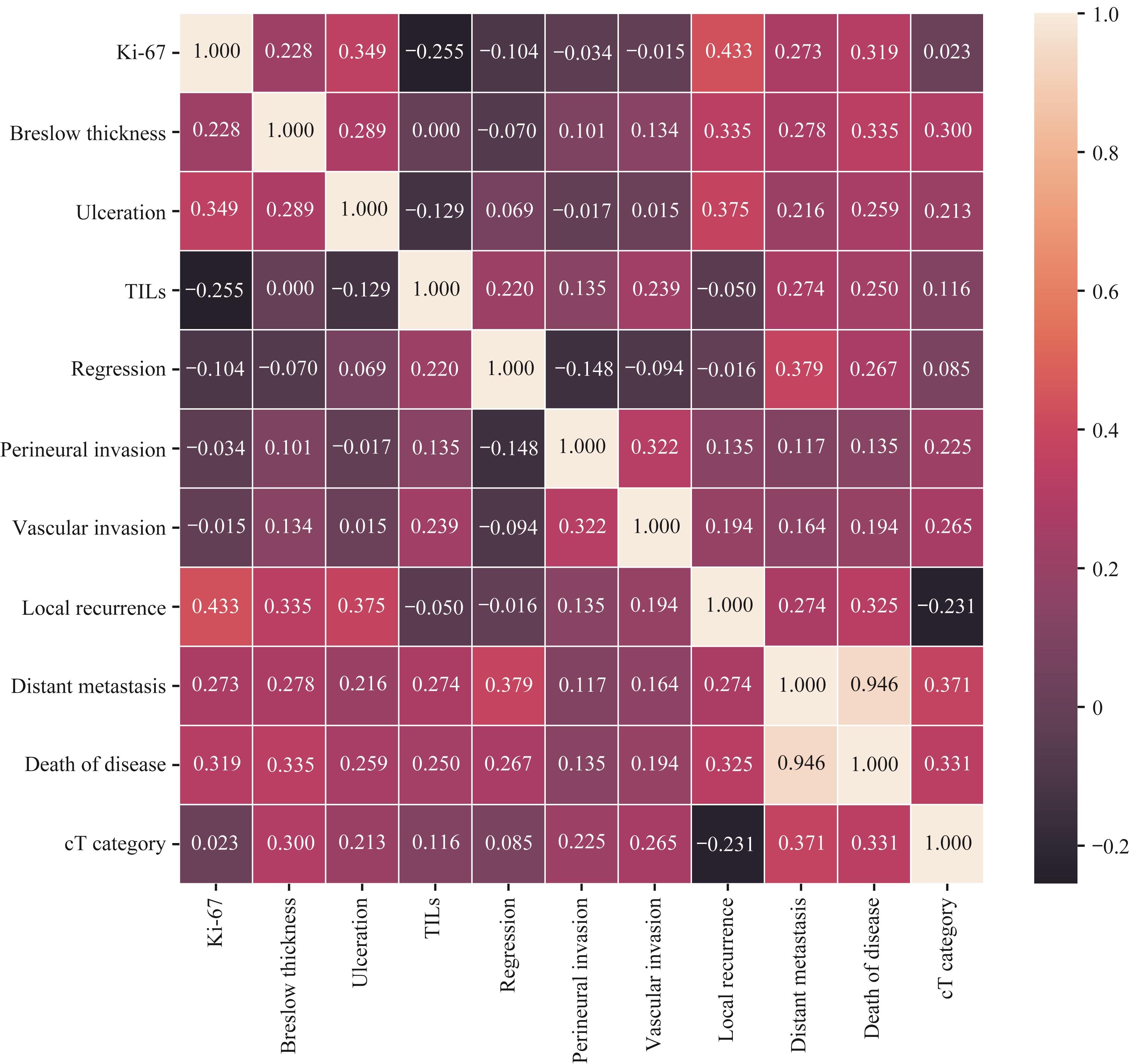
Fig 1 Heat map of the correlation between histopathological factors and prognosis
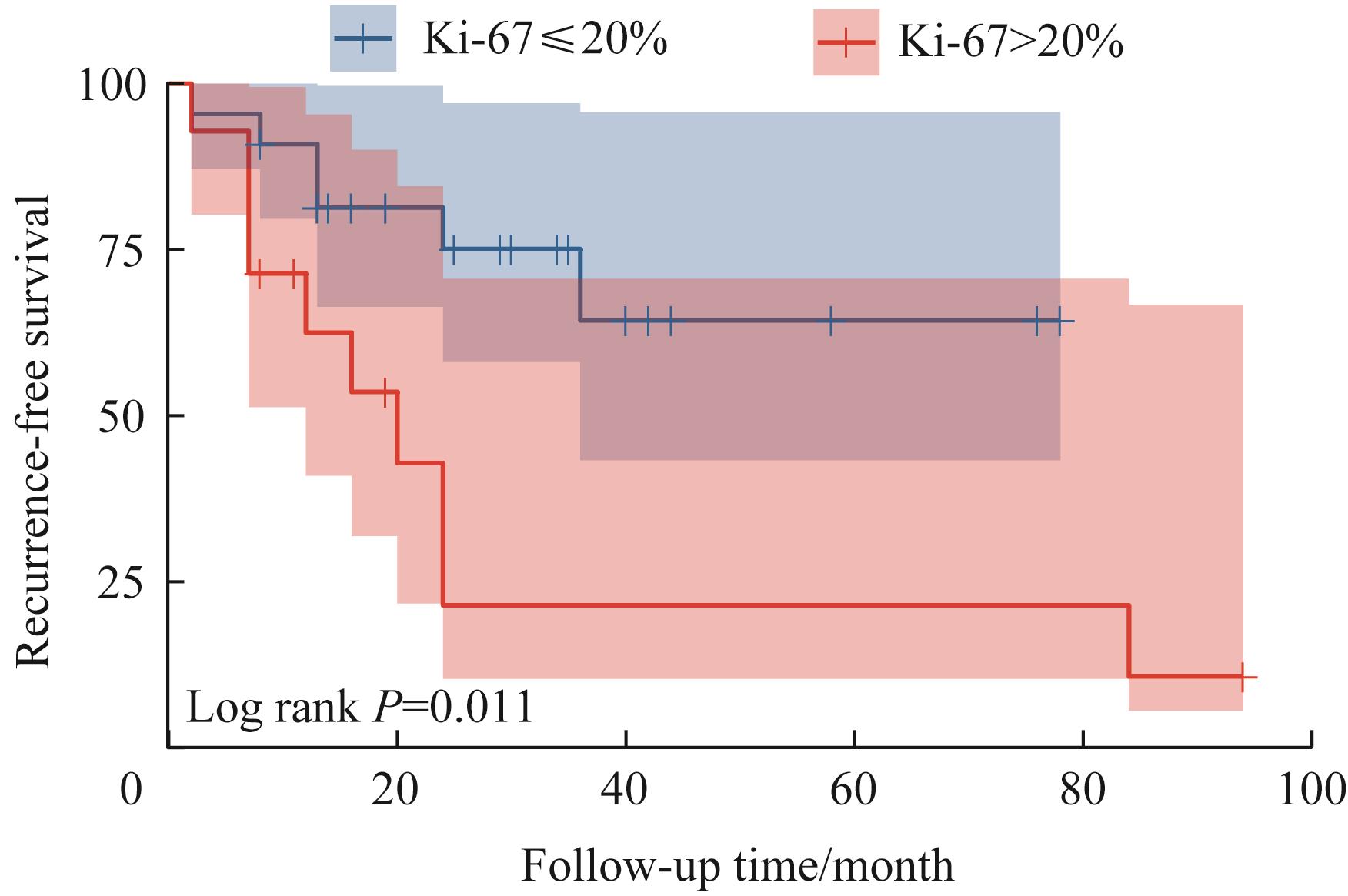
Fig 2 Analysis of recurrence-free survival curve of Ki-67≤20% and >20%
| 1 | ZHOU C, WANG Y, JIA R, et al. Conjunctival melanoma in Chinese patients: local recurrence, metastasis, mortality, and comparisons with caucasian patients[J]. Invest Ophthalmol Vis Sci, 2017, 58(12): 5452-5459. |
| 2 | GRIMES J M, SHAH N V, SAMIE F H, et al. Conjunctival melanoma: current treatments and future options[J]. Am J Clin Dermatol, 2020, 21(3): 371-381. |
| 3 | JAIN P, FINGER P T, DAMATO B, et al. Multicenter, international assessment of the eighth edition of the American Joint Committee on Cancer: cancer staging manual for conjunctival melanoma[J]. JAMA Ophthalmol, 2019, 137(8): 905-911. |
| 4 | ESMAELI B, RUBIN M L, XU S Q, et al. Greater tumor thickness, ulceration, and positive sentinel lymph node are associated with worse prognosis in patients with conjunctival melanoma: implications for future AJCC classifications[J]. Am J Surg Pathol, 2019, 43(12): 1701-1710. |
| 5 | ESMAELI B, ROBERTS D, ROSS M, et al. Histologic features of conjunctival melanoma predictive of metastasis and death (an American Ophthalmological thesis)[J]. Trans Am Ophthalmol Soc, 2012, 110: 64-73. |
| 6 | MENON S S, GURUVAYOORAPPAN C, SAKTHIVEL K M, et al. Ki-67 protein as a tumour proliferation marker[J]. Clin Chimica Acta, 2019, 491: 39-45. |
| 7 | SCOLYER R A, RAWSON R V, GERSHENWALD J E, et al. Melanoma pathology reporting and staging[J]. Mod Pathol, 2020, 33(Suppl 1): 15-24. |
| 8 | JIA S, ZHU T, SHI H, et al. American Joint Committee on Cancer (AJCC) tumor staging system predicts the outcome and metastasis pattern in conjunctival melanoma[J]. Ophthalmology, 2022, S0161-6420(22)00168-3. DOI: 10.1016/j.ophtha.2022.02.029. |
| 9 | SHIELDS C L, SHIELDS J A. Tumors of the conjunctiva and cornea[J]. Indian J Ophthalmol, 2019, 67(12): 1930-1948. |
| 10 | SHIELDS C L, MARKOWITZ J S, BELINSKY I, et al. Conjunctival melanoma: outcomes based on tumor origin in 382 consecutive cases[J]. Ophthalmology, 2011, 118(2): 389-395.e1-2. |
| 11 | WONG J R, NANJI A A, GALOR A, et al. Management of conjunctival malignant melanoma: a review and update[J]. Expert Rev Ophthalmol, 2014, 9(3): 185-204. |
| 12 | KARIM R, CONWAY R M. Conservative resection and adjuvant plaque brachytherapy for early-stage conjunctival melanoma [J]. Clin Exp Ophthalmol, 2011, 39(4): 293-298. |
| 13 | FINGER P T, PAVLICK A C. Checkpoint inhibition immunotherapy for advanced local and systemic conjunctival melanoma: a clinical case series[J]. J Immunother Cancer, 2019, 7(1): 83. |
| 14 | THOMPSON J F, SOONG S J, BALCH C M, et al. Prognostic significance of mitotic rate in localized primary cutaneous melanoma: an analysis of patients in the multi-institutional American Joint Committee on Cancer melanoma staging database[J]. J Clin Oncol, 2011, 29(16): 2199-2205. |
| 15 | NAGARAJAN P, CURRY J L, NING J, et al. Tumor thickness and mitotic rate robustly predict melanoma-specific survival in patients with primary vulvar melanoma: a retrospective review of 100 cases[J]. Clin Cancer Res, 2017, 23(8): 2093-2104. |
| 16 | PIÑERO-MADRONA A, RUIZ-MERINO G, CEREZUELA FUENTES P, et al. Mitotic rate as an important prognostic factor in cutaneous malignant melanoma[J]. Clin Transl Oncol, 2019, 21(10): 1348-1356. |
| 17 | MAURICHI A, MICELI R, CAMERINI T, et al. Prediction of survival in patients with thin melanoma: results from a multi-institution study[J]. J Clin Oncol, 2014, 32(23): 2479-2485. |
| 18 | AIVAZIAN K, AHMED T, EL SHAROUNI M A, et al. Histological regression in melanoma: impact on sentinel lymph node status and survival[J]. Mod Pathol, 2021, 34(11): 1999-2008. |
| 19 | EL SHAROUNI M A, AIVAZIAN K, WITKAMP A J, et al. Association of histologic regression with a favorable outcome in patients with stage 1 and stage 2 cutaneous melanoma[J]. JAMA Dermatol, 2021, 157(2): 166-173. |
| 20 | BASTIAN B C. Hypothesis: a role for telomere crisis in spontaneous regression of melanoma[J]. Arch Dermatol, 2003, 139(5): 667-668. |
| 21 | SUN X M, KAUFMAN P D. Ki-67: more than a proliferation marker[J]. Chromosoma, 2018, 127(2): 175-186. |
| 22 | JAKOBIEC F A, BHAT P, COLBY K A. Immunohistochemical studies of conjunctival nevi and melanomas[J]. Arch Ophthalmol, 2010, 128(2): 174-183. |
| [1] | Wang Yiran, Zhang Zhe, Shen Jianfeng. Comprehensive analysis of the function, prognosis, and immune infiltration characteristics of SF3B1 mutations in uveal melanoma [J]. Journal of Shanghai Jiao Tong University (Medical Science), 2026, 46(4): 475-485. |
| [2] | Mei Zixian, Meng Xuchen, Su Wenjing, Zhong Weijie, Tang Dingzhong, Li Yi. Clinical-inflammatory combined model for predicting poor prognosis in male patients with anterior circulation acute ischemic stroke with large vessel occlusion after mechanical thrombectomy [J]. Journal of Shanghai Jiao Tong University (Medical Science), 2026, 46(3): 332-339. |
| [3] | YANG Xiaoyu, HUANG Rui, WU Yijia, ZHANG Zhe, FANG Yan, SHEN Jianfeng. Mechanistic study of targeting melanoma with STING pathway deficiencies via PIKfyve inhibitor [J]. Journal of Shanghai Jiao Tong University (Medical Science), 2025, 45(9): 1126-1137. |
| [4] | CHEN Siyuan, SHI Qing, FU Di, WANG Li, CHENG Shu, XU Pengpeng, ZHAO Weili. Clinicopathologic characteristics, gene mutation profile, and prognostic analysis of diffuse large B-cell lymphoma with lung involvement [J]. Journal of Shanghai Jiao Tong University (Medical Science), 2025, 45(9): 1214-1220. |
| [5] | YAN Zhi, WU Xingyue, YAO Weiqin, YAN Lingzhi, JIN Song, SHANG Jingjing, SHI Xiaolan, WU Depei, FU Chengcheng. Dynamic changes and prognostic significance of immunoparesis in newly diagnosed multiple myeloma patients [J]. Journal of Shanghai Jiao Tong University (Medical Science), 2025, 45(7): 807-814. |
| [6] | XU Tianyun, SHEN Yiming, JIANG Meng. Clinical management of heart failure with improved ejection fraction: treatment and maintenance [J]. Journal of Shanghai Jiao Tong University (Medical Science), 2025, 45(4): 493-499. |
| [7] | LUO Wen, LÜ Mingjun, ZHANG Zhen, ZHANG Xue, YAO Zhirong. Research progress on the dual effects of autophagy in cutaneous melanoma and its role in drug resistance [J]. Journal of Shanghai Jiao Tong University (Medical Science), 2025, 45(2): 233-240. |
| [8] | HAO Meiling, MA Yanni, MA Xuhui, ZHANG Yanjie, ZENG Hanlin, CHEN Shanshuang. CD10+ neutrophils promote CD8+ T-cell depletion in mucosal malignant melanoma through the SELPG-SELL pathway [J]. Journal of Shanghai Jiao Tong University (Medical Science), 2025, 45(11): 1466-1479. |
| [9] | WANG Boen, CHEN Siyuan, SHI Qing, ZHANG Muchen, YI Hongmei, DONG Lei, WANG Li, CHENG Shu, XU Pengpeng, ZHAO Weili. Clinicopathologic characteristics of patients with kidney-involved diffuse large B-cell lymphoma [J]. Journal of Shanghai Jiao Tong University (Medical Science), 2024, 44(9): 1162-1168. |
| [10] | SONG Chenlu, XIANG Jun, YANG Huizhong. Early alarming effect of serum heparin-binding protein on prognosis and occurrence of sepsis in severely burned patients [J]. Journal of Shanghai Jiao Tong University (Medical Science), 2024, 44(4): 474-481. |
| [11] | WANG Guijie, DU Chuanchong, LU Ye, ZHAO Jian, SHEN Xie, JIN Donglin, GENG Jiacai. Changes of serum high mobility group box 1 and soluble triggering receptor expressed on myeloid cells-1 in patients with multiple injuries and their prognostic significance [J]. Journal of Shanghai Jiao Tong University (Medical Science), 2024, 44(3): 350-357. |
| [12] | LUO Mengxing, ZOU Xin, GAO Yaxian, WU Xiaocui, YU Fangyou, HU Yang, ZENG Qibing, LIU Zhonghua. Analysis of the effect of anti-tuberculosis treatment and lung injury in patients with tuberculosis combined with underlying disease [J]. Journal of Shanghai Jiao Tong University (Medical Science), 2023, 43(8): 1017-1023. |
| [13] | ZHOU Xiaowen, LI Qian, ZHANG Zhe, SHEN Jianfeng, FAN Xianqun. RBX1 regulates uveal melanoma immune-related genes via STAT1 [J]. Journal of Shanghai Jiao Tong University (Medical Science), 2023, 43(6): 709-717. |
| [14] | LI Ying, TAN Yangxia, YIN Hongxin, JIANG Yanling, CHEN Li, MENG Guoyu. Research progress in the pathogenesis and prognosis of ZNF384 fusion subtype acute leukemia [J]. Journal of Shanghai Jiao Tong University (Medical Science), 2023, 43(5): 631-640. |
| [15] | MEN Ru, ZHU Minxia, ZHANG Weiming. Serum potassium level in maintenance hemodialysis patients and its effect on outcome [J]. Journal of Shanghai Jiao Tong University (Medical Science), 2023, 43(4): 507-513. |
| Viewed | ||||||
|
Full text |
|
|||||
|
Abstract |
|
|||||