Journal of Shanghai Jiao Tong University (Medical Science) ›› 2022, Vol. 42 ›› Issue (8): 1062-1069.doi: 10.3969/j.issn.1674-8115.2022.08.011
• Clinical research • Previous Articles Next Articles
ZHAO Keke( ), JIANG Beibei(), ZHANG Lu, WANG Lingyun, ZHANG Yaping, XIE Xueqian()
Received:2022-04-17
Accepted:2022-07-25
Online:2022-08-28
Published:2022-10-08
Contact:
XIE Xueqian
E-mail:1797673460@qq.com;jennifer.chiang@hot mail.com;xiexueqian@hotmail.com
Supported by:CLC Number:
ZHAO Keke, JIANG Beibei, ZHANG Lu, WANG Lingyun, ZHANG Yaping, XIE Xueqian. Feasibility of ultra-low-dose noncontrast CT based on deep learning image reconstruction to evaluate chest lesions[J]. Journal of Shanghai Jiao Tong University (Medical Science), 2022, 42(8): 1062-1069.
Add to citation manager EndNote|Ris|BibTeX
URL: https://xuebao.shsmu.edu.cn/EN/10.3969/j.issn.1674-8115.2022.08.011
| Variable | Included patient | P value | |
|---|---|---|---|
0.07 mSv (n=40) | 0.14 mSv (n=40) | ||
| Age/year | 64±9 | 61±12 | 0.165 |
| Gender/n (%) | |||
| Male | 28 (52) | 26 (48) | 0.633 |
| Female | 12 (46) | 14 (54) | |
| BMI/(kg·m-2) | 23.14±3.61 | 22.30±3.03 | 0.173 |
| <18.5 | 3 | 4 | 0.243 |
| ≥18.5 and <25.0 | 26 | 31 | |
| ≥25.0 | 11 | 5 | |
| Lung target tumor lesion/n | |||
| Malignant | 13 | 15 | 0.377 |
| Benign or no histological result | 9 | 17 | |
| Mediastinal lymph node/n | |||
| Malignant | 8 | 3 | 0.793 |
| Benign or no histological result | 4 | 2 | |
| Hilar lymph node/n | |||
| Malignant | 2 | 3 | 0.294 |
| Benign or no histological result | 3 | 1 | |
Tab 1 Basic characteristics of the included patients
| Variable | Included patient | P value | |
|---|---|---|---|
0.07 mSv (n=40) | 0.14 mSv (n=40) | ||
| Age/year | 64±9 | 61±12 | 0.165 |
| Gender/n (%) | |||
| Male | 28 (52) | 26 (48) | 0.633 |
| Female | 12 (46) | 14 (54) | |
| BMI/(kg·m-2) | 23.14±3.61 | 22.30±3.03 | 0.173 |
| <18.5 | 3 | 4 | 0.243 |
| ≥18.5 and <25.0 | 26 | 31 | |
| ≥25.0 | 11 | 5 | |
| Lung target tumor lesion/n | |||
| Malignant | 13 | 15 | 0.377 |
| Benign or no histological result | 9 | 17 | |
| Mediastinal lymph node/n | |||
| Malignant | 8 | 3 | 0.793 |
| Benign or no histological result | 4 | 2 | |
| Hilar lymph node/n | |||
| Malignant | 2 | 3 | 0.294 |
| Benign or no histological result | 3 | 1 | |
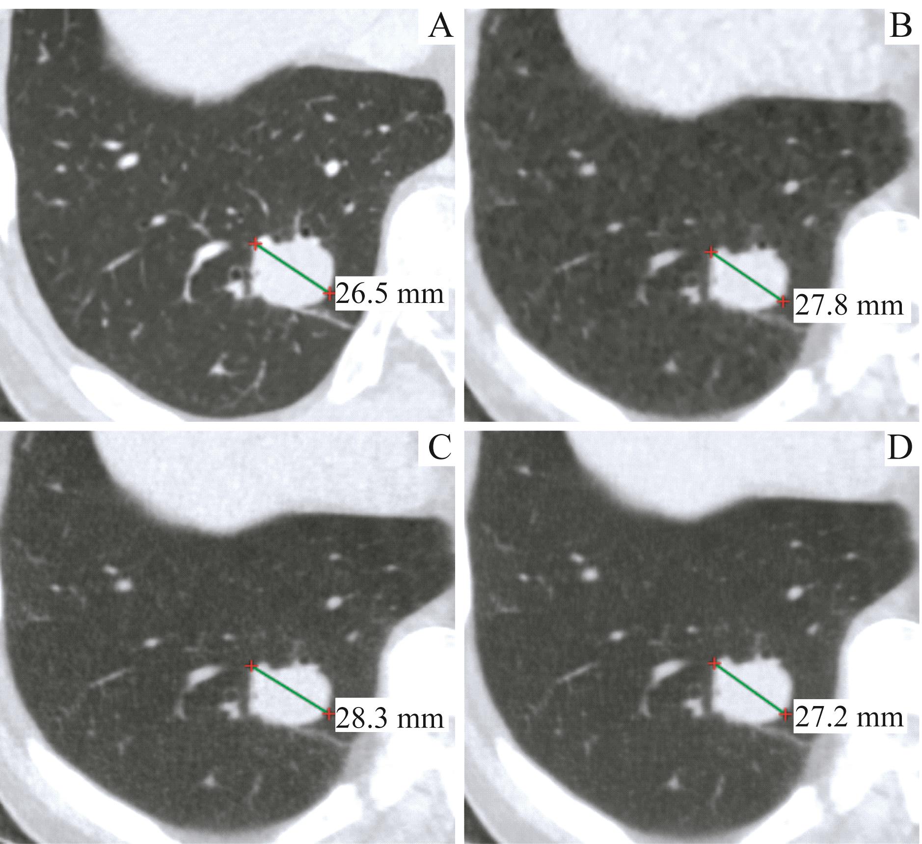
Fig 1 Measured values of lung target lesions in different image reconstruction kernels

Fig 2 Measured values of lymph node target lesions in different image reconstruction kernels
| Item | r | ||
|---|---|---|---|
| ASIR-V-80% vs enhanced CT | DLIR-M vs enhanced CT | DLIR-H vs enhanced CT | |
| All lung target tumor lesion | 0.988 | 0.987 | 0.990 |
| Malignant | 0.985 | 0.980 | 0.986 |
| Benign or no histological result | 0.991 | 0.993 | 0.994 |
| GGN (diameter≤1 cm) | 0.905 | 0.906 | 0.969 |
| Mediastinal lymph node | 0.969 | 0.957 | 0.977 |
| Malignant | 0.952 | 0.930 | 0.955 |
| Benign or no histological result | 0.999 | 0.997 | 1.000 |
| Hilar lymph node | 0.972 | 0.994 | 0.994 |
Tab 2 Pearson's correlation coefficients of target lesions measured on ultra-low-dose CT and enhanced CT images
| Item | r | ||
|---|---|---|---|
| ASIR-V-80% vs enhanced CT | DLIR-M vs enhanced CT | DLIR-H vs enhanced CT | |
| All lung target tumor lesion | 0.988 | 0.987 | 0.990 |
| Malignant | 0.985 | 0.980 | 0.986 |
| Benign or no histological result | 0.991 | 0.993 | 0.994 |
| GGN (diameter≤1 cm) | 0.905 | 0.906 | 0.969 |
| Mediastinal lymph node | 0.969 | 0.957 | 0.977 |
| Malignant | 0.952 | 0.930 | 0.955 |
| Benign or no histological result | 0.999 | 0.997 | 1.000 |
| Hilar lymph node | 0.972 | 0.994 | 0.994 |
| Item | Arithmetic mean ( | ||
|---|---|---|---|
| ASIR-V-80% vs enhanced CT | DLIR-M vs enhanced CT | DLIR-H vs enhanced CT | |
| All lung target tumor lesion | 8.5% (-3.3%‒20.3%) | 8.5% (-4.2%‒21.3%) | 4.3% (-5.7%‒14.3%) |
| Malignant | 8.7% (-2.4%‒19.7%) | 10.3% (-3.7%‒24.3%) | 4.8% (-5.5%‒15.2%) |
| Benign or no histological result | 8.3% (-4.4%‒21.0%) | 6.7% (-3.6%‒17.0%) | 3.8% (-5.9%‒13.5%) |
| GGN (diameter≤1 cm) | 14.4% (-4.4%‒33.2%) | 16.3% (-4.1%‒36.7%) | 7.0% (-5.7%‒19.7%) |
| Mediastinal lymph node | 9.7% (-6.0%‒25.3%) | 8.8% (-9.9%‒27.5%) | 5.1% (-9.1%‒19.3%) |
| Malignant | 11.8% (-6.1%‒29.7%) | 10.5% (-11.3%‒32.4%) | 6.3% (-10.9%‒23.6%) |
| Benign or no histological result | 5.8% (-0.4%‒11.9%) | 5.7% (-3.9%‒15.2%) | 2.8% (-0.2%‒5.8%) |
| Hilar lymph node | 20.2% (-1.2%‒41.5%) | 23.4% (13.5%‒33.2%) | 18.3% (8.8%‒27.9%) |
Tab 3 Bland-Altman analysis of the variability of measured values of target lesions on ultra-low-dose CT and enhanced CT images
| Item | Arithmetic mean ( | ||
|---|---|---|---|
| ASIR-V-80% vs enhanced CT | DLIR-M vs enhanced CT | DLIR-H vs enhanced CT | |
| All lung target tumor lesion | 8.5% (-3.3%‒20.3%) | 8.5% (-4.2%‒21.3%) | 4.3% (-5.7%‒14.3%) |
| Malignant | 8.7% (-2.4%‒19.7%) | 10.3% (-3.7%‒24.3%) | 4.8% (-5.5%‒15.2%) |
| Benign or no histological result | 8.3% (-4.4%‒21.0%) | 6.7% (-3.6%‒17.0%) | 3.8% (-5.9%‒13.5%) |
| GGN (diameter≤1 cm) | 14.4% (-4.4%‒33.2%) | 16.3% (-4.1%‒36.7%) | 7.0% (-5.7%‒19.7%) |
| Mediastinal lymph node | 9.7% (-6.0%‒25.3%) | 8.8% (-9.9%‒27.5%) | 5.1% (-9.1%‒19.3%) |
| Malignant | 11.8% (-6.1%‒29.7%) | 10.5% (-11.3%‒32.4%) | 6.3% (-10.9%‒23.6%) |
| Benign or no histological result | 5.8% (-0.4%‒11.9%) | 5.7% (-3.9%‒15.2%) | 2.8% (-0.2%‒5.8%) |
| Hilar lymph node | 20.2% (-1.2%‒41.5%) | 23.4% (13.5%‒33.2%) | 18.3% (8.8%‒27.9%) |

Fig 3 Bland-Altman analysis of the variability of measured values of target lesions on ultra-low-dose CT and enhanced CT images
| Influential factor | Difference of measured values (ASIR-V-80% and enhanced CT) | Difference of measured values (DLIR-M and enhanced CT) | Difference of measured values (DLIR-H and enhanced CT) | |||
|---|---|---|---|---|---|---|
| β | P value | β | P value | β | P value | |
| BMI | -0.003 | 0.976 | -0.013 | 0.913 | -0.042 | 0.717 |
| Age | -0.099 | 0.392 | -0.014 | 0.631 | -0.049 | 0.675 |
| Gender | -0.135 | 0.251 | -0.095 | 0.416 | -0.105 | 0.370 |
| CT dose | -0.084 | 0.463 | -0.073 | 0.525 | -0.077 | 0.506 |
| Lesion type | 0.256 | 0.034 | 0.257 | 0.033 | 0.287 | 0.018 |
| Lesion type (without hilar lymph node) | 0.013 | 0.919 | -0.049 | 0.702 | -0.027 | 0.839 |
| Histological result | 0.175 | 0.143 | 0.203 | 0.088 | 0.142 | 0.233 |
Tab 4 Multiple linear regression analysis of the influential factors on the differences between the measured values of ultra-low-dose CT and enhanced CT of target lesions
| Influential factor | Difference of measured values (ASIR-V-80% and enhanced CT) | Difference of measured values (DLIR-M and enhanced CT) | Difference of measured values (DLIR-H and enhanced CT) | |||
|---|---|---|---|---|---|---|
| β | P value | β | P value | β | P value | |
| BMI | -0.003 | 0.976 | -0.013 | 0.913 | -0.042 | 0.717 |
| Age | -0.099 | 0.392 | -0.014 | 0.631 | -0.049 | 0.675 |
| Gender | -0.135 | 0.251 | -0.095 | 0.416 | -0.105 | 0.370 |
| CT dose | -0.084 | 0.463 | -0.073 | 0.525 | -0.077 | 0.506 |
| Lesion type | 0.256 | 0.034 | 0.257 | 0.033 | 0.287 | 0.018 |
| Lesion type (without hilar lymph node) | 0.013 | 0.919 | -0.049 | 0.702 | -0.027 | 0.839 |
| Histological result | 0.175 | 0.143 | 0.203 | 0.088 | 0.142 | 0.233 |
| 1 | EISENHAUER E A, THERASSE P, BOGAERTS J, et al. New response evaluation criteria in solid tumours: revised RECIST guideline (version 1.1)[J]. Eur J Cancer, 2009, 45(2): 228-247. |
| 2 | SHI L, TASHIRO S. Estimation of the effects of medical diagnostic radiation exposure based on DNA damage[J]. J Radiat Res, 2018, 59(suppl_2): ii121-ii129. |
| 3 | WOOD D E, KAZEROONI E A, BAUM S L, et al. Lung cancer screening, version 3.2018, NCCN clinical practice guidelines in oncology[J]. J Natl Compr Canc Netw, 2018, 16(4): 412-441. |
| 4 | MAZLOUMI M, VAN GOMPEL G, KERSEMANS V, et al. The presence of contrast agent increases organ radiation dose in contrast-enhanced CT[J]. Eur Radiol, 2021, 31(10): 7540-7549. |
| 5 | PERISINAKIS K, SEIMENIS I, TZEDAKIS A, et al. Radiation burden and associated cancer risk for a typical population to be screened for lung cancer with low-dose CT: a phantom study[J]. Eur Radiol, 2018, 28(10): 4370-4378. |
| 6 | KIM Y, KIM Y K, LEE B E, et al. Ultra-low-dose CT of the thorax using iterative reconstruction: evaluation of image quality and radiation dose reduction[J]. AJR Am J Roentgenol, 2015, 204(6): 1197-1202. |
| 7 | JIANG B, LI N, SHI X, et al. Deep learning reconstruction shows better lung nodule detection for ultra-low-dose chest CT[J]. Radiology, 2022, 303(1): 202-212. |
| 8 | SHIRI I, AKHAVANALLAF A, SANAAT A, et al. Ultra-low-dose chest CT imaging of COVID-19 patients using a deep residual neural network[J]. Eur Radiol, 2021, 31(3): 1420-1431. |
| 9 | CABALLERO B. Humans against obesity: who will win?[J]. Adv Nutr, 2019, 10(suppl_1): S4-S9. |
| 10 | SUN J, LI H, WANG B, et al. Application of a deep learning image reconstruction (DLIR) algorithm in head CT imaging for children to improve image quality and lesion detection[J]. BMC Med Imaging, 2021, 21(1): 108. |
| 11 | KIM J H, YOON H J, LEE E, et al. Validation of deep-learning image reconstruction for low-dose chest computed tomography scan: emphasis on image quality and noise[J]. Korean J Radiol, 2021, 22(1): 131-138. |
| 12 | PARAKH A, CAO J, PIERCE T T, et al. Sinogram-based deep learning image reconstruction technique in abdominal CT: image quality considerations[J]. Eur Radiol, 2021, 31(11): 8342-8353. |
| 13 | NAM J G, AHN C, CHOI H, et al. Image quality of ultralow-dose chest CT using deep learning techniques: potential superiority of vendor-agnostic post-processing over vendor-specific techniques[J]. Eur Radiol, 2021, 31(7): 5139-5147. |
| 14 | JENSEN C T, LIU X, TAMM E P, et al. Image quality assessment of abdominal CT by use of new deep learning image reconstruction: initial experience[J]. AJR Am J Roentgenol, 2020, 215(1): 50-57. |
| 15 | BENZ D C, BENETOS G, RAMPIDIS G, et al. Validation of deep-learning image reconstruction for coronary computed tomography angiography: impact on noise, image quality and diagnostic accuracy[J]. J Cardiovasc Comput Tomogr, 2020, 14(5): 444-451. |
| 16 | NODA Y, KAGA T, KAWAI N, et al. Low-dose whole-body CT using deep learning image reconstruction: image quality and lesion detection[J]. Br J Radiol, 2021, 94(1121): 20201329. |
| 17 | JENSEN C T, GUPTA S, SALEH M M, et al. Reduced-dose deep learning reconstruction for abdominal CT of liver metastases[J]. Radiology, 2022, 303(1): 90-98. |
| 18 | 蒋蓓蓓, 张亚平, 张琳, 等. 深度卷积神经网络对≤3 cm的亚实性肺腺癌CT图像病理学分型预测的可视化研究[J]. 上海交通大学学报(医学版), 2019, 39(9): 1045-1051. |
| JIANG B B, ZHANG Y P, ZHANG L, et al. A visualization study of deep convolutional neural network to classify the pathological type of sub-soild pulmonary adenocarcinoma of ≤3 cm based on CT images [J]. J Shanghai Jiao Tong Univ (Med Sci), 2019, 39(9): 1045-1051. |
| [1] | Peng Qianqian, Song Jinghan, Xu Xingyi, Xiao Hui. Bioinformatic analysis and validation of the RNA-binding protein HuR promoting non-small cell lung cancer progression via ITGB1 [J]. Journal of Shanghai Jiao Tong University (Medical Science), 2026, 46(4): 451-466. |
| [2] | Zhu Menglin, Liu Xiao, Xu Xiaodan, Wang Ganhong, Xia Kaijian, Chen Jian. Prediction of delayed post-polypectomy bleeding using a multimodal model [J]. Journal of Shanghai Jiao Tong University (Medical Science), 2026, 46(4): 509-520. |
| [3] | YIN Ziming, WANG Rongqin, YANG Ziyi, LIU Yingbin, CHEN Tao, SHU Yijun, GONG Wei. Graph neural network-based auxiliary diagnostic model for gallbladder cancer on CT imaging [J]. Journal of Shanghai Jiao Tong University (Medical Science), 2025, 45(9): 1221-1231. |
| [4] | HUANG Xin, LIU Jiahui, YE Jingwen, QIAN Wenli, XU Wanxing, WANG Lin. Development and clinical application of a machine learning-driven model for metabolite-based diagnosis of small cell lung cancer [J]. Journal of Shanghai Jiao Tong University (Medical Science), 2025, 45(8): 1009-1016. |
| [5] | ZHANG Yuqin, AIHEMAITI Yilixiati, WANG Yanli, YANG Zhi, HUANG Jian. Ubiquitination and degradation of RPTPα mediated by MARCH9 [J]. Journal of Shanghai Jiao Tong University (Medical Science), 2025, 45(8): 957-968. |
| [6] | ZOU Peichen, LIU Hongyu, AIHEMAITI· Ayinazhaer, ZHU Liang, TANG Yabin, LEI Huimin. Metabolic profiling of lung cancer cells with acquired resistance to sotorasib [J]. Journal of Shanghai Jiao Tong University (Medical Science), 2025, 45(2): 138-149. |
| [7] | ZHANG Xianzhou, DU Fenglin, WU Lei, REN Yizhe, ZHAO Mingna, LOU Jiatao. Mechanistic study of OGT-promoted non-small cell lung cancer proliferation via the ERK signaling pathway [J]. Journal of Shanghai Jiao Tong University (Medical Science), 2025, 45(10): 1288-1297. |
| [8] | ZHU Mingyang, XU Yuanyuan, REN Jianghao, HUANG Jiazheng, LI Ruonan, TAN Qiang. Review of sublobar resection for lung adenocarcinoma with ground-glass presence [J]. Journal of Shanghai Jiao Tong University (Medical Science), 2024, 44(7): 922-927. |
| [9] | CHEN Jian, WANG Zhenni, XIA Kaijian, WANG Ganhong, LIU Luojie, XU Xiaodan. Comparative study on methods for colon polyp endoscopic image segmentation and classification based on deep learning [J]. Journal of Shanghai Jiao Tong University (Medical Science), 2024, 44(6): 762-772. |
| [10] | WANG Mengting, CHEN Yinan, XUANYUAN Xinyang, YUAN Haihua. Construction and experimental validation of mouse PDX model by malignant pleural effusion-derived tumor cells from lung cancer [J]. Journal of Shanghai Jiao Tong University (Medical Science), 2024, 44(4): 435-443. |
| [11] | LIU Chenxi, HAN Lin, YANG Yi, ZHOU Han, LIU Yayun, SHENG Deqiao. GPR87 promotes invasion and migration through the RHO/ROCK pathway in non-small cell lung cancer [J]. Journal of Shanghai Jiao Tong University (Medical Science), 2024, 44(12): 1514-1525. |
| [12] | LIU Yang, WU Mengyi, HU Yao, QI Kun, WANG Yubin, ZHAO Yue, SONG Jinlin. Preliminary application of a cervical vertebra segmentation method based on Transformer and diffusion model for lateral cephalometric radiographs in orthodontic clinical practice [J]. Journal of Shanghai Jiao Tong University (Medical Science), 2024, 44(12): 1579-1586. |
| [13] | HUANG Huayan, XU-ZHANG Wendi, XIA Liliang, YU Yongfeng, LU Shun. Advances in immunotherapy of advanced non-small cell lung cancer with EGFR mutation [J]. Journal of Shanghai Jiao Tong University (Medical Science), 2023, 43(5): 611-618. |
| [14] | ZHAO Zhuoming, LIU Zhenhao, LU Manman, ZHANG Yu, XU Linfeng, XIE Lu. Analysis of tumor-related features of non-small cell lung cancer based on TCR repertoire workflow [J]. Journal of Shanghai Jiao Tong University (Medical Science), 2023, 43(12): 1520-1528. |
| [15] | LIAO Yahui, LIU Liyun, ZHU Hongrui, LIN Houwen, YAN Jizhou, SUN Fan. Marine sponge-derived smenospongine overcomes resistance of cisplatin via inhibiting EGFR-Akt-ABCG2 pathway in NSCLC cells [J]. Journal of Shanghai Jiao Tong University (Medical Science), 2022, 42(8): 997-1007. |
| Viewed | ||||||
|
Full text |
|
|||||
|
Abstract |
|
|||||