Journal of Shanghai Jiao Tong University (Medical Science)
WANG Lanxi1,2( ), MA Guanrong1,2, JIANG Yongzhu1,2, CHANG Xiulin1,2, FANG Liaoqiong1, BAI Jin1,2()
Received:2022-12-08
Accepted:2023-06-01
Online:2023-08-30
Contact:
BAI Jin
E-mail:2962911834@qq.com;sajinbai@cqmu.edu.cn
CLC Number:
WANG Lanxi, MA Guanrong, JIANG Yongzhu, CHANG Xiulin, FANG Liaoqiong, BAI Jin. Effects of Escherichia coli outer membrane vesicles on proliferation of breast cancer cells and tumor growth of tumor-bearing mice[J]. Journal of Shanghai Jiao Tong University (Medical Science).
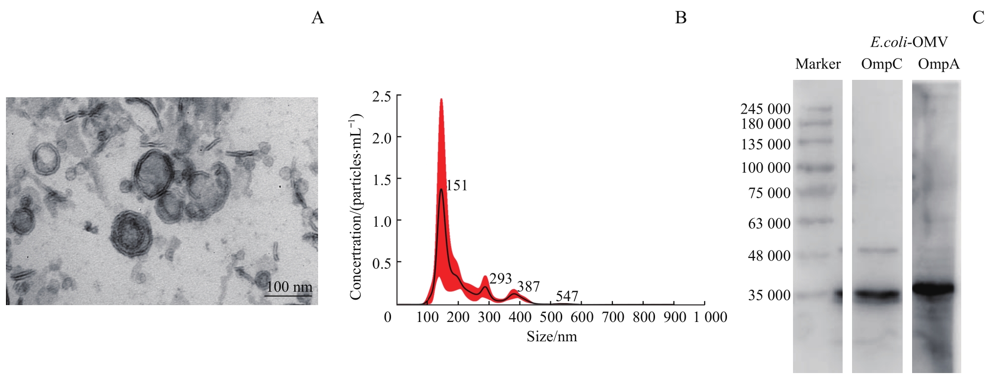
Fig 1 Analysis of the characterization of E.coli-OMV
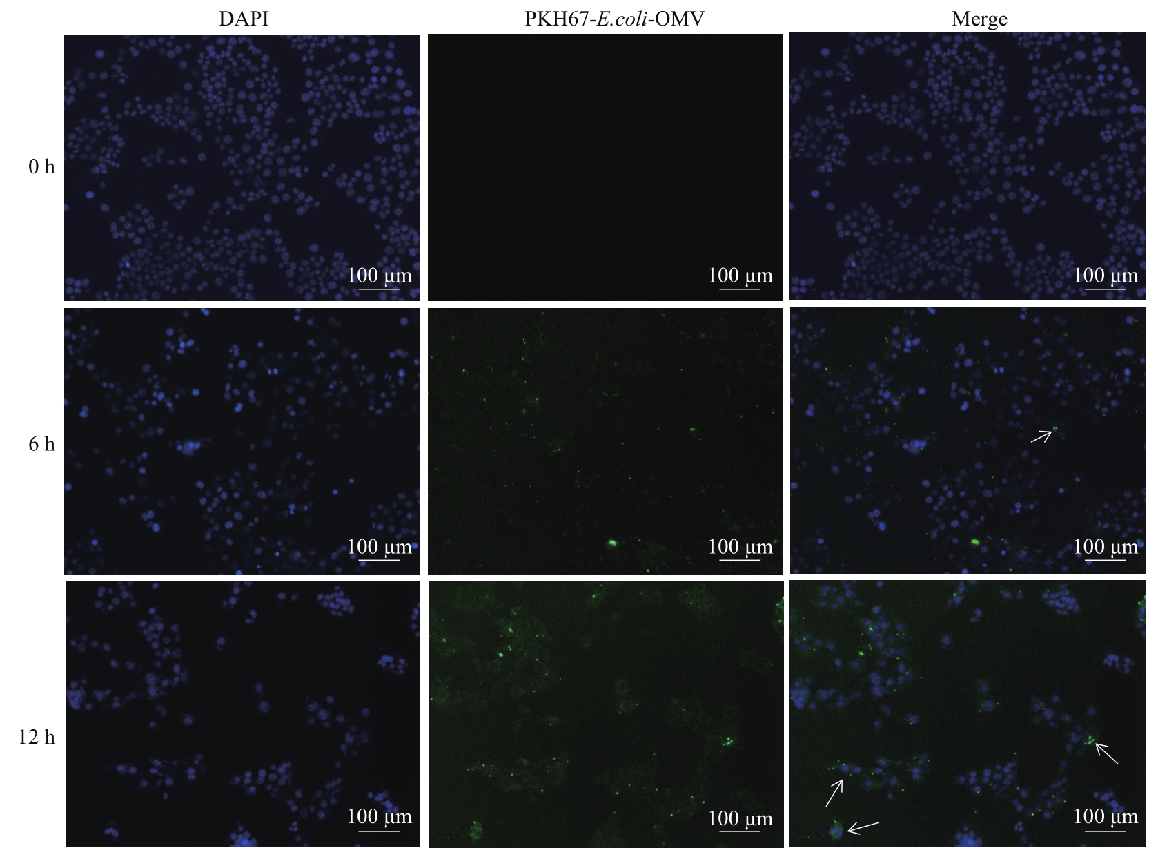
Fig 2 Observation on the uptake of E.coli-OMV by 4T1 cells (×100)
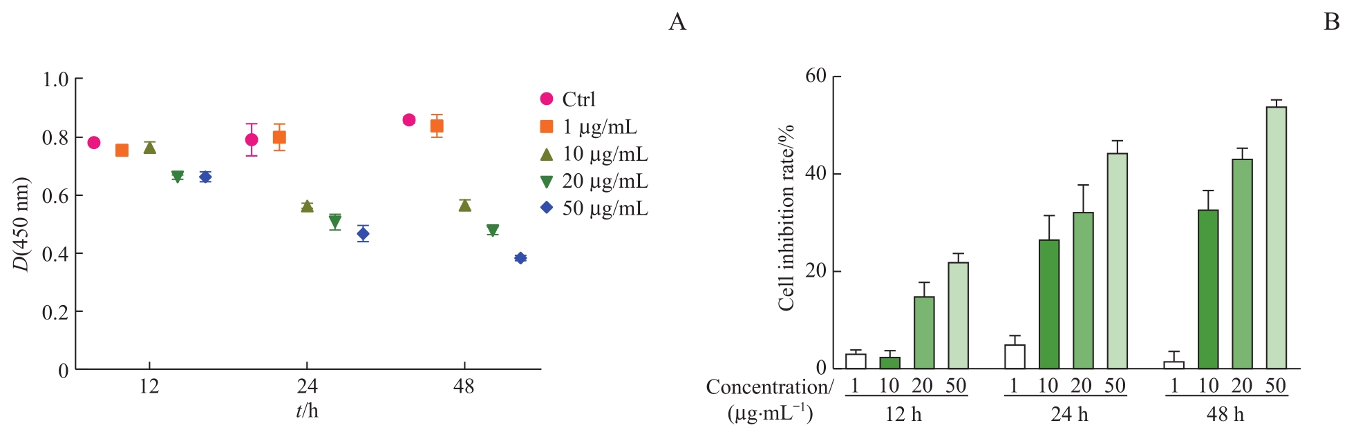
Fig 3 Effect of E. coli-OMV on 4T1 cells proliferation by CCK-8 method
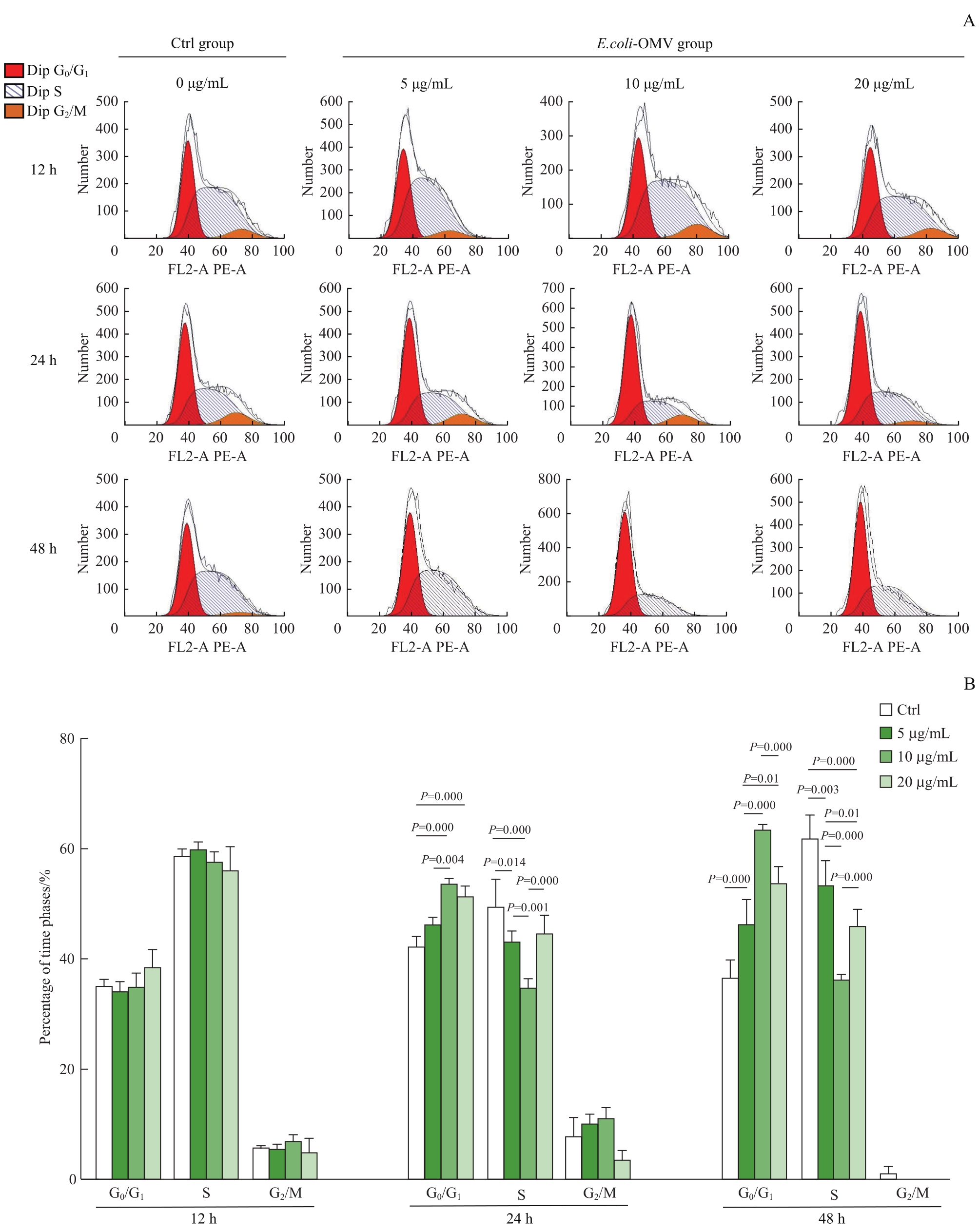
Fig 4 Effect of E. coli-OMV on 4T1 cell cycle distribution by flow cytometry

Fig 5 Changes in body weight of BALB/c-4T1 tumor-bearing mice in the two groups
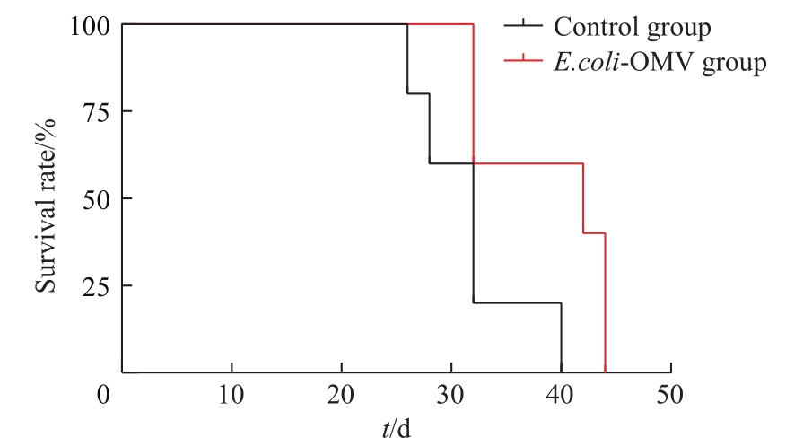
Fig 6 Survival rate analysis of BALB/c-4T1 tumor-bearing mice in the two groups

Fig 7 Changes in tumor volume growth of BALB/c-4T1 tumor-bearing mice in the two groups
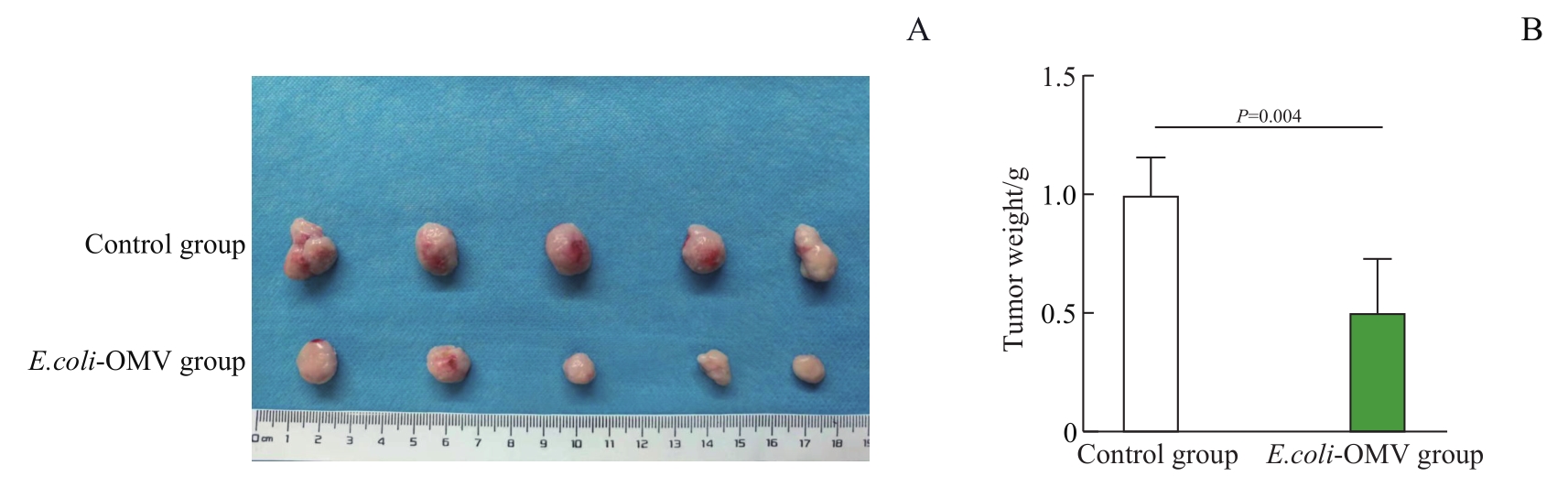
Fig 8 Changes in tumor weight of BALB/c-4T1 tumor-bearing mice in the two groups
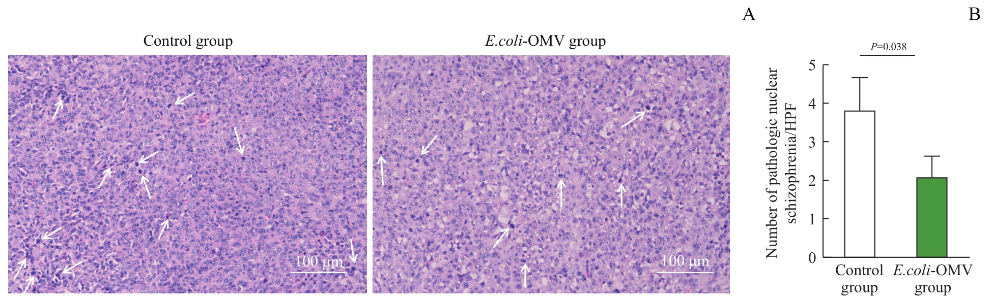
Fig 9 Analysis of HE staining of tumor tissues of BALB/c-4T1 tumor-bearing mice in the two groups (×200)
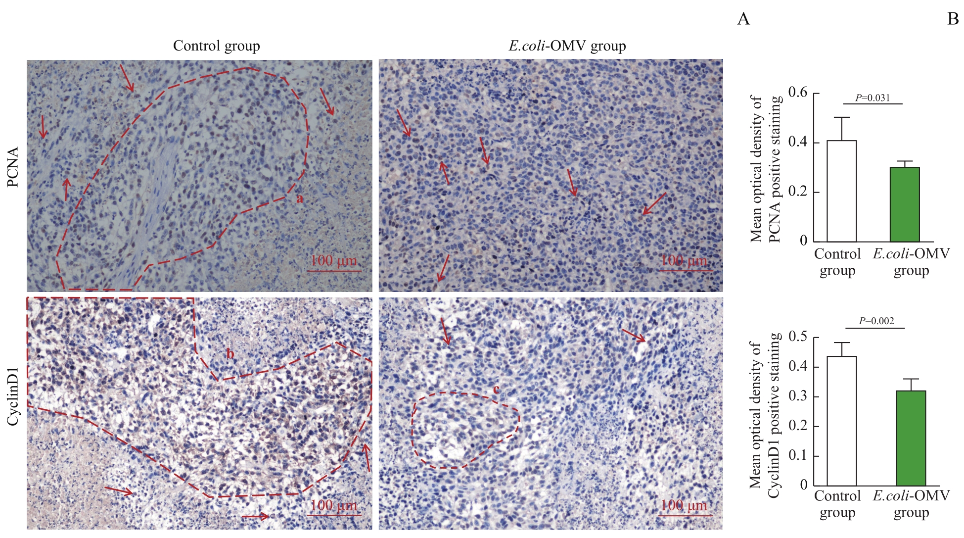
Fig 10 Analysis of the expression of PCNA and CyclinD1 in tumor tissues of BALB/c-4T1 tumor-bearing mice in the two groups by immunohistochemical staining (×200)
| 1 | KATSURA C, OGUNMWONYI I, KANKAM H K, et al. Breast cancer: presentation, investigation and management[J]. Br J Hosp Med (Lond), 2022, 83(2): 1-7. |
| 2 | AKRAM M, IQBAL M, DANIYAL M, et al. Awareness and current knowledge of breast cancer[J]. Biol Res, 2017, 50(1): 33. |
| 3 | LIANG S Y, WANG C, SHAO Y C, et al. Recent advances in bacteria-mediated cancer therapy[J]. Front Bioeng Biotechnol, 2022, 10: 1026248. |
| 4 | FISUSI F A, AKALA E O. Drug combinations in breast cancer therapy[J]. Pharm Nanotechnol, 2019, 7(1): 3-23. |
| 5 | SONG S, VUAI M S, ZHONG M. The role of bacteria in cancer therapy: enemies in the past, but allies at present[J]. Infect Agent Cancer, 2018, 13: 9. |
| 6 | 王敏, 苏乌云. 治疗癌症的新型武器: 细菌[J]. 世界最新医学信息文摘(连续型电子期刊), 2019, 19(66): 102-103, 105. |
| WANG M, SU W Y, et al. Bacteria: a new weapon against cancer[J]. World Latest Medicine Information, 2019, 19(66): 102-103, 105. | |
| 7 | SEDIGHI M, ZAHEDI BIALVAEI A, HAMBLIN M R, et al. Therapeutic bacteria to combat cancer; current advances, challenges, and opportunities[J]. Cancer Med, 2019, 8(6): 3167-3181. |
| 8 | KIKUCHI Y, OBANA N, TOYOFUKU M, et al. Diversity of physical properties of bacterial extracellular membrane vesicles revealed through atomic force microscopy phase imaging[J]. Nanoscale, 2020, 12(14): 7950-7959. |
| 9 | 邱晓涵, 李泳江, 吴军勇, 等. 细菌外膜囊泡: 疾病治疗的新途径[J]. 药学学报, 2021, 56(12): 3441-3450. |
| QIU X H, LI Y J, WU J Y, et al. Bacterial outer membrane vesicles: a new approach to diseases therapy[J]. Acta Pharmaceutica Sinica, 2021, 56(12): 3441-3450. | |
| 10 | CHEN Q, BAI H Z, WU W T, et al. Bioengineering bacterial vesicle-coated polymeric nanomedicine for enhanced cancer immunotherapy and metastasis prevention[J]. Nano Lett, 2020, 20(1): 11-21. |
| 11 | CHEN Y, LIU L G, FU H, et al. Comparative proteomic analysis of outer membrane vesicles from Shigella flexneri under different culture conditions[J]. Biochem Biophys Res Commun, 2014, 453(4): 696-702. |
| 12 | TOYOFUKU M, NOMURA N, EBERL L. Types and origins of bacterial membrane vesicles[J]. Nat Rev Microbiol, 2019, 17(1): 13-24. |
| 13 | RUDNICKA M, NOSZCZYŃSKA M, MALICKA M, et al. Outer membrane vesicles as mediators of plant-bacterial interactions[J]. Front Microbiol, 2022, 13: 902181. |
| 14 | 胡慧冰, 侯昕宇, 贺牧野, 等. 细菌外膜囊泡包覆的载药纳米粒的制备及其小鼠鼻腔免疫效果评价[J]. 上海交通大学学报(医学版), 2018, 38(2): 155-160. |
| HU H B, HOU X Y, HE M Y, et al. Preparation of bacterial outer membrane vesicle coated nanoparticle loaded with drug and evaluation of its nasal immune effect in mice[J]. Journal of Shanghai Jiao Tong University (Medical Science), 2018, 38(2): 155-160. | |
| 15 | YAGHOUBI A, KHAZAEI M, HASANIAN S, et al. Bacteriotherapy in breast cancer[J]. Int J Mol Sci, 2019, 20(23): 5880. |
| 16 | FARKAS-HIMSLEY H, CHEUNG R. Bacterial proteinaceous products (bacteriocins) as cytotoxic agents of neoplasia[J]. Cancer Res, 1976, 36(10): 3561-3567. |
| 17 | STRZALKA W, ZIEMIENOWICZ A. Proliferating cell nuclear antigen (PCNA): a key factor in DNA replication and cell cycle regulation[J]. Ann Bot, 2011, 107(7): 1127-1140. |
| 18 | GOLIAS C H, CHARALABOPOULOS A, CHARALABOPOULOS K. Cell proliferation and cell cycle control: a mini review[J]. Int J Clin Pract, 2004, 58(12): 1134-1141. |
| [1] | WANG Jingyi, DENG Jiali, ZHU Yi, DING Xinyi, GUO Jiajing, WANG Zhongling. Experimental study on novel pH-responsive manganese-based nanoprobes for ferroptosis and magnetic resonance imaging in breast cancer [J]. Journal of Shanghai Jiao Tong University (Medical Science), 2025, 45(9): 1183-1193. |
| [2] | DENG Jiali, GUO Jiajing, WANG Jingyi, DING Xinyi, ZHU Yi, WANG Zhongling. Self -assembled drug -loaded nanoprobes for pyroptosis sensitization and chemical exchange saturation transfer imaging in breast cancer [J]. Journal of Shanghai Jiao Tong University (Medical Science), 2025, 45(3): 271-281. |
| [3] | WU Shiyi, CHEN Si, LIU Bohan, LIU Yuting, LIU Yiwen, HE Yiqing, DU Yan, ZHANG Guoliang, GUO Qian, GAO Feng, YANG Cuixia. Role of "HA coat" in modulating stemness and endocrine resistance in ER+ breast cancer [J]. Journal of Shanghai Jiao Tong University (Medical Science), 2025, 45(10): 1298-1307. |
| [4] | WU Qizhen, LIU Qiming, CHAI Yezi, TAO Zhengyu, WANG Yinan, GUO Xinning, JIANG Meng, PU Jun. Evaluation of machine learning prediction of altered inflammatory metabolic state after neoadjuvant therapy for breast cancer [J]. Journal of Shanghai Jiao Tong University (Medical Science), 2024, 44(9): 1169-1181. |
| [5] | HAN Yishan, XU Ziqi, TAO Mengyu, FAN Guangjian, YU Bo. PRMT6 promotes the proliferation and migration of breast cancer cells [J]. Journal of Shanghai Jiao Tong University (Medical Science), 2024, 44(8): 999-1010. |
| [6] | XUE Yu, ZHANG Hailong, LEI Ming. Expression of cancer-testis antigen SPANXB and its mechanism in affecting hepatocellular carcinoma progress [J]. Journal of Shanghai Jiao Tong University (Medical Science), 2024, 44(7): 801-813. |
| [7] | WANG Wei, WANG Hongli, ALIBIYATI·i Ain, YILIYAER· Rousu, AYI NUER, YANG Liang. Function of vasohibin-2 and the mechanism of alternative splicing in triple-negative breast cancer [J]. Journal of Shanghai Jiao Tong University (Medical Science), 2024, 44(12): 1526-1535. |
| [8] | TAN Chen, XU Zhangrun, XUE Yang, CHEN Jiayu, YAO Lijun. Research progress in drug repurposing in the treatment of breast cancer [J]. Journal of Shanghai Jiao Tong University (Medical Science), 2024, 44(11): 1454-1459. |
| [9] | DU Shaoqian, TAO Mengyu, CAO Yuan, WANG Hongxia, HU Xiaoqu, FAN Guangjian, ZANG Lijuan. CXCL9 expression in breast cancer and its correlation with the characteristics of tumor immunoinfiltration [J]. Journal of Shanghai Jiao Tong University (Medical Science), 2023, 43(7): 860-872. |
| [10] | CAO Yuan, WANG Hongxia, ZHU Ying, LI Junjian. Expression of tetraspanin 1 in breast cancer and its mechanism in promoting the progression of breast cancer [J]. Journal of Shanghai Jiao Tong University (Medical Science), 2023, 43(3): 293-300. |
| [11] | YANG Xiaoxuan, ZHU Shan, QIAN Cheng, CHU Xiaoying. Effect of intraoperative use of low-dose dexmedetomidine on the prognosis of patients undergoing breast cancer surgery [J]. Journal of Shanghai Jiao Tong University (Medical Science), 2023, 43(2): 194-200. |
| [12] | WANG Lanxi, MA Guanrong, JIANG Yongzhu, CHANG Xiulin, FANG Liaoqiong, BAI Jin. Effects of Escherichia coli outer membrane vesicles on proliferation of breast cancer cells and tumor growth of tumor-bearing mice [J]. Journal of Shanghai Jiao Tong University (Medical Science), 2023, 43(10): 1245-1254. |
| [13] | ZONG Chunyan, HE Jie, ZHANG Zhe, JIA Renbing, SHEN Jianfeng. Role of APOBEC3B in regulating replication stress of uveal melanoma [J]. Journal of Shanghai Jiao Tong University (Medical Science), 2022, 42(8): 1034-1044. |
| [14] | XU Feixiang, WANG Sheng, XUE Mingming, TONG Chaoyang, CHEN Yumei. Effect of altered expression of long non-coding RNA-B230352I09 on proliferation and cycle of H9C2 cardiomyocytes [J]. Journal of Shanghai Jiao Tong University (Medical Science), 2022, 42(5): 578-582. |
| [15] | XIA Kunjian, DENG Linlin, WANG Lin. Construction and evaluation of a prediction model for liver injury induced by chemotherapy for breast cancer [J]. Journal of Shanghai Jiao Tong University (Medical Science), 2022, 42(4): 502-509. |
| Viewed | ||||||
|
Full text |
|
|||||
|
Abstract |
|
|||||