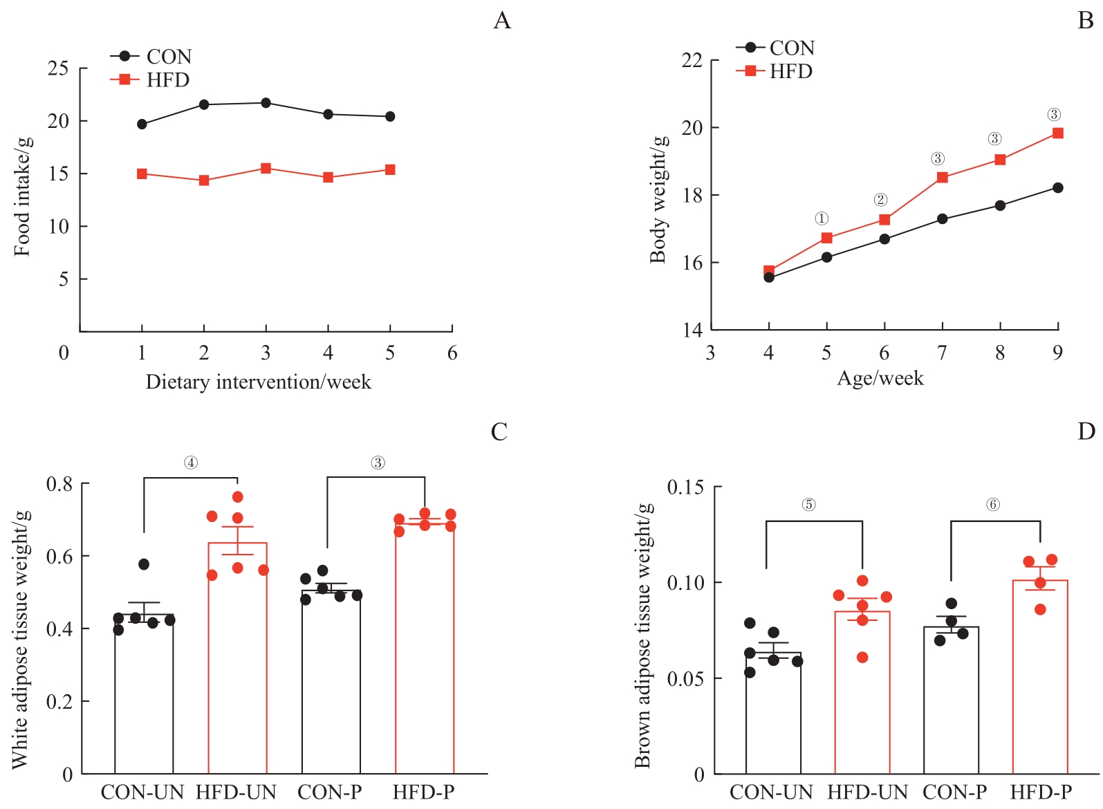
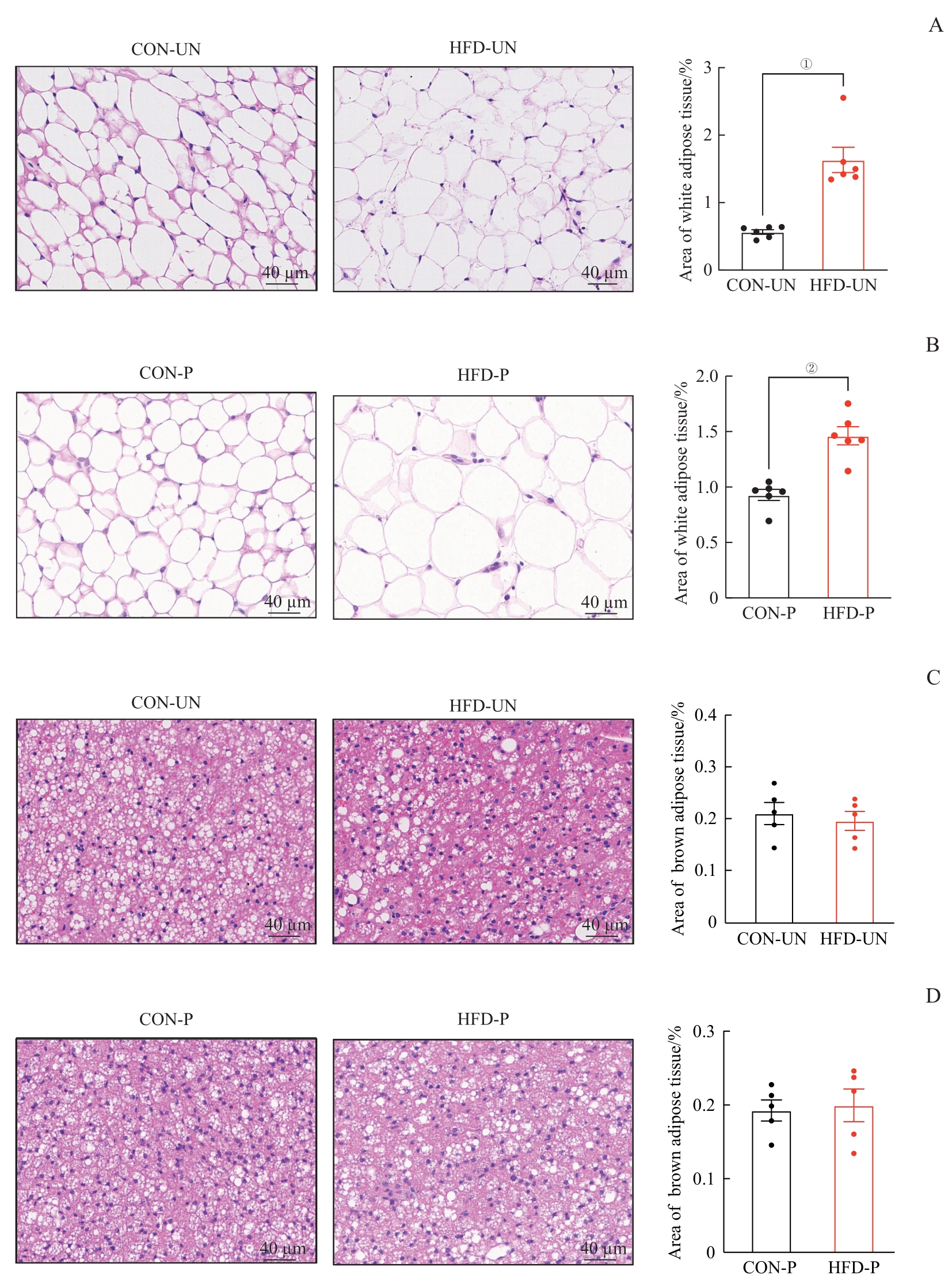
| 1 |
BLÜHER M. Obesity: global epidemiology and pathogenesis[J]. Nat Rev Endocrinol, 2019, 15: 288-298.
|
| 2 |
BLÜHER M. Metabolically healthy obesity[J]. Endocr Rev, 2020, 41(3): bnaa004.
|
| 3 |
MARQUARD K L, STEPHENS S M, JUNGHEIM E S, et al. Polycystic ovary syndrome and maternal obesity affect oocyte size in in vitro fertilization/intracytoplasmic sperm injection cycles[J]. Fertil Steril, 2011, 95(6): 2146-9, 2149.e1.
|
| 4 |
CHEN C, XU X, YAN Y, et al. Estimated global overweight and obesity burden in pregnant women based on panel data model[J]. PLoS One, 2018, 13(8): e0202183.
|
| 5 |
GOLDSTEIN R F, ABELL S K, RANASINHA S, et al. Association of gestational weight gain with maternal and infant outcomes: a systematic review and meta-analysis[J]. JAMA, 2017, 317(21): 2207-2225.
|
| 6 |
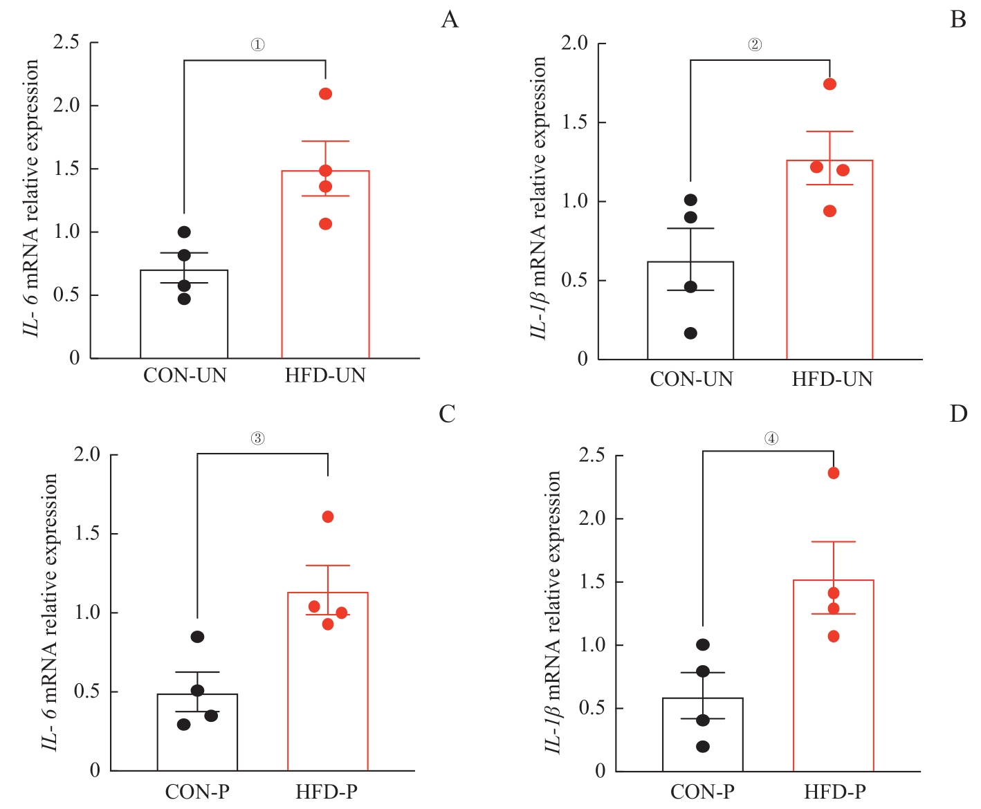
CHENG L, WANG J, DAI H, et al. Brown and beige adipose tissue: a novel therapeutic strategy for obesity and type 2 diabetes mellitus[J]. Adipocyte, 2021, 10(1): 48-65.
|
| 7 |
ROSEN E D, SPIEGELMAN B M. What we talk about when we talk about fat[J]. Cell, 2014, 156(1/2): 20-44.
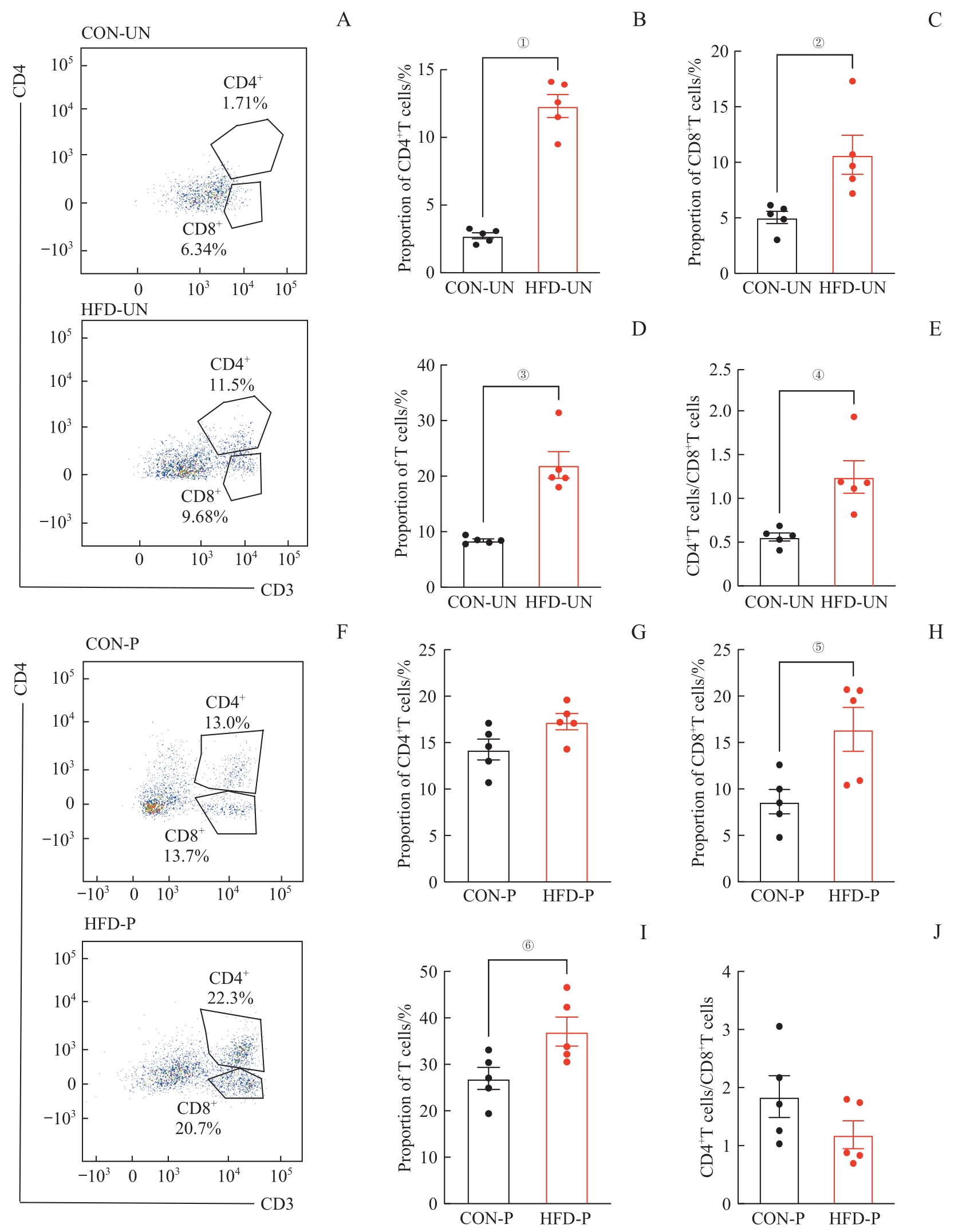
|
| 8 |
MONTANARI T, POŠĆIĆ N, COLITTI M. Factors involved in white-to-brown adipose tissue conversion and in thermogenesis: a review[J]. Obes Rev, 2017, 18(5): 495-513.
|
| 9 |
WANG Q A, TAO C, GUPTA R K, et al. Tracking adipogenesis during white adipose tissue development, expansion and regeneration[J]. Nat Med, 2013, 19: 1338-1344.
|
| 10 |
LEE J H, PARK A, OH K J, et al. The role of adipose tissue mitochondria: regulation of mitochondrial function for the treatment of metabolic diseases[J]. Int J Mol Sci, 2019, 20(19): E4924.
|
| 11 |
VISHVANATH L, GUPTA R K. Contribution of adipogenesis to healthy adipose tissue expansion in obesity[J]. J Clin Invest, 2019, 129(10): 4022-4031.
|
| 12 |
HAGSTRÖM H, SIMON T G, ROELSTRAETE B, et al. Maternal obesity increases the risk and severity of NAFLD in offspring[J]. J Hepatol, 2021, 75(5): 1042-1048.
|
| 13 |
CAO B, LIU C, ZHANG Q, et al. Maternal high-fat diet leads to non-alcoholic fatty liver disease through upregulating hepatic SCD1 expression in neonate rats[J]. Front Nutr, 2020, 7: 581723.
|
| 14 |
World Health Organization. Obesity and overweight[EB/OL]. [2024-03-01]. https://www.who.int/news-room/fact-sheets/detail/obesity-and-overweight.
|
| 15 |
GREEN M, ARORA K, PRAKASH S. Microbial medicine: prebiotic and probiotic functional foods to target obesity and metabolic syndrome[J]. Int J Mol Sci, 2020, 21(8): E2890.
|
| 16 |
ROHM T V, MEIER D T, OLEFSKY J M, et al. Inflammation in obesity, diabetes, and related disorders[J]. Immunity, 2022, 55(1): 31-55.
|
| 17 |
MEDZHITOV R. The spectrum of inflammatory responses[J]. Science, 2021, 374(6571): 1070-1075.
|
| 18 |
SUTHERLAND T E, DYER D P, ALLEN J E. The extracellular matrix and the immune system: a mutually dependent relationship[J]. Science, 2023, 379(6633): eabp8964.
|
| 19 |
GREENBERG A S, OBIN M S. Obesity and the role of adipose tissue in inflammation and metabolism[J]. Am J Clin Nutr, 2006, 83(2): 461S-465S.
|
| 20 |
NISHIMURA S, MANABE I, NAGASAKI M, et al. CD8+ effector T cells contribute to macrophage recruitment and adipose tissue inflammation in obesity[J]. Nat Med, 2009, 15: 914-920.
|
| 21 |
STRISSEL K J, DEFURIA J, SHAUL M E, et al. T-cell recruitment and Th1 polarization in adipose tissue during diet-induced obesity in C57BL/6 mice[J]. Obesity (Silver Spring), 2010, 18(10): 1918-1925.
|
| 22 |
POPOV Y, SCHUPPAN D. CD8+ T cells drive adipose tissue inflammation: a novel clue for NASH pathogenesis?[J]. J Hepatol, 2010, 52(1): 130-132.
|
| 23 |
TAO Y, LI Y H, ZHANG D, et al. Decidual CXCR4+ CD56bright NK cells as a novel NK subset in maternal-foetal immune tolerance to alleviate early pregnancy failure[J]. Clin Transl Med, 2021, 11(10): e540.
|
| 24 |
WU X, JIN L P, YUAN M M, et al. Human first-trimester trophoblast cells recruit CD56brightCD16- NK cells into decidua by way of expressing and secreting of CXCL12/stromal cell-derived factor 1[J]. J Immunol, 2005, 175(1): 61-68.
|
| 25 |
HAUGSTØYL M E, CORNILLET M, STRAND K, et al. Phenotypic diversity of human adipose tissue-resident NK cells in obesity[J]. Front Immunol, 2023, 14: 1130370.
|
| 26 |
BARKER D J. The origins of the developmental origins theory[J]. J Intern Med, 2007, 261(5): 412-417.
|
 ), XU Yidan1, LIU Yiqin1, ZHANG Qianren2, DONG Yan1,3(