Journal of Shanghai Jiao Tong University (Medical Science) ›› 2025, Vol. 45 ›› Issue (8): 969-980.doi: 10.3969/j.issn.1674-8115.2025.08.004
• Basic research • Previous Articles Next Articles
JIANG Qianyu, YAO Chengcheng, JI Ping, WANG Ying( )
)
Received:2025-02-17
Accepted:2025-04-03
Online:2025-08-28
Published:2025-08-26
Contact:
WANG Ying
E-mail:ywangssm@shsmu.edu.cn;ywangssmu@shsmu.edu.cn
Supported by:CLC Number:
JIANG Qianyu, YAO Chengcheng, JI Ping, WANG Ying. Microenvironmental profiles of wound tissues with accelerated healing properties by HAMA hydrogel[J]. Journal of Shanghai Jiao Tong University (Medical Science), 2025, 45(8): 969-980.
Add to citation manager EndNote|Ris|BibTeX
URL: https://xuebao.shsmu.edu.cn/EN/10.3969/j.issn.1674-8115.2025.08.004
| Name | Forward (5'→3' ) | Reverse (3'→5' ) |
|---|---|---|
| Arg1 | CTCCAAGCCAAAGTCCTTAGAG | AGGAGCTGTCATTAGGGACATC |
| Nos2 | GTTCTCAGCCCAACAATACAAGA | GTGGACGGGTCGATGTCAC |
| Itgam | CCATGACCTTCCAAGAGAATGC | ACCGGCTTGTGCTGTAGTC |
| Itgb2 | AGGAGCATCGCTAATCCTGAG | CCTGGTCGCAAGTAAAGTGTC |
| Gapdh | AGGTCGGTGTGAACGGATTTG | TGTAGACCATGTAGTTGAGGTCA |
Tab 1 Primer sequences
| Name | Forward (5'→3' ) | Reverse (3'→5' ) |
|---|---|---|
| Arg1 | CTCCAAGCCAAAGTCCTTAGAG | AGGAGCTGTCATTAGGGACATC |
| Nos2 | GTTCTCAGCCCAACAATACAAGA | GTGGACGGGTCGATGTCAC |
| Itgam | CCATGACCTTCCAAGAGAATGC | ACCGGCTTGTGCTGTAGTC |
| Itgb2 | AGGAGCATCGCTAATCCTGAG | CCTGGTCGCAAGTAAAGTGTC |
| Gapdh | AGGTCGGTGTGAACGGATTTG | TGTAGACCATGTAGTTGAGGTCA |
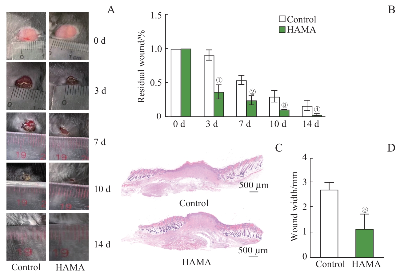
Fig 1 Promotive effects of HAMA hydrogel on wound healing
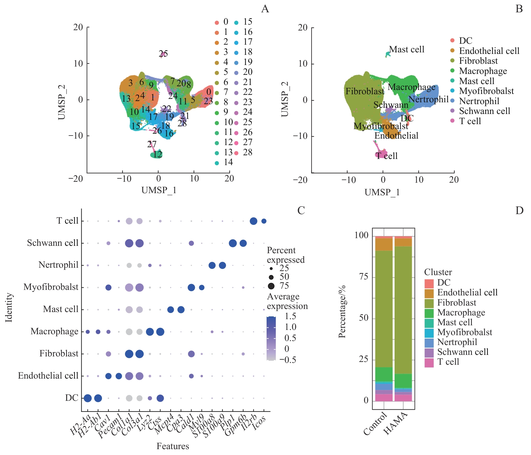
Fig 2 Single-Cell RNA sequencing analysis of cell composition at mouse skin wound sites
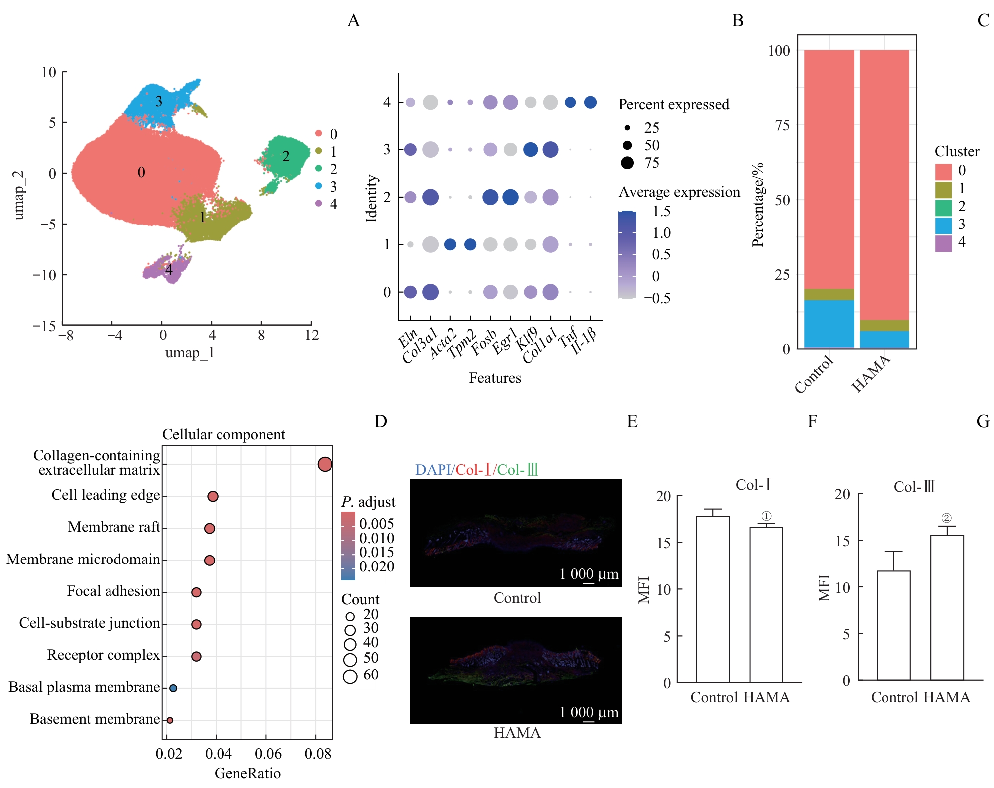
Fig 3 Analysis on composition and function of fibroblasts in wound sites
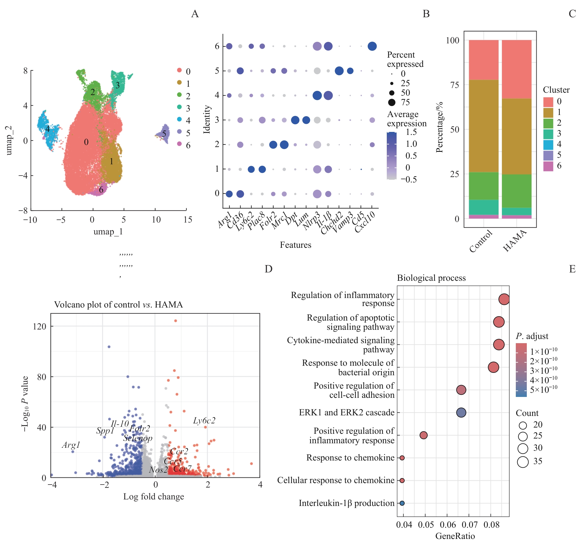
Fig 4 Analysis on composition and function of macrophages in wound sites
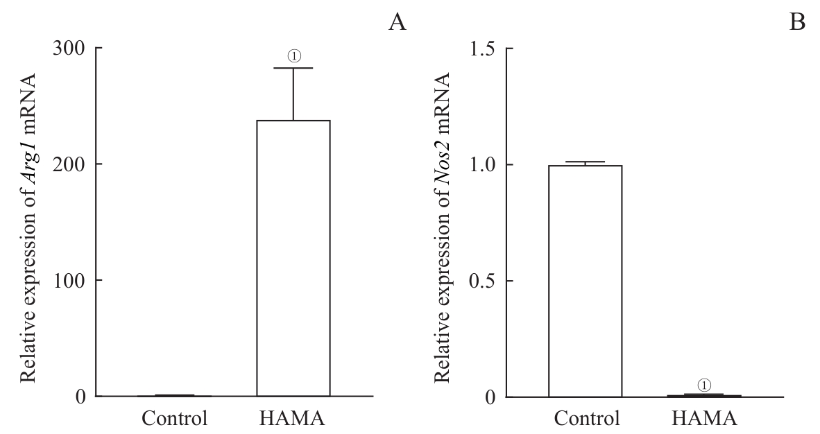
Fig 5 Effects of HAMA hydrogel on the expression of polarization-related genes in the macrophages
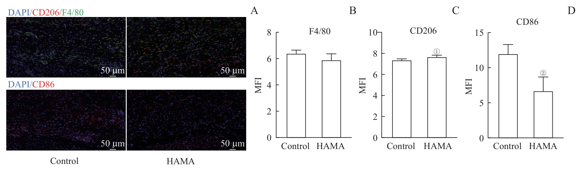
Fig 6 Analysis of local macrophage polarization characteristics in wound sites treated with HAMA hydrogel
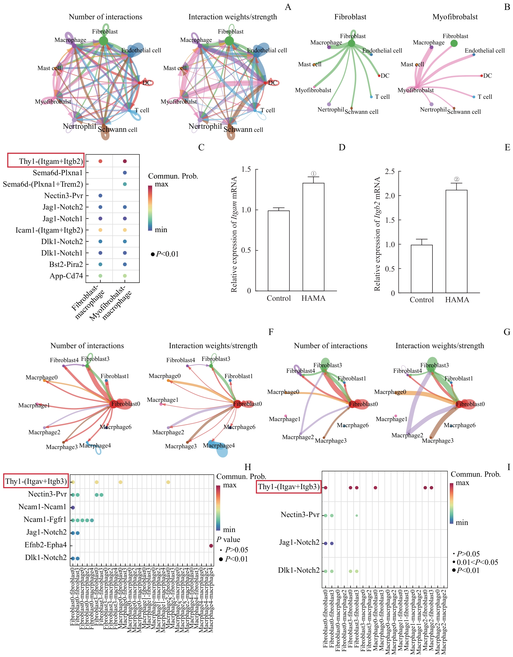
Fig 7 Interaction patterns between fibroblasts and macrophages in wound tissues
| [1] | MASCHALIDI S, MEHROTRA P, KEÇELI B N, et al. Targeting SLC7A11 improves efferocytosis by dendritic cells and wound healing in diabetes[J]. Nature, 2022, 606(7915): 776-784. |
| [2] | LU Y Z, NAYER B, SINGH S K, et al. CGRP sensory neurons promote tissue healing via neutrophils and macrophages[J]. Nature, 2024, 628(8008): 604-611. |
| [3] | GURTNER G C, WERNER S, BARRANDON Y, et al. Wound repair and regeneration[J]. Nature, 2008, 453(7193): 314-321. |
| [4] | FISCHER A, WANNEMACHER J, CHRIST S, et al. Neutrophils direct preexisting matrix to initiate repair in damaged tissues[J]. Nat Immunol, 2022, 23(4): 518-531. |
| [5] | GALLI S J, BORREGAARD N, WYNN T A. Phenotypic and functional plasticity of cells of innate immunity: macrophages, mast cells and neutrophils[J]. Nat Immunol, 2011, 12(11): 1035-1044. |
| [6] | SINHA S, SPARKS H D, LABIT E, et al. Fibroblast inflammatory priming determines regenerative versus fibrotic skin repair in reindeer[J]. Cell, 2022, 185(25): 4717-4736.e25. |
| [7] | JIANG D S, GUO R J, MACHENS H G, et al. Diversity of fibroblasts and their roles in wound healing[J]. Cold Spring Harb Perspect Biol, 2023, 15(3): a041222. |
| [8] | ZHAO L, FENG Z P, LYU Y, et al. Electroactive injectable hydrogel based on oxidized sodium alginate and carboxymethyl chitosan for wound healing[J]. Int J Biol Macromol, 2023, 230: 123231. |
| [9] | SETHI S, THAKUR S, SHARMA D, et al. Malic acid cross-linked chitosan based hydrogel for highly effective removal of chromium (Ⅵ) ions from aqueous environment[J]. React Funct Polym, 2022, 177: 105318. |
| [10] | HELMECKE T, HAHN D, MATZKE N, et al. Inflammation-controlled anti-inflammatory hydrogels[J]. Adv Sci (Weinh), 2023, 10(7): e2206412. |
| [11] | CHENG L, CAI Z W, YE T J, et al. Injectable polypeptide-protein hydrogels for promoting infected wound healing[J]. Adv Funct Materials, 2020, 30(25): 2001196. |
| [12] | XU Z, LIU G, LIU P, et al. Hyaluronic acid-based glucose-responsive antioxidant hydrogel platform for enhanced diabetic wound repair [J]. Acta Biomater, 2022, 147: 147-157. |
| [13] | HONG S, YANG K, KANG B, et al. Hyaluronic acid catechol: a biopolymer exhibiting a pH-dependent adhesive or cohesive property for human neural stem cell engineering[J]. Adv Funct Materials, 2013, 23(14): 1774-1780. |
| [14] | SINGH A, CORVELLI M, UNTERMAN S A, et al. Enhanced lubrication on tissue and biomaterial surfaces through peptide-mediated binding of hyaluronic acid[J]. Nat Mater, 2014, 13(10): 988-995. |
| [15] | CAI Z X, ZHANG H B, WEI Y, et al. Reduction- and pH-sensitive hyaluronan nanoparticles for delivery of iridium (Ⅲ) anticancer drugs[J]. Biomacromolecules, 2017, 18(7): 2102-2117. |
| [16] | SHU X Z, LIU Y C, LUO Y, et al. Disulfide cross-linked hyaluronan hydrogels[J]. Biomacromolecules, 2002, 3(6): 1304-1311. |
| [17] | SERBAN M A, PRESTWICH G D. Synthesis of hyaluronan haloacetates and biology of novel cross-linker-free synthetic extracellular matrix hydrogels[J]. Biomacromolecules, 2007, 8(9): 2821-2828. |
| [18] | LIU B, KONG Y F, ALIMI O A, et al. Multifunctional microgel-based cream hydrogels for postoperative abdominal adhesion prevention[J]. ACS Nano, 2023, 17(4): 3847-3864. |
| [19] | ZHOU K, YANG C L, SHI K, et al. Activated macrophage membrane-coated nanoparticles relieve osteoarthritis-induced synovitis and joint damage[J]. Biomaterials, 2023, 295: 122036. |
| [20] | LEE J, KIM D, JANG C H, et al. Highly elastic 3D-printed gelatin/HA/placental-extract scaffolds for bone tissue engineering[J]. Theranostics, 2022, 12(9): 4051-4066. |
| [21] | LIU N B, ZHU S J, DENG Y Z, et al. Construction of multifunctional hydrogel with metal-polyphenol capsules for infected full-thickness skin wound healing[J]. Bioact Mater, 2022, 24: 69-80. |
| [22] | FARAHANI M, SHAFIEE A. Wound healing: from passive to smart dressings[J]. Adv Healthc Mater, 2021, 10(16): e2100477. |
| [23] | GAO S Y, CHEN T, WANG Z, et al. Immuno-activated mesenchymal stem cell living electrospun nanofibers for promoting diabetic wound repair[J]. J Nanobiotechnology, 2022, 20(1): 294. |
| [24] | WANG X, ZHAO D H, LI Y T, et al. Collagen hydrogel with multiple antimicrobial mechanisms as anti-bacterial wound dressing[J]. Int J Biol Macromol, 2023, 232: 123413. |
| [25] | PENG Z W, XUE H, LIU X, et al. Tough, adhesive biomimetic hyaluronic acid methacryloyl hydrogels for effective wound healing[J]. Front Bioeng Biotechnol, 2023, 11: 1222088. |
| [26] | AHMED M K, ZAYED M A, EL-DEK S I, et al. Nanofibrous ε-polycaprolactone scaffolds containing Ag-doped magnetite nanoparticles: physicochemical characterization and biological testing for wound dressing applications in vitro and in vivo[J]. Bioact Mater, 2021, 6(7): 2070-2088. |
| [27] | YAMASAKI S, ISHIKAWA E, SAKUMA M, et al. Mincle is an ITAM-coupled activating receptor that senses damaged cells[J]. Nat Immunol, 2008, 9(10): 1179-1188. |
| [28] | CHEN J P, CHEN D F, CHEN J L, et al. An all-in-one CO gas therapy-based hydrogel dressing with sustained insulin release, anti-oxidative stress, antibacterial, and anti-inflammatory capabilities for infected diabetic wounds[J]. Acta Biomater, 2022, 146: 49-65. |
| [29] | FU Y J, SHI Y F, WANG L Y, et al. All-natural immunomodulatory bioadhesive hydrogel promotes angiogenesis and diabetic wound healing by regulating macrophage heterogeneity[J]. Adv Sci (Weinh), 2023, 10(13): e2206771. |
| [30] | PEÑA O A, MARTIN P. Cellular and molecular mechanisms of skin wound healing[J]. Nat Rev Mol Cell Biol, 2024, 25(8): 599-616. |
| [31] | HINZ B. Formation and function of the myofibroblast during tissue repair[J]. J Invest Dermatol, 2007, 127(3): 526-537. |
| [32] | WAN R, WEISSMAN J P, GRUNDMAN K, et al. Diabetic wound healing: the impact of diabetes on myofibroblast activity and its potential therapeutic treatments[J]. Wound Repair Regen, 2021, 29(4): 573-581. |
| [33] | YOUNESI F S, MILLER A E, BARKER T H, et al. Fibroblast and myofibroblast activation in normal tissue repair and fibrosis[J]. Nat Rev Mol Cell Biol, 2024, 25(8): 617-638. |
| [34] | MOTZ K, LINA I, MURPHY M K, et al. M2 macrophages promote collagen expression and synthesis in laryngotracheal stenosis fibroblasts[J]. Laryngoscope, 2021, 131(2): E346-E353. |
| [35] | HE J H, FANG B, SHAN S Z, et al. Mechanical stretch promotes hypertrophic scar formation through mechanically activated cation channel Piezo1[J]. Cell Death Dis, 2021, 12(3): 226. |
| [36] | LI S Y, LI C, ZHANG Y T, et al. Targeting mechanics-induced fibroblast activation through CD44-RhoA-YAP pathway ameliorates crystalline silica-induced silicosis[J]. Theranostics, 2019, 9(17): 4993-5008. |
| [37] | AMUSO V M, HAAS M R, COOPER P O, et al. Fibroblast-mediated macrophage recruitment supports acute wound healing[J]. J Investig Dermatol, 2025, 145(7): 1781-1797.e8. |
| [38] | SHEN L Y, LI Y S, ZHAO H K. Fibroblast growth factor signaling in macrophage polarization: impact on health and diseases[J]. Front Immunol, 2024, 15: 1390453. |
| [39] | CHEN C, YANG J C, SHANG R Y, et al. Orchestration of macrophage polarization dynamics by fibroblast-secreted exosomes during skin wound healing[J]. J Invest Dermatol, 2025, 145(1): 171-184.e6. |
| [1] | Du Xin, Li Xuebing, Li Yongwei. Research progress of macrophage metabolic reprogramming in recurrent spontaneous abortion [J]. Journal of Shanghai Jiao Tong University (Medical Science), 2026, 46(4): 537-544. |
| [2] | Chen Jiayu, Zhang Huili. Role and mechanism of fibroblast mitochondrial dysfunction in pulmonary arterial hypertension [J]. Journal of Shanghai Jiao Tong University (Medical Science), 2026, 46(3): 291-300. |
| [3] | HUANG Yinghe, ZHAO Guanyu, SUN Yang, HOU Jianji, ZUO Yong. Research progress on macrophage metabolic regulation in wound healing of diabetes mellitus type 2 [J]. Journal of Shanghai Jiao Tong University (Medical Science), 2025, 45(6): 792-799. |
| [4] | WEI Lanyi, XUE Xiaochuan, CHEN Junjun, YANG Quanjun, WANG Mengyue, HAN Yonglong. Research progress of tumor-associated macrophages in immune microenvironment and targeted therapy of osteosarcoma [J]. Journal of Shanghai Jiao Tong University (Medical Science), 2023, 43(5): 624-630. |
| [5] | SHA Pan, ZHAO Xuewen, ZHU Haotian, GAO Chongzhou, LIU Shen. Research progress in the mechanism and intervention of tendon adhesion [J]. Journal of Shanghai Jiao Tong University (Medical Science), 2022, 42(8): 1116-1121. |
| [6] | QI Yangyang, XIONG Ying. Phenotype, function and clinical significance of galectin-9 positive tumor-associated macrophages in muscle-invasive bladder cancer [J]. Journal of Shanghai Jiao Tong University (Medical Science), 2022, 42(12): 1666-1676. |
| [7] | Lin-xiu-mei GUO, Yi-xin ZHANG. Application of skin autofluorescence detection technique to diagnosis of diseases [J]. JOURNAL OF SHANGHAI JIAOTONG UNIVERSITY (MEDICAL SCIENCE), 2021, 41(2): 251-256. |
| [8] | ZHAO Wei-guang, LIU Zhi-hong. Advances in study of regulation of tumor immune inflammatory microenvironment by cancer-associated fibroblasts [J]. JOURNAL OF SHANGHAI JIAOTONG UNIVERSITY (MEDICAL SCIENCE), 2020, 40(9): 1288-1293. |
| [9] | LU Shi-yuan1, HONG Jie1, CHEN Ying-xuan1, CHEN Jin-xian2, ZHONG Ming2, FANG Jing-yuan1. Study of Fusobacterium nucleatum-related bacterial biofilm promoting M2 polarization of macrophages and chemoresistance in colon cancer [J]. JOURNAL OF SHANGHAI JIAOTONG UNIVERSITY (MEDICAL SCIENCE), 2020, 40(8): 1018-1029. |
| [10] | WANG Qi, ZHU Guan-ya, XIE Ting, GE Kui, NIU Yi-wen. Changes of ATP metabolism and purinergic receptors in inflammatory response stage of diabetic wound healing [J]. , 2020, 40(1): 10-. |
| [11] | ZHANG Wen, FU Xiu-jun, YAO Min. Promotion of 640 nm red light on keratinocyte migration via CD26 [J]. , 2019, 39(8): 843-. |
| [12] | LI Xue-chuan,TENG Pei-min,YUAN Bo,QIAO Liang,YANG Hui-zhong. Effect of shaped polyurethane foam dressing on healing of scalp donor sites in the patients with extensive burn [J]. , 2019, 39(5): 514-. |
| [13] | SHI Yong-ping, YUAN Bo. Real-time effect of adipose stem cells on fibrogenesis of dermal fibroblast co-stimulatedtransforming growth factor-β1 [J]. , 2019, 39(12): 1382-. |
| [14] | JIANG Chun-lan, CHENG Hui-juan, JIAO Ting. In vitroand in vivo study of apoptotic macrophages inducedmethylene-blue-mediated photodynamic therapy in periodontitis [J]. , 2018, 38(12): 1429-. |
| [15] | YANG Chuan-feng, PENG Yin-bo, HAO Jian, SONG Chen-lu, HU Yan-ge, YAO Min. Effect of a novel chitosan-silver nitrate gel dressing on anti-septic and wound healing#br# [J]. , 2017, 37(7): 1004-. |
| Viewed | ||||||
|
Full text |
|
|||||
|
Abstract |
|
|||||