上海交通大学学报(医学版), 2025, 45(1): 42-50 doi: 10.3969/j.issn.1674-8115.2025.01.005

论著 · 临床研究

腹部脂肪面积与2型糖尿病患者胰岛β细胞第一时相分泌功能的关系

陆佳萍,1,, 刘醒,2,, 张林杉2, 赵琳2, 张敏1, 李小英2, 刘玥隽,2

1.复旦大学附属中山医院青浦分院内分泌科,上海 201700

2.复旦大学附属中山医院内分泌科,上海 200032

Relationship between abdominal fat area and first-phase insulin secretion function of pancreatic β-cells in patients with type 2 diabetes

LU Jiaping,1,, LIU Xing,2,, ZHANG Linshan2, ZHAO Lin2, ZHANG Min1, LI Xiaoying2, LIU Yuejun,2

1.Department of Endocrinology, Qingpu Branch of Zhongshan Hospital, Fudan University, Shanghai 201700, China

2.Department of Endocrinology and Metabolism, Zhongshan Hospital, Fudan University, Shanghai 200032, China

通讯作者: 刘玥隽,主治医师,博士;电子信箱:liu.yuejun@zs-hospital.sh.cn

第一联系人: *为共同第一作者

编委: 包玲

收稿日期: 2024-07-09   接受日期: 2024-10-15   网络出版日期: 2025-01-28

基金资助: 国家自然科学基金.  81900771
中山医院优秀青年项目.  2019ZSYQ19
青浦区卫生健康委员会项目.  QWJ2022-01

Corresponding authors: LIU Yuejun, E-mail:liu.yuejun@zs-hospital.sh.cn.

Received: 2024-07-09   Accepted: 2024-10-15   Online: 2025-01-28

作者简介 About authors

陆佳萍(1993—),女,主治医师,硕士;电子信箱:lujiaping_1993@hotmail.com E-mail:lujiaping_1993@hotmail.com

刘 醒(1988—),女,主治医师,博士;电子信箱:liu.xing@zs-hospital.sh.cn。 E-mail:liu.xing@zs-hospital.sh.cn

摘要

目的·探讨2型糖尿病患者腹部脂肪面积与胰岛β细胞第一时相分泌功能之间的关系,并建立列线图预测模型。方法·选取2020年10月—2024年2月在复旦大学附属中山医院内分泌科住院并完善精氨酸刺激试验以评估胰岛β细胞第一时相分泌功能的2型糖尿病患者120例。根据试验结果,将患者分为胰岛素分泌功能残存组和功能衰竭组。收集患者的一般资料及实验室指标,通过内脏脂肪检测仪检测患者腹部皮下脂肪面积(subcutaneous fat area,SFA)和内脏脂肪面积(visceral fat area,VFA)。经单因素分析筛选变量,使用多因素Logistic回归确定影响因素,从而建立列线图预测模型。采用受试者操作特征曲线(receiver operating characteristic curve,ROC曲线)分析的曲线下面积(area under the curve,AUC)、一致性指数(concordance index,C-index)评估模型预测效能。结果·纳入的74例患者(61.7%)胰岛功能残存,46例(38.3%)胰岛功能衰竭。胰岛功能衰竭组患者糖尿病病程更长,腰围、臀围、体质量指数(body mass index,BMI)、尿酸、游离三碘甲状腺原氨酸(free triiodothyronine,FT3)、脂肪组织胰岛素抵抗指数(adipose tissue insulin resistance,Adipo-IR)、踝肱指数(ankle brachial index,ABI)、SFA和VFA水平较低,臂踝脉搏波速度(brachial ankle pulse wave velocity,baPWV)较快。多因素Logistic回归分析显示,SFA、VFA、FT3、baPWV和ABI是胰岛功能衰竭的独立危险因素。基于以上因素构建列线图模型,其中纳入VFA、FT3、ABI、baPWV的模型预测能力最佳,C-index为0.81。结论·胰岛β细胞第一时相分泌功能衰竭患者SFA和VFA均较低,纳入SFA或VFA的列线图可用于预测2型糖尿病患者β细胞第一时相分泌功能。

关键词: 2型糖尿病 ; 腹部脂肪面积 ; 胰岛功能 ; 列线图

Abstract

Objective ·To explore the relationship between abdominal fat area and the first-phase insulin secretion function of pancreatic β-cells in patients with type 2 diabetes, and to establish predictive models of nomogram. Methods ·From October 2020 to February 2024, a total of 120 patients with type 2 diabetes, who were hospitalized in the Department of Endocrinology, Zhongshan Hospital, Fudan University, and underwent the arginine stimulation test, were recruited for the study. Patients were categorized into an insulin secretion function-preserved group (i.e. preserved group) and a depleted group according to the results of the arginine stimulation test. General information and laboratory parameters were collected. Subcutaneous fat area (SFA) and visceral fat area (VFA) were non-invasively measured by abdominal fat detector. The variables were screened by univariate analysis, and multivariate Logistic regression was used to identify the influencing factors, followed by the establishment of predictive models of nomogram. The area under the receiver operating characteristic curve (ROC curve) and concordance index (C-index) were used to evaluate the predictive performance of the models. Results ·Seventy-four patients (61.7%) were assigned to the preserved group, and 46 patients (38.3%) to the depleted group. Patients in the depleted group had a longer diabetes duration, lower waist circumference, hip circumference, body mass index (BMI), uric acid, free triiodothyronine (FT3), adipose tissue insulin resistance (Adipo-IR), ankle brachial index (ABI), SFA and VFA, and higher brachial ankle pulse wave velocity (baPWV). Multivariate Logistic regression showed that SFA, VFA, FT3, baPWV, and ABI were independent risk factors for the depleted insulin secretion function. Nomogram models were constructed based on the above risk factors. Among them, the model comprising VFA, FT3, ABI, and baPWV showed the best predictive performance with a C-index of 0.81. Conclusion ·SFA and VFA are lower in patients with depleted first-phase insulin secretion function of pancreatic β-cells. The nomogram model, including SFA or VFA, can be used to predict first-phase insulin secretion function of pancreatic β-cells in patients with type 2 diabetes.

Keywords: type 2 diabetes ; abdominal fat area ; insulin secretion function ; nomogram

PDF (2215KB) 元数据 多维度评价 相关文章 导出 EndNote| Ris| Bibtex  收藏本文

本文引用格式

陆佳萍, 刘醒, 张林杉, 赵琳, 张敏, 李小英, 刘玥隽. 腹部脂肪面积与2型糖尿病患者胰岛β细胞第一时相分泌功能的关系. 上海交通大学学报(医学版)[J], 2025, 45(1): 42-50 doi:10.3969/j.issn.1674-8115.2025.01.005

LU Jiaping, LIU Xing, ZHANG Linshan, ZHAO Lin, ZHANG Min, LI Xiaoying, LIU Yuejun. Relationship between abdominal fat area and first-phase insulin secretion function of pancreatic β-cells in patients with type 2 diabetes. Journal of Shanghai Jiao Tong University (Medical Science)[J], 2025, 45(1): 42-50 doi:10.3969/j.issn.1674-8115.2025.01.005

2型糖尿病患病率逐年增加。国际糖尿病联盟(International Diabetes Federation,IDF)2021年发布的结果显示,全球成年糖尿病患者有5.366亿人,2045年预计达到7.832亿人1,将造成严重的社会经济负担,故糖尿病防治具有重要的临床意义。2型糖尿病的病理生理特征是胰岛素抵抗伴β细胞分泌功能障碍2。然而随着疾病的发展,胰岛β细胞分泌功能逐渐下降直至最终衰竭,需要启动胰岛素治疗。因此,评价胰岛分泌功能并及时启动胰岛素治疗在2型糖尿病诊治中有重要意义。精氨酸刺激试验能有效评估β细胞第一时相分泌功能3,但该方法的实施需要经过培训的专科护理人员,以及对患者短时间内多次抽血,无法在所有2型糖尿病患者中施行。此外,并不是所有的医院都能够检测血清胰岛素和C肽水平,或无法当日出具检测结果。因此,需要建立其他快速简便的方法来评估胰岛分泌功能。

脂肪组织是人体重要的内分泌器官。腹部皮下脂肪扩张是胰岛素抵抗和2型糖尿病发展过程中的关键变化4。研究5发现,计算机断层成像(computed tomography,CT)测量的内脏脂肪面积(visceral fat area,VFA)和内脏/皮下脂肪比值(visceral-to-subcutaneous fat ratio,VSR)增加与2型糖尿病发病风险显著相关,也是2型糖尿病的独立危险因素。磁共振成像(magnetic resonance imaging,MRI)评估的腹部皮下脂肪面积(subcutaneous fat area,SFA)与β细胞分泌功能呈正相关6。通过测量VFA、SFA有助于评估胰岛功能。然而,CT检查具有放射性,MRI检查耗时长,临床应用有限。内脏脂肪检测仪利用生物电阻法可无创测量SFA和VFA,无放射性、操作简便、耗时短,可在临床广泛应用。然而,通过内脏脂肪检测仪检测的SFA、VFA与2型糖尿病患者β细胞分泌功能之间的关系需要进一步评估。

本研究拟运用内脏脂肪检测仪无创测量的腹部脂肪面积,分析腹部脂肪面积与2型糖尿病患者胰岛β细胞第一时相分泌功能的关系,建立预测模型。并采用受试者操作特征曲线(receiver operating characteristic curve,ROC曲线)的曲线下面积(area under the curve,AUC)和一致性指数(concordance index,C-index)评估模型的预测效能。本研究旨在协助临床医师快速简便评估2型糖尿病患者的胰岛功能,从而及时启动胰岛素治疗。

1 对象与方法

1.1 研究对象

回顾性选取2020年10月—2024年2月在复旦大学附属中山医院内分泌科住院的2型糖尿病患者。纳入标准:① 年龄18~75岁。② 符合中华医学会糖尿病学分会《中国2型糖尿病防治指南(2020年版)》的糖尿病诊断标准7。③ 完成精氨酸刺激试验。④ 完成内脏脂肪检测仪测量。排除标准:① 患有胰岛素自身免疫综合征等影响血清胰岛素、C肽检测结果的疾病。② 妊娠期妇女。③ 存在严重的慢性疾病,如肿瘤等。④ 精氨酸刺激试验时空腹血糖(fasting plasma glucose,FPG)>11 mmol/L。根据精氨酸刺激试验结果,将患者分为胰岛功能正常组、胰岛功能受损组和胰岛功能衰竭组。由于识别胰岛分泌功能衰竭的患者并及时开始胰岛素治疗具有重要临床意义,本研究将功能正常和受损的患者合并为胰岛功能残存组。

1.2 资料收集

收集患者年龄、性别,测量其身高、体质量、腰围、臀围,计算体质量指数(body mass index,BMI)、腰臀比。并收集患者入院后空腹实验室检查结果,包括FPG、胰岛素、C肽、糖化白蛋白(glycated albumin,GA)、糖化血红蛋白(glycated hemoglobin,HbA1c)、丙氨酸转氨酶(alanine aminotransferase,ALT)、门冬氨酸转氨酶(aspartate aminotransferase,AST)、γ-谷氨酰转移酶(γ-glutamyltransferase,GGT)、总胆汁酸(total bile acid,TBA)、血肌酐(serum creatinine,Scr)、尿酸(uric acid,UA)、总胆固醇(total cholesterol,TC)、三酰甘油(triacylglycerol,TAG)、低密度脂蛋白胆固醇(low-density lipoprotein cholesterol,LDL-C)、高密度脂蛋白胆固醇(high-density lipoprotein cholesterol,HDL-C)、非高密度脂蛋白胆固醇(non-high-density lipoprotein cholesterol,NHDL-C)、游离脂肪酸(free fatty acid,FFA)、游离三碘甲状腺原氨酸(free triiodothyronine,FT3)、游离甲状腺素(free thyroxine,FT4)、促甲状腺激素(thyroid stimulating hormone,TSH)、C反应蛋白(C-reactive protein,CRP)、血清淀粉样蛋白A(serum amyloid A protein,SAA)、红细胞沉降率(erythrocyte sedimentation rate,ESR),以及餐后2 h血糖(2-hour postprandial blood glucose,2 h-PG)、胰岛素(2-hour postprandial serum insulin,2 h-insulin)、C肽(2-hour postprandial serum C-peptide,2 h-C-peptide)水平。采用慢性肾脏病流行病学合作研究(chronic kidney disease epidemiology collaboration,CKD-EPI)方程计算估算的肾小球滤过率(estimated glomerular filtration rate,eGFR)。留取3次24 h尿蛋白定量(24-hour urinary albumin excretion,24 h-UAE)并取平均值。对未使用胰岛素治疗的患者计算如下指标:稳态模型胰岛素抵抗指数(homeostasis model assessment-insulin resistance,HOMA-IR)=空腹胰岛素(μU/mL)×FPG(mmol/L)/22.5(血清胰岛素1 pmol/L=0.144 μU/mL),稳态模型β细胞功能指数(homeostatic model assessment of β-cell function,HOMA-β)=20×空腹胰岛素(μU/mL)/[空腹血糖(mmol/L)-3.5],脂肪组织胰岛素抵抗指数(adipose tissue insulin resistance,Adipo-IR)=游离脂肪酸(mmol/L)×空腹胰岛素(μU/mL)×FPG(mmol/L)8。代谢综合征(metabolic syndrome,MetS)诊断标准7为研究对象满足以下条件中的一项计1分,累积分数(MetS score)≥3分:① 男性腰围≥90 cm或女性腰围≥85 cm。② TAG≥1.7 mmol/L或已接受针对此脂质异常的治疗。③ 男性HDL-C≤1.03 mmol/L,女性HDL-C≤1.30 mmol/L,或已接受针对此脂质异常的治疗。④ 血压≥130/85 mmHg(1 mmHg=0.133 kPa)或已服用降压药治疗。⑤ 先前诊断为2型糖尿病。

1.3 精氨酸刺激试验

为避免糖毒性对β细胞分泌功能的影响,所有受试者均在FPG≤11 mmol/L下进行精氨酸刺激试验。受试者于试验前一日晚10点后禁食,试验日先采集其血样测定即刻葡萄糖、胰岛素、C肽水平。随后夹管接生理盐水推注,确定无外渗后予精氨酸试剂5 g(20 mL/支)推注,在30 s内全部推完。推时记时并于推后2、4、6 min采集血样测定葡萄糖、胰岛素、C肽水平。胰岛β细胞第一时相分泌功能评估方法:胰岛素增值(ΔI)或C肽增值(ΔCP)为2、4、6 min时胰岛素或C肽均值与空腹时的差值。ΔI≥23.09 mU/L或ΔCP≥1.63 ng/mL为β细胞第一时相分泌功能正常,12.23 mU/L≤ΔI<23.09 mU/L或0.97 ng/mL≤ΔCP<1.63 ng/mL为功能受损,ΔI<12.23 mU/L或ΔCP<0.97 ng/mL为功能衰竭9。已使用胰岛素治疗的患者根据ΔCP评估。

1.4 腹部脂肪测定等无创检查

采用基于生物电阻法的内脏脂肪检测仪DUALSCAN HDS-2000(日本欧姆龙公司)测定SFA、VFA。采用FibroScan®(法国Echosens公司)测定肝脏硬度值(liver stiffness measurement,LSM)和受控衰减参数(controlled attenuation parameter,CAP)10。采用动脉硬化检测装置/血压脉波检查装置BP-203RPEⅢ(日本欧姆龙公司)测定踝臂指数(ankle brachial index,ABI)、臂踝脉搏波速度(brachial ankle pulse wave velocity,baPWV)。采用SD800彩色多普勒超声显像仪(荷兰飞利浦公司)测定颈动脉内膜中层厚度(intima-media thickness,IMT)。

1.5 统计学分析

采用SPSS 19.0软件和R 4.1.2软件进行数据分析。定量资料进行正态性分布检验,正态分布的以x±s表示,非正态分布的以MQ1Q3 )表示。2组间均值比较采用Wilcoxon符号秩检验。定性资料使用n(%)表示,并采用χ2检验。以P<0.05挑选变量纳入多因素Logistic回归筛选胰岛功能衰竭的独立风险因子,根据纳入因子个数确定最小样本量。建立β细胞第一时相分泌功能衰竭列线图预测模型,用R语言中的rms包进行列线图可视化输出。采用AUC、C-index评估模型预测效能。P<0.05表示差异具有统计学意义。

2 结果

2.1 患者一般基线特征比较

本研究共纳入120例患者,平均年龄(56.6±11.1)岁,其中男性78例、女性42例,42例(35.0%)患者接受了胰岛素治疗。根据精氨酸刺激试验结果,74例受试者β细胞第一时相分泌功能残存,46例功能衰竭。由表1可见,衰竭组腹型肥胖患者的比例较低、糖尿病病程更长,代谢综合征的患病率更低,腰围、臀围、体质量、BMI、空腹胰岛素、空腹C肽、HOMA-IR、尿酸、FT3、Adipo-IR、ABI、VFA和SFA较低,baPWV较快(均P<0.05)。

表1   胰岛分泌功能残存组和衰竭组临床资料及生化指标比较

Tab 1  Comparison of clinical and biological parameters between the insulin secretion function-preserved group and the depleted group

ItemPreserved group (n=74)Depleted group (n=46)P value
Age/year54.0 (47.8, 64.0)60.0 (50.5, 67.3)0.096
Male/n(%)52 (70.3)26 (56.5)0.125
Hypertension/n(%)36 (48.6)26 (56.5)0.401
Dyslipidemia/n(%)64 (86.5)37 (80.4)0.529
Abdominal obesity/n(%)57 (77.0)22 (47.8)<0.001
MetS score/n(%)0.036
1 point or 2 points15 (20.3)19 (41.3)
3 points31 (41.9)12 (26.1)
4 points28 (37.8)15 (32.6)
Diabetes duration/year5.00 (0, 10.00)10.00 (4.00, 20.00)<0.001
Treated with insulin/n(%)21 (28.4)21 (45.7)0.054
Waist circumference/cm93.20±8.4688.47±8.750.004
Hip circumference/cm99.74±6.0594.98±7.83<0.001
Waist-to-hip ratio0.93±0.060.93±0.060.848
Weight/kg74.22±10.7567.40±11.330.001
BMI/(kg·m-2)25.46 (24.10, 27.75)23.71 (22.70, 25.99)0.002
FPG/(mmol·L-1)7.60 (6.30, 9.90)8.15 (6.28, 10.90)0.492
Fasting insulin/(pmol·L-1)64.58 (38.19, 88.89)42.36 (18.40, 64.24)0.006
Fasting C-peptide/(ng·mL-1)2.31 (1.62, 2.86)1.78 (1.09, 2.26)0.003
HOMA-IR2.73 (1.96, 4.60)1.82 (1.19, 2.63)0.016
HOMA-β44.64 (25.29, 73.03)26.32 (8.80, 66.71)0.082
2 h-PG/(mmol·L-1)11.10 (8.90, 14.20)11.20 (9.05, 14.45)0.779
2 h-insulin/(pmol·L-1)165.58 (81.94, 254.17)138.89 (57.99, 211.46)0.268
2 h-C-peptide/(ng·mL-1)4.61 (2.89, 6.43)3.29 (2.10, 5.03)0.120
GA/%22.05 (17.93, 28.55)25.80 (20.20, 31.20)0.086
HbA1C/%9.35 (7.68, 10.78)9.90 (8.45, 11.40)0.089
ALT/(U·L-1)18.50 (13.75, 33.50)17.00 (10.75, 25.25)0.075
AST/(U·L-1)18.00 (14.00, 27.35)15.00 (12.00, 23.25)0.068
GGT/(U·L-1)26.50 (18.75, 47.50)22.00 (15.00, 32.50)0.091
TBA/(μmol·L-1)3.30 (2.15, 5.20)3.10 (1.80, 4.33)0.444
Scr/(μmol·L-1)68.00 (60.00, 81.00)68.50 (56.75, 82.00)0.885
eGFR/[mL·(min·1.73m2)-1]98.00 (83.00, 108.00)92.00 (80.00, 102.75)0.232
UA/(μmol·L-1)337.00 (274.00, 403.00)284.00 (241.75, 354.00)0.049
24 h-UAE/[mg·(24 h)-1]15.85 (8.99, 24.19)19.75 (11.26, 32.99)0.125
TC/(mmol·L-1)4.57±1.114.56±0.920.956
TAG/(mmol·L-1)1.62 (1.27, 2.26)1.55 (1.05, 2.17)0.473
LDL-C/(mmol·L-1)2.67±0.962.66±0.860.934
NHDL-C/(mmol·L-1)3.34 (2.82, 4.17)3.52 (2.64, 4.04)0.987
HDL-C/(mmol·L-1)1.05 (0.91, 1.18)1.08 (0.96, 1.24)0.308
FFA/(mmol·L-1)0.54 (0.43, 0.61)0.47 (0.33, 0.62)0.088
Adipo-IR4.22 (2.71, 7.11)3.11 (1.45,6.25)0.040
FT3/(pmol·L-1)4.50 (4.10, 4.90)4.20 (3.65, 4.55)0.001
FT4/(pmol·L-1)16.59±2.7816.99±2.100.405
TSH/(mIU·L-1)1.76 (1.10, 2.08)1.43 (1.06, 2.36)0.359
CRP/(mg·L-1)1.05 (0.53, 2.08)0.90 (0.58, 2.25)0.871
SAA/(mg·L-1)3.50 (3.33, 4.25)3.50 (3.20, 5.43)0.993
ESR/(mm·h-1)7.00 (4.00, 13.00)10.00 (3.50, 15.00)0.385
IMT≥1.0 mm/n(%)14 (22.2)10 (26.3)0.640
baPWV/(m·s-1)15.41 (13.57, 17.83)17.63 (14.18, 22.19)0.009
ABI1.16 (1.08, 1.22)1.12 (1.06, 1.17)0.033
CAP/(dB·m-1)275.68±52.84267.00±59.180.478
LSM/kPa5.65 (4.50, 6.55)5.25 (4.33, 6.23)0.415
VFA/cm281.91±30.8164.50±29.210.003
SFA/cm2186.30 (156.40, 227.70)151.95 (127.15, 190.55)<0.001

新窗口打开| 下载CSV


2.2 多因素Logistic回归筛选胰岛功能衰竭的独立风险因子

为了进一步探索与胰岛功能衰竭密切相关的独立风险因子,我们首先以表1P<0.05为切点挑选变量,随后将共线性诊断指标方差膨胀因子(variance inflation factor,VIF)<5的变量纳入回归模型。模型1纳入SFA和无创指标,模型2纳入SFA、无创指标和实验室检查结果,模型3纳入VFA和无创指标,模型4纳入VFA、无创指标和实验室检查结果。表2提示,SFA、VFA、FT3、ABI、baPWV是胰岛功能衰竭的独立风险因子。根据多因素Logistic回归结果构建预测模型,AUC结果提示纳入VFA的模型3和4具有较好的预测效能(图1)。

表2   β细胞第一时相分泌功能衰竭的多因素Logistic分析

Tab 2  Multivariate Logistic regression analysis of depleted first-phase insulin secretion function of β-cells

ItemβOR (95%CI)P value
Model 1
Diabetes duration0.0521.053 (0.986‒1.124)0.121
ABI×10-0.6000.549 (0.321‒0.939)0.028
baPWV0.1491.160 (1.011‒1.331)0.034
SFA-0.0120.988 (0.978‒0.997)0.012
Model 2
Diabetes duration0.0521.054 (0.979‒1.135)0.166
Fasting C-peptide-0.3960.673 (0.385‒1.177)0.165
UA-0.0020.998 (0.993‒1.003)0.425
FT3-1.0980.333 (0.145‒0.769)0.010
ABI×10-0.6220.537 (0.303‒0.951)0.033
baPWV0.1851.203 (1.029‒1.406)0.020
SFA-0.0100.990 (0.980‒1.001)0.076
Model 3
Diabetes duration0.0561.057 (0.990‒1.129)0.096
ABI×10-0.6950.499 (0.285‒0.874)0.015
baPWV0.1591.172 (1.020‒1.347)0.025
VFA-0.0260.974 (0.957‒0.992)0.005
Model 4
Diabetes duration0.0581.060 (0.983‒1.143)0.130
Fasting C-peptide-0.4590.632 (0.361‒1.108)0.109
UA-0.0010.999 (0.994‒1.004)0.753
FT3-1.0570.348 (0.154‒0.783)0.011
ABI×10-0.6890.502 (0.279‒0.903)0.021
baPWV0.1981.219 (1.040‒1.428)0.015
VFA-0.0220.978 (0.957‒0.999)0.036

Note: For gender, males were assigned a value of 1 and females 0. ABI was multiplied by 10. OR—odds ratio; CI—confidence interval.

新窗口打开| 下载CSV


图1

图1   预测β细胞第一时相分泌功能衰竭的ROC曲线

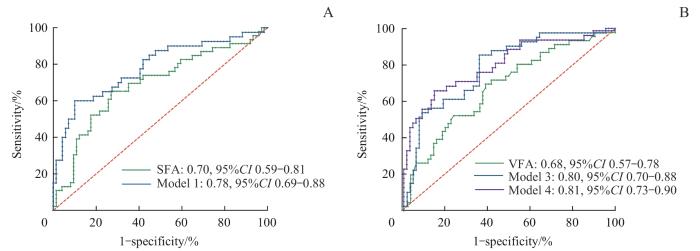
Note: A. Models including SFA for predicting depleted insulin secretion function. B. Models including VFA for predicting depleted insulin secretion function.

Fig 1   ROC curve of models for predicting depleted first-phase insulin secretion function of β-cells


2.3 列线图模型的构建与评价

为了更直观运用多因素Logistic回归结果在临床上快速评估2型糖尿病患者胰岛素功能是否衰竭,我们根据以上有较好预测价值的模型1、3、4构建了预测胰岛功能衰竭的列线图。列线图1由ABI、baPWV和SFA组成(图2A),列线图2由ABI、baPWV和VFA组成(图2B),列线图3由FT3、ABI、baPWV和VFA组成(图2C)。校准曲线(图3)显示胰岛功能衰竭的实际值和预测值之间有较好的一致性。3个模型的C-index分别为0.78、0.80和0.81。

图2

图2   预测β细胞第一时相分泌功能衰竭的列线图

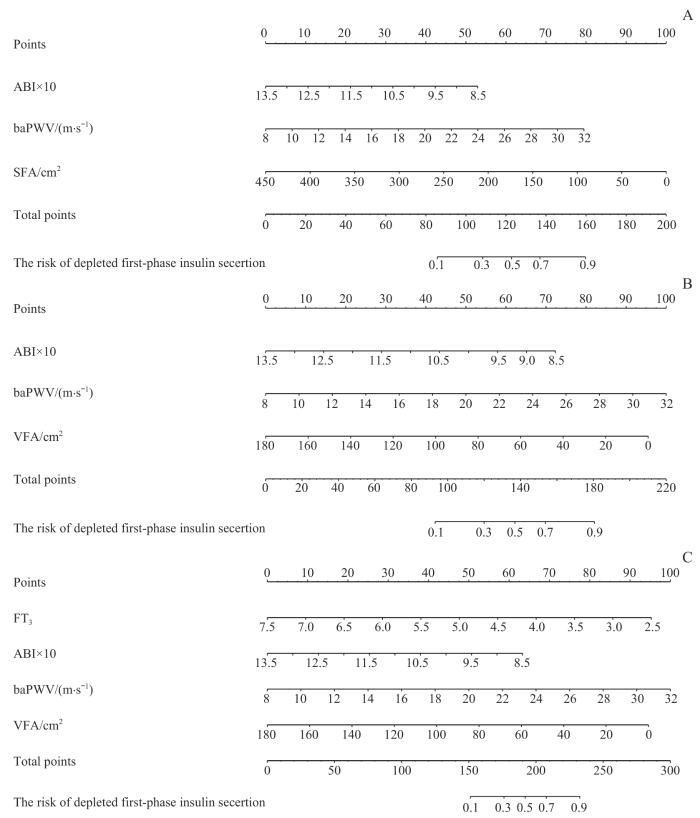
Note: A. SFA-simple model involved SFA and non-invasive markers. B. VFA-simple model involved VFA and non-invasive markers. C. VFA-invasive model involved VFA and biomarkers.

Fig 2   Nomogram models for predicting depleted first-phase insulin secretion function of β-cells


图3

图3   列线图的校准曲线

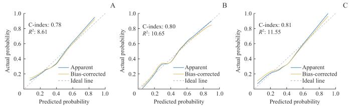
Note: A. SFA-simple model. B. VFA-simple model. C. VFA-invasive model. The dashed grey lines represent perfect prediction (ideal line); the blue lines represent the entire cohort (apparent); the yellow lines, bias-corrected via bootstrapping (with 1 000 replicates), indicate observed nomogram performance (bias-corrected).

Fig 3   Calibration plots of the nomogram models


3 讨论

评估2型糖尿病患者胰岛功能并及时启动胰岛素治疗具有重要意义。2型糖尿病发生发展时胰岛素抵抗、胰岛功能衰竭存在较多影响因素,腹部脂肪组织是重要影响因素之一。本研究通过无创方法测量SFA和VFA,发现2型糖尿病患者SFA和VFA减少是胰岛功能衰竭独立风险因子,为糖尿病腹部脂肪与胰岛素分泌功能的关系提供了新见解。此外,本研究建立了仅需临床资料和无创测量SFA及VFA的简易列线图,以及纳入实验室检查结果的复合列线图,有助于各级医院临床医师快速评估2型糖尿病患者的胰岛功能。

本研究选取SFA和VFA 2个腹部脂肪面积参数评估胰岛分泌功能,是因为脂肪组织是糖代谢中起重要作用的内分泌器官。脂肪过多导致肥胖/超重引起2型糖尿病、脂肪萎缩导致胰岛素抵抗引起脂肪萎缩性糖尿病,反映了脂肪与胰岛素抵抗、糖代谢的2种截然不同的病理状况。生理状态下,皮下脂肪通过细胞增生和肥大储存多余能量4。当脂肪储存能力达到极限时,脂肪在肝脏、胰腺和肌肉等组织中异位沉积,导致非酒精性脂肪性肝病/肝炎(nonalcoholic fatty liver disease/nonalcoholic steatohepatitis,NAFLD/NASH)、胰岛素分泌功能受损和胰岛素抵抗。胰岛素抵抗会诱发胰岛素过度分泌,从而进一步加剧胰岛素分泌功能的受损,最终导致胰岛素分泌逐渐衰竭。脂肪储存能力与脂肪生成能力受限或受损有关,并在一定程度上受基因调控11。高加索人和亚洲人超重和肥胖的标准不同,亚洲人对脂肪扩张能力受限的影响更为敏感12。此外,胰腺中的脂肪异位沉积还会引发局部炎症,进而影响胰岛素分泌13。通过应用成纤维细胞生长因子21(fibroblast growth factor 21,FGF21)治疗,可以增加皮下脂肪组织的扩张能力,从而改善胰岛素敏感性14。最新研究15发现,2型糖尿病患者骨骼肌质量与胰岛功能、胰岛素抵抗密切相关,提高骨骼肌质量及相应减少脂肪含量对2型糖尿病患者胰岛功能改善具有重要意义。结合本研究结果,骨骼肌、脂肪比例增加可能与2型糖尿病患者胰岛功能有关,这一观点有待于进一步研究证实。此外,脂肪因子在葡萄糖稳态中也发挥着重要作用。既往研究11提示,脂联素具有抗炎作用,能提高胰岛素敏感性。羟基硬脂酸棕榈酸酯(palmitic acid ester of hydroxystearic acid,PAHSA)是一种新发现的由脂肪细胞合成的脂肪因子,能增强β细胞增殖和存活能力,提高胰高血糖素样肽-1(glucagon-like peptide-1,GLP-1)分泌,从而增强GLP1诱导的β细胞胰岛素分泌16。因此,脂肪组织减少伴随这类脂肪因子的减少可能导致胰岛素分泌受损。

本研究多因素Logistic回归提示脂肪面积联合baPWV和ABI能预测胰岛分泌功能。BaPWV和ABI是2个评估动脉粥样硬化简单、实用的无创指标,但也与糖代谢密切相关。文献报告,baPWV与空腹和2 h C肽水平正相关,这表明内源性胰岛素分泌与动脉硬化之间存在密切联系17。此外,baPWV的增加先于空腹血糖升高,也是糖尿病发生的危险因素18。糖尿病患者中,ABI与心血管结局和死亡独立相关,与中层动脉钙化引起的动脉硬度增加有关19。机制上,糖尿病时的低度炎症破坏内皮功能,导致动脉壁硬度增加,从而加重毛细血管损伤。动脉硬度增加会损害高流量低阻力器官的功能,例如胰腺,从而影响胰岛分泌功能。毛细血管功能障碍引起的组织灌流减少也会损害肌肉等组织的功能,进一步影响糖代谢。此外,低度炎症和氧化应激是糖尿病和动脉硬度的共同病理特征,彼此相互影响18。因此,baPWV和ABI可以作为胰岛分泌功能的良好预测指标。

本研究还发现甲状腺激素在预测胰岛分泌功能方面起重要作用。甲状腺激素是参与新陈代谢的关键激素,其作用于碳水化合物调节基础代谢,增加循环中的游离脂肪酸,并通过促进脂肪分解加速胰岛素降解。葡萄糖刺激的胰岛素分泌和降解也受到甲状腺激素的调节20,而甲状腺激素抵抗综合征的患者胰岛素敏感性降低21。最新研究22表明,肝脏中的甲状腺激素通过调节GLP-1的分泌,改善胰岛素分泌和糖代谢。此外,FT3具有类似有丝分裂的作用,可以直接激活Akt通路,促进β细胞增殖、减少凋亡,从而保留β细胞数量,改善其蛋白质合成并增加胰岛素分泌23-24。因此,甲状腺激素与胰岛分泌功能密切相关。

基于以上与胰岛素分泌密切相关的风险因子,我们构建了列线图模型。列线图是将回归结果进行可视化输出,相较于Logistic回归模型更直观,在临床上常用于多指标联合诊断或预测疾病发生发展。我们首次将列线图模型用于2型糖尿病患者胰岛功能预测,建立了仅需临床资料和无创检查结果的简易列线图,以及纳入实验室检查结果的复合列线图,以契合不同医院的实际需求,未来有望在社区医院、二级医院广泛使用,为临床医师快速评估2型糖尿病患者的胰岛功能提供有力支持。

本研究仍存在一定局限性。研究未检测脂肪细胞因子,且为单中心研究,样本量较小,列线图仅进行了内部验证。因此,未来需扩大样本量,增加检测项目,并进行外部验证。

综上,本研究用无创手段检测SFA、VFA并构建列线图预测2型糖尿病患者胰岛功能。该列线图预测模型具有良好的临床实用价值,能够帮助临床医师更加直观地预判2型糖尿病患者胰岛功能衰竭。

作者贡献声明

刘玥隽设计研究。刘醒、张林杉和赵琳招募受试者。陆佳萍收集数据。刘玥隽、陆佳萍、刘醒分析数据并撰写文稿。张敏和李小英为文章提供指导。陆佳萍、刘醒对本研究有同等贡献。所有作者均阅读并同意最终稿件的提交。

AUTHOR's CONTRIBUTIONS

LIU Yuejun designed the study. LIU Xing, ZHANG Linshan, and ZHAO Lin recruited the participants. LU Jiaping collected the data. LIU Yuejun, LU Jiaping, and LIU Xing analyzed the data and wrote the manuscript. ZHANG Min and LI Xiaoying provided guidance on the article. LU Jiaping and LIU Xing contributed equally to this study. All the authors have read the final version of paper and consented for submission.

利益冲突声明

所有作者声明不存在利益冲突。

COMPETING INTERESTS

All authors disclare no relevant conflict of interests.

参考文献

SUN H, SAEEDI P, KARURANGA S, et al. IDF Diabetes Atlas: global, regional and country-level diabetes prevalence estimates for 2021 and projections for 2045[J]. Diabetes Res Clin Pract, 2022, 183: 109119.

[本文引用: 1]

NAUCK M A, WEFERS J, MEIER J J. Treatment of type 2 diabetes: challenges, hopes, and anticipated successes[J]. Lancet Diabetes Endocrinol, 2021, 9(8): 525-544.

[本文引用: 1]

HANNON T S, KAHN S E, UTZSCHNEIDER K M, et al. Review of methods for measuring β-cell function: design considerations from the Restoring Insulin Secretion (RISE) Consortium[J]. Diabetes Obes Metab, 2018, 20(1): 14-24.

[本文引用: 1]

CĂTOI A F, BUSETTO L. Metabolically healthy obesity and bariatric surgery[J]. Obes Surg, 2019, 29(9): 2989-3000.

[本文引用: 2]

KIM E H, KIM H K, LEE M J, et al. Sex differences of visceral fat area and visceral-to-subcutaneous fat ratio for the risk of incident type 2 diabetes mellitus[J]. Diabetes Metab J, 2022, 46(3): 486-498.

[本文引用: 1]

GYLLENHAMMER L E, ALDERETE T L, TOLEDO-CORRAL C M, et al. Saturation of subcutaneous adipose tissue expansion and accumulation of ectopic fat associated with metabolic dysfunction during late and post-pubertal growth[J]. Int J Obes (Lond), 2016, 40(4): 601-606.

[本文引用: 1]

中华医学会糖尿病学分会. 中国2型糖尿病防治指南(2020年版)[J]. 中华糖尿病杂志, 2021, 13(4): 315-409.

[本文引用: 2]

Chinese Diabetes Society. Guideline for the prevention and treatment of type 2 diabetes mellitus in China (2020 edition)[J]. Chinese Journal of Diabetes, 2021, 13(4): 315-409.

[本文引用: 2]

TER HORST K W, VAN GALEN K A, GILIJAMSE P W, et al. Methods for quantifying adipose tissue insulin resistance in overweight/obese humans[J]. Int J Obes (Lond), 2017, 41(8): 1288-1294.

[本文引用: 1]

朱小鹏, 颜红梅, 常薪霞, 等. 精氨酸刺激试验评估胰岛β细胞1相分泌功能的价值及对2型糖尿病治疗的指导作用[J]. 中国临床医学, 2017, 24(4): 548-553.

[本文引用: 1]

ZHU X P, YAN H M, CHANG X X, et al. The value of arginine stimulation test in evaluating the first-phase insulin secretion and its guiding role for the treatment of type 2 diabetes mellitus[J]. Chinese Journal of Clinical Medicine, 2017, 24(4): 548-553.

[本文引用: 1]

SASSO M, BEAUGRAND M, DE LEDINGHEN V, et al. Controlled attenuation parameter (CAP): a novel VCTE™ guided ultrasonic attenuation measurement for the evaluation of hepatic steatosis: preliminary study and validation in a cohort of patients with chronic liver disease from various causes[J]. Ultrasound Med Biol, 2010, 36(11): 1825-1835.

[本文引用: 1]

SMITH U, KAHN B B. Adipose tissue regulates insulin sensitivity: role of adipogenesis, de novo lipogenesis and novel lipids[J]. J Intern Med, 2016, 280(5): 465-475.

[本文引用: 2]

LI R, LU W, JIA J, et al. Relationships between indices of obesity and its cardiovascular comorbidities in a Chinese population[J]. Circ J, 2008, 72(6): 973-978.

[本文引用: 1]

NEELAND I J, TURER A T, AYERS C R, et al. Dysfunctional adiposity and the risk of prediabetes and type 2 diabetes in obese adults[J]. JAMA, 2012, 308(11): 1150-1159.

[本文引用: 1]

LI H T, WU G Y, FANG Q C, et al. Fibroblast growth factor 21 increases insulin sensitivity through specific expansion of subcutaneous fat[J]. Nat Commun, 2018, 9(1): 272.

[本文引用: 1]

陈姝, 储爱卉, 卞文轩, 等. 2型糖尿病患者骨骼肌质量与胰岛功能相关性研究[J]. 中国临床研究, 2024, 37(7): 1045-1050.

[本文引用: 1]

CHEN S, CHU A H, BIAN W X, et al. Correlation between skeletal muscle mass and islet function in patients with type 2 diabetes mellitus[J]. Chinese Journal of Clinical Research, 2024, 37(7): 1045-1050.

[本文引用: 1]

SYED I, RUBIN DE CELIS M F, MOHAN J F, et al. PAHSAs attenuate immune responses and promote β cell survival in autoimmune diabetic mice[J]. J Clin Invest, 2019, 129(9): 3717-3731.

[本文引用: 1]

SUN Y C, ZHU Y Q, ZHANG L, et al. Relationship between insulin secretion and arterial stiffness in essential hypertension[J]. Int J Hypertens, 2021, 2021: 5015797.

[本文引用: 1]

ZHENG M Y, ZHANG X Y, CHEN S H, et al. Arterial stiffness preceding diabetes: a longitudinal study[J]. Circ Res, 2020, 127(12): 1491-1498.

[本文引用: 2]

NIE F Z, HE J A, CAO H, et al. Predictive value of abnormal ankle-brachial index in patients with diabetes: a meta-analysis[J]. Diabetes Res Clin Pract, 2021, 174: 108723.

[本文引用: 1]

TEIXEIRA P F D S, DOS SANTOS P B, PAZOS-MOURA C C. The role of thyroid hormone in metabolism and metabolic syndrome[J]. Ther Adv Endocrinol Metab, 2020, 11: 2042018820917869.

[本文引用: 1]

MORAN C, MCENIERY C M, SCHOENMAKERS N, et al. Dyslipidemia, insulin resistance, ectopic lipid accumulation, and vascular function in resistance to thyroid hormone β[J]. J Clin Endocrinol Metab, 2021, 106(5): e2005-e2014.

[本文引用: 1]

YAN Y, NIU Z M, SUN C, et al. Hepatic thyroid hormone signalling modulates glucose homeostasis through the regulation of GLP-1 production via bile acid-mediated FXR antagonism[J]. Nat Commun, 2022, 13(1): 6408.

[本文引用: 1]

VERGA FALZACAPPA C, PETRUCCI E, PATRIARCA V, et al. Thyroid hormone receptor TRβ1 mediates Akt activation by T3 in pancreatic β cells[J]. J Mol Endocrinol, 2007, 38(1/2): 221-233.

[本文引用: 1]

VERGA FALZACAPPA C, PATRIARCA V, BUCCI B, et al. The TRβ1 is essential in mediating T3 action on Akt pathway in human pancreatic insulinoma cells[J]. J Cell Biochem, 2009, 106(5): 835-848.

[本文引用: 1]

/