| 1 |
KUGELBERG C F. Impacted lower third molars and periodontal health. An epidemiological, methodological, retrospective and prospective clinical, study[J]. Swed Dent J Suppl, 1990, 68: 1-52.
|
| 2 |
TRYBEK G, ANIKO-WŁODARCZYK M, JAROŃ A. Assessment of electrosensitivity of the pulp of the mandibular second molar after surgical removal of an impacted mandibular third molar[J]. J Clin Med, 2021, 10(16) : 3614.
|
| 3 |
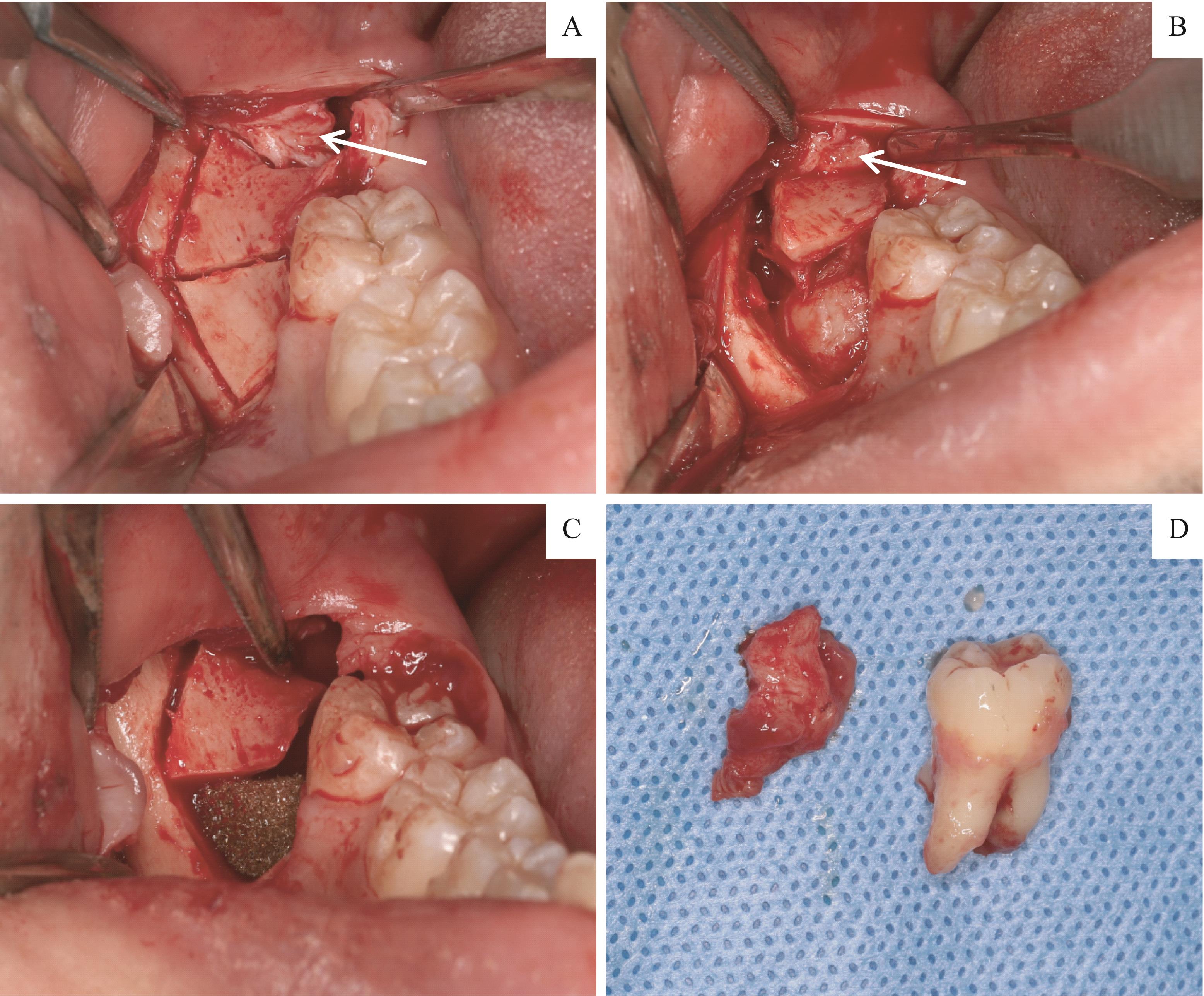
ARENAZ-BUA J, LUACES-REY R, SIRONVALLE-SOLIVA S, et al. A comparative study of platelet-rich plasma, hydroxyapatite, demineralized bone matrix and autologous bone to promote bone regeneration after mandibular impacted third molar extraction[J]. Med Oral, 2010, 15(3): e483-e489.
|
| 4 |
BARBATO L, KALEMAJ Z, BUTI J, et al. Effect of surgical intervention for removal of mandibular third molar on periodontal healing of adjacent mandibular second molar: a systematic review and Bayesian network meta-analysis[J]. J Periodontol, 2016, 87(3): 291-302.
|
| 5 |
SAMMARTINO G, TIA M, MARENZI G, et al. Use of autologous platelet-rich plasma (PRP) in periodontal defect treatment after extraction of impacted mandibular third molars[J]. J Oral Maxillofac Surg, 2005, 63(6): 766-770.
|
| 6 |
JUNG R E, IOANNIDIS A, HÄMMERLE C H F, et al. Alveolar ridge preservation in the esthetic zone[J]. Periodontol, 2000, 2018, 77(1): 165-175.
|
| 7 |
戈旌, 杨驰, 郑家伟, 等. 阻生第三磨牙拔除术后自体骨回植第二磨牙远中骨缺损的单中心随机对照临床研究[J]. 中国口腔颌面外科杂志, 2017, 15(4): 334-340.
|
|
GE J,YANG C, ZHENG J W, et al. Autogenous bone grafting for treatment of osseous defect after impacted mandibular third molar extraction: a single-center randomized controlled trial[J]. Chin J Oral Maxillofac Surg, 2017, 15(4): 334-340.
|
| 8 |
ZHOU J, HONG H Y, ZHOU H, et al. Orthodontic extraction of a high-risk impacted mandibular third molar contacting the inferior alveolar nerve, with the aid of a ramus mini-screw[J]. Quintessence Int, 2021, 52(6): 538-546.
|
| 9 |
DAWARE S N, BALAKRISHNA R, DEOGADE S C, et al. Assessment of postoperative discomfort and nerve injuries after surgical removal of mandibular third molar: a prospective study[J]. J Family Med Prim Care, 2021, 10(4): 1712-1717.
|
| 10 |
Couso-Queiruga E, Mansouri C J, Alade A A, et al. Alveolar ridge preservation reduces the need for ancillary bone augmentation in the context of implant therapy.[J]. J Periodontol, 2022.DOI: 10.1002/JPER.22-0030.
|
| 11 |
Sakkas A, Wilde F, Heufelder M, et al. Autogenous bone grafts in oral implantology: is it still a “gold standard”? A consecutive review of 279 patients with 456 clinical procedures[J]. Int J Implant Dent, 2017, 3(1): 23.
|
| 12 |
梁德凤, 周鑫才, 李艳芬, 等. 自体骨即刻移植对阻生第三磨牙拔除术后第二磨牙远中骨质缺损的疗效分析[J]. 口腔医学, 2019, 39(7): 624-627, 631.
|
|
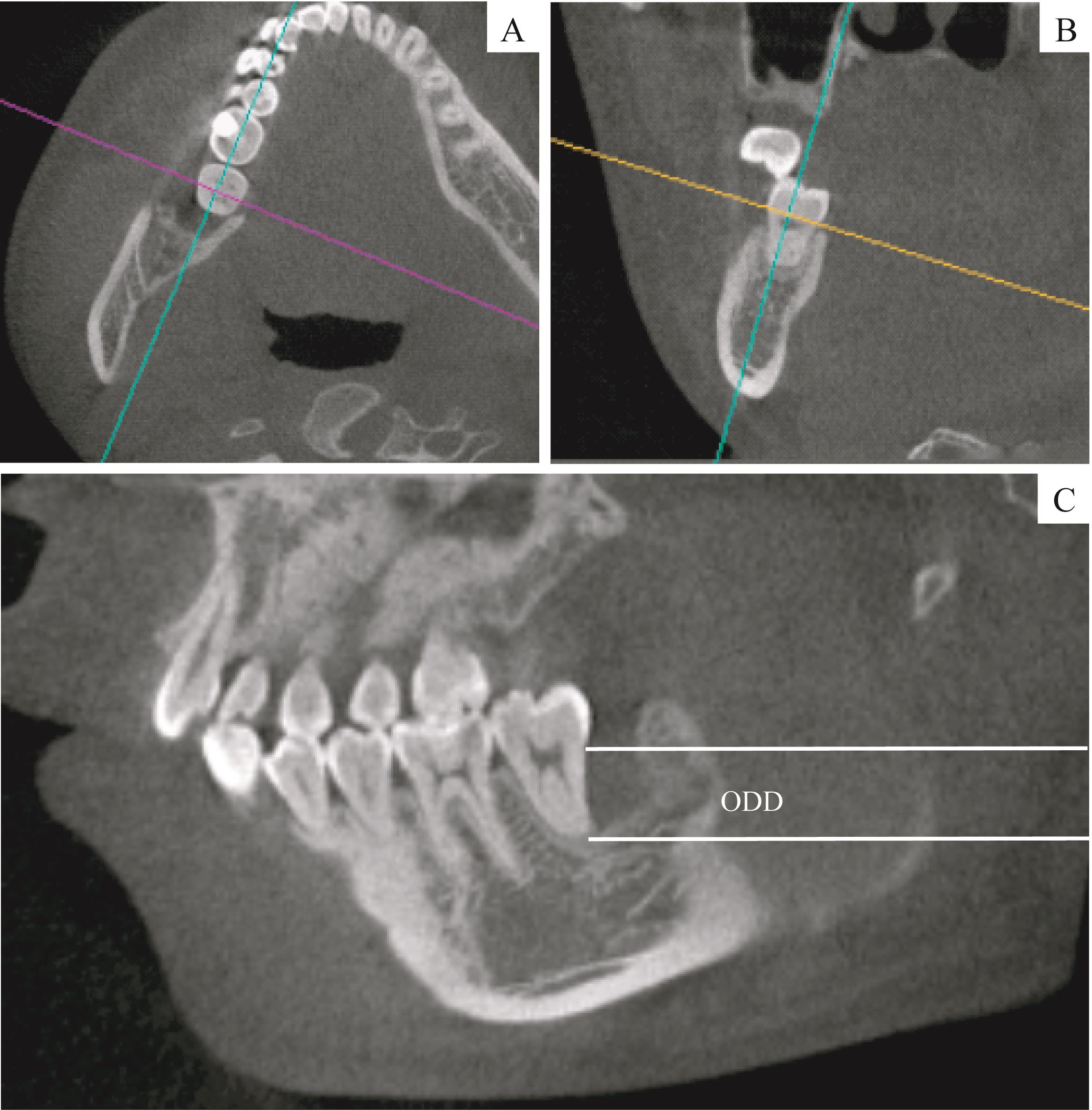
LIANG D F, ZHOU X C, LI Y F, et al. Effect of autogenous bone graft on distal bone defect of the second molar after the extraction of the impacted third molar[J]. Stomatology, 2019, 39(7): 624-627, 631.
|
| 13 |
Elgali I, Omar O,Dahlin C, et al. Guided bone regeneration: materials and biological mechanisms revisited[J]. Eur J Oral Sci, 2017, 125(5) : 315-337.
|
| 14 |
IŞıK G, YÜCE M Ö, KOÇAK-TOPBAŞ N, et al. Guided bone regeneration simultaneous with implant placement using bovine-derived xenograft with and without liquid platelet-rich fibrin: a randomized controlled clinical trial[J]. Clin Oral Investig, 2021, 25(9): 5563-5575.
|
 ), 陈敏洁(