上海交通大学学报(医学版) ›› 2023, Vol. 43 ›› Issue (3): 278-292.doi: 10.3969/j.issn.1674-8115.2023.03.003

• 论著 · 基础研究 • 上一篇    

分选链接蛋白1在胰腺导管腺癌中的表达及其促进胰腺导管腺癌进展的机制研究

潘泓(), 廖颖娜, 盖严支, 钱立恒, 聂惠贞()   

  1. 上海交通大学医学院附属仁济医院上海市肿瘤研究所,癌基因及相关基因国家重点实验室,上海 200240
  • 收稿日期:2022-09-06 接受日期:2023-03-15 出版日期:2023-03-28 发布日期:2023-03-28
  • 通讯作者: 聂惠贞 E-mail:panhong123@sjtu.edu.cn;hznie@shsci.org
  • 作者简介:潘 泓(1998—),男,硕士生;电子信箱:panhong123@sjtu.edu.cn
  • 基金资助:
    上海市自然科学基金(21ZR1461400)

Expression of sorting nexin 1 in pancreatic ductal adenocarcinoma and its mechanism in promoting PDAC progress

PAN Hong(), LIAO Yingna, GAI Yanzhi, QIAN Liheng, NIE Huizhen()   

  1. State Key Laboratory of Oncogenes and Related Genes, Shanghai Cancer Institute, Renji Hospital, Shanghai Jiao Tong University School of Medicine, Shanghai 200240, China
  • Received:2022-09-06 Accepted:2023-03-15 Online:2023-03-28 Published:2023-03-28
  • Contact: NIE Huizhen E-mail:panhong123@sjtu.edu.cn;hznie@shsci.org
  • Supported by:
    Natural Science Foundation of Shanghai(21ZR1461400)

摘要:

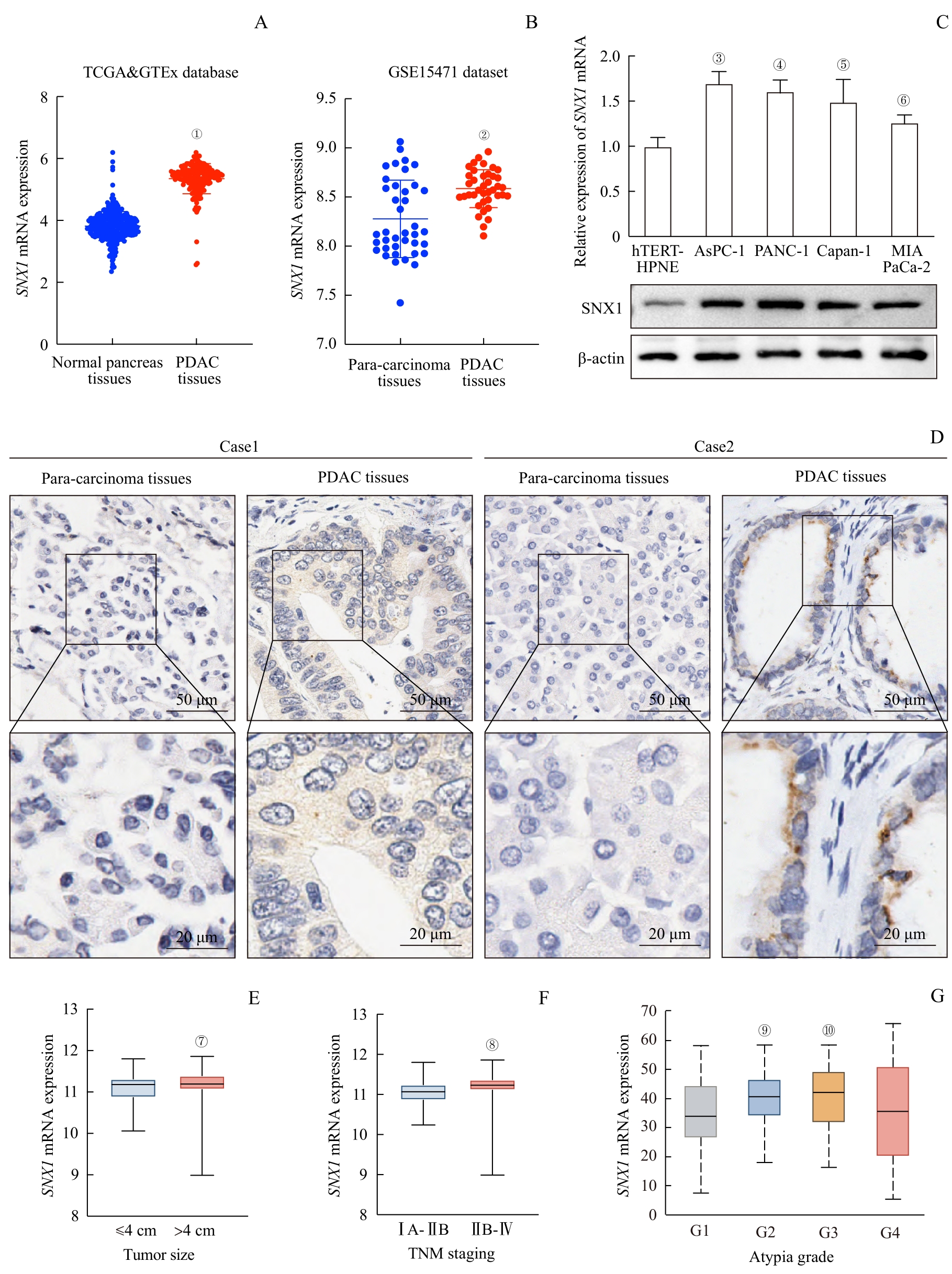
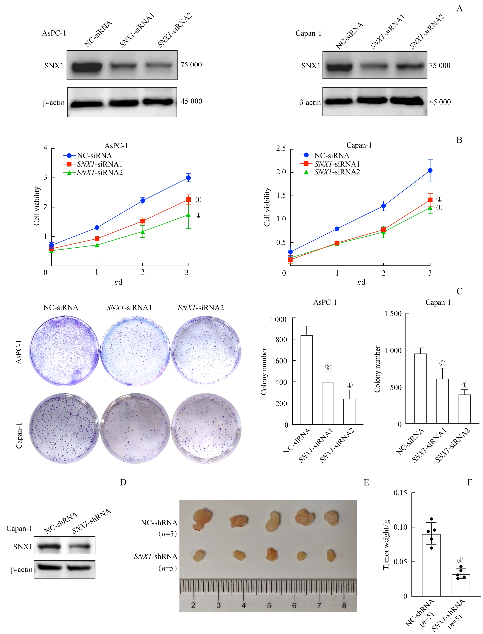
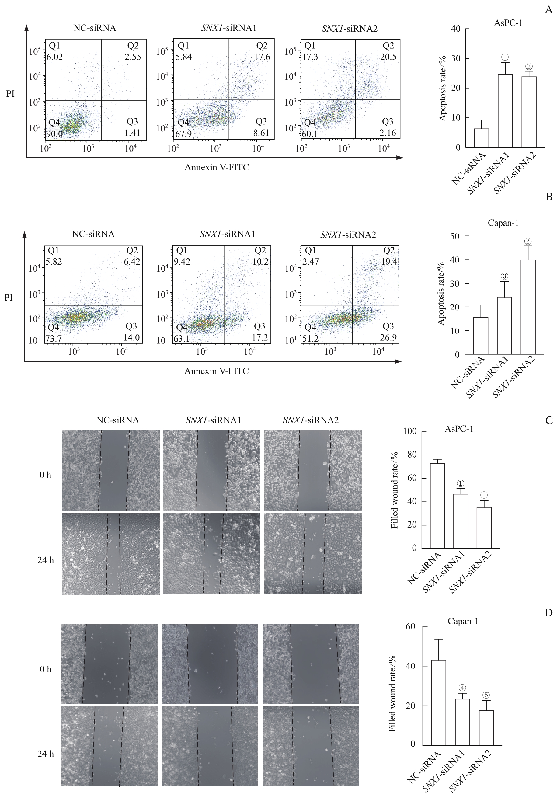
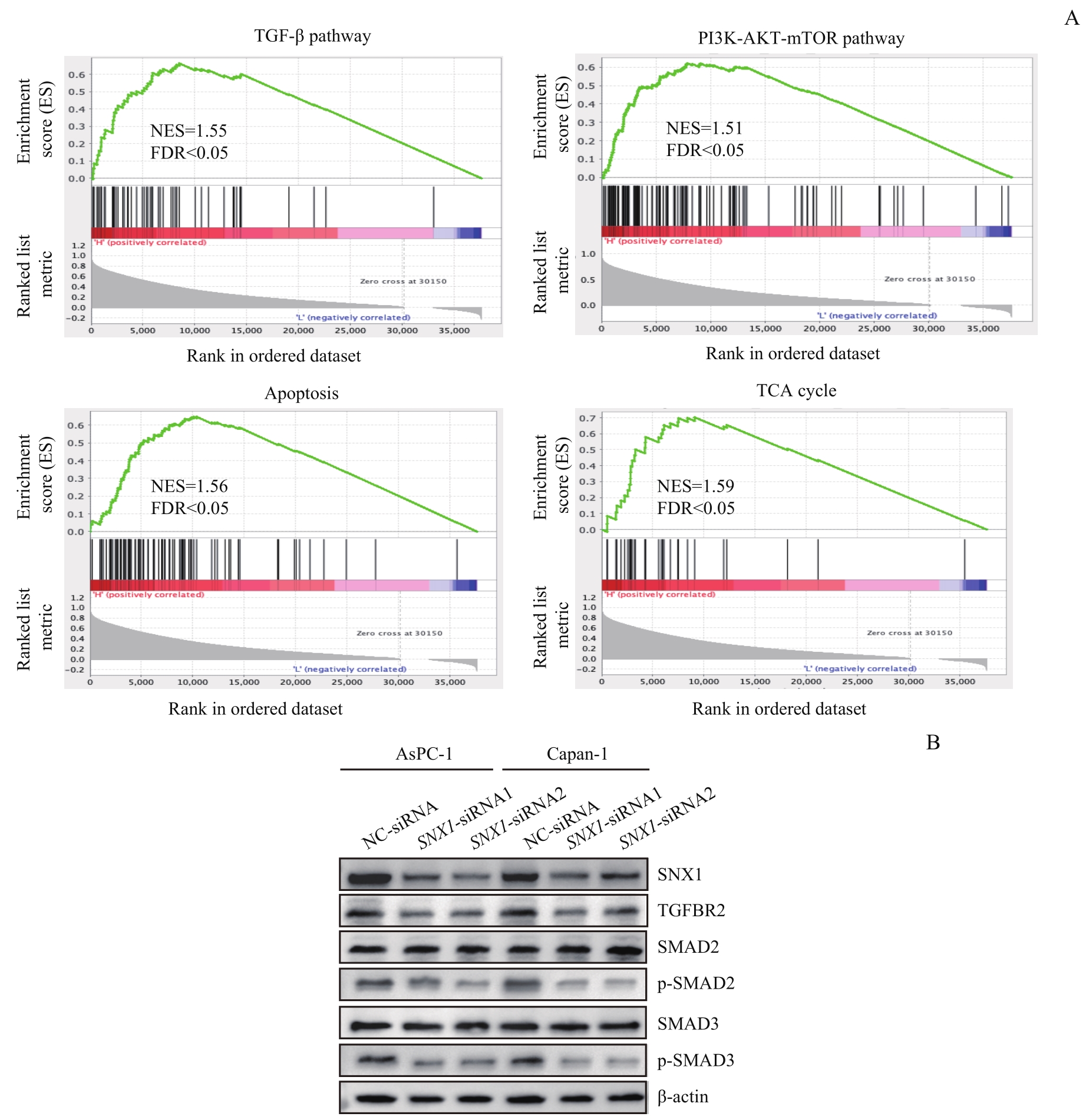
目的·探究分选链接蛋白1(sorting nexin 1,SNX1)在胰腺导管腺癌(pancreatic ductal adenocarcinoma,PDAC)发生和发展过程中表达水平的变化,及其对PDAC细胞增殖、迁移、凋亡和巨胞饮的影响,并分析其促进PDAC进展的分子机制。方法·分别在癌症基因组图谱(The Cancer Genome Atlas,TCGA)数据库和基因型-组织表达(Genotype-Tissue Expression,GTEx)数据库、基因表达综合(Gene Expression Omnibus,GEO)数据库中的GSE15471数据集分析SNX1 mRNA在PDAC及正常胰腺组织中的表达量变化。采用免疫组织化学染色(immunohistochemistry staining,IHC)检测PDAC患者的癌组织及癌旁组织中SNX1的表达变化。利用实时荧光定量PCR(quantitative real-time PCR,qPCR)和蛋白质印迹法(Western blotting)检测SNX1在hTERT-HPNE细胞及PDAC细胞中的表达水平。分别采用CCK8实验、平板克隆形成实验、划痕实验和流式细胞术检测由转染siRNA引起SNX1下调导致的AsPC-1、Capan-1细胞的增殖能力、迁移能力、凋亡水平的变化。构建稳定敲除SNX1的Capan-1细胞株,使用裸鼠皮下成瘤实验检测下调SNX1对细胞在裸鼠体内增殖能力的影响。利用免疫荧光染色(immunofluorescence,IF)确定SNX1在PDAC细胞中的分布,并用四甲基罗丹明-葡聚糖(TMR-dextran)检测由转染siRNA引起SNX1下调导致的AsPC-1、Capan-1细胞巨胞饮水平的变化。利用基因集富集分析(Gene Set Enrichment Analysis,GSEA)软件预测与SNX1相关的信号通路,并选取转化生长因子-β(transforming growth factor-β,TGF-β)通路进行后续分析及实验验证。构建稳定过表达SNX1的Capan-1、AsPC-1细胞株,使用TGF-β通路抑制剂氧化苦参碱(oxymatrine,Oxy)处理上述过表达细胞,并采用CCK8实验、平板克隆形成实验、划痕实验、流式细胞术和TMR-dextran检测Oxy处理对过表达SNX1引起的细胞的增殖、迁移、凋亡、巨胞饮水平变化的影响。结果·TCGA和GTEx数据库的合并分析的结果显示SNX1 mRNA在PDAC组织中的表达高于正常胰腺组织,GSE15471数据集分析的结果显示SNX1 mRNA在PDAC组织中的表达高于癌旁组织(均P=0.000)。IHC的结果显示SNX1在PDAC患者的癌组织中的表达亦高于癌旁组织。qPCR和Western blotting的结果显示,与hTERT-HPNE细胞相比,SNX1在PDAC细胞中的mRNA、蛋白水平均有上调(均P<0.05)。下调SNX1能够抑制AsPC-1、Capan-1细胞的增殖、迁移能力,下调其巨胞饮水平,促进其凋亡;过表达SNX1则可产生相反的结果。同时,敲除SNX1能够抑制Capan-1细胞在裸鼠体内的增殖能力。IF的结果显示SNX1在PDAC细胞中与溶酶体存在共定位。GSEA的结果显示,SNX1的表达与细胞凋亡、三羧酸循环、TGF-β和磷脂酰肌醇3-激酶-丝氨酸/苏氨酸蛋白激酶-哺乳动物雷帕霉素靶蛋白信号通路相关;在PDAC细胞中下调SNX1能够抑制TGF-β信号通路的激活,过表达SNX1则可促进该通路的激活,且加入该通路抑制剂可抑制由过表达SNX1引起的细胞表型变化。结论·SNX1在PDAC细胞和组织中高表达,其可通过激活TGF-β信号通路增强PDAC细胞的增殖、迁移能力和巨胞饮水平,并抑制其凋亡。

关键词: 胰腺导管腺癌, 分选链接蛋白1, 巨胞饮, 转化生长因子-β信号通路

Abstract:

Objective Methods ·The Cancer Genome Atlas (TCGA) and Genotype-Tissue Expression (GTEx) database and the GSE15471 dataset in Gene Expression Omnibus (GEO) database were used to analyze the SNX1 mRNA expression in PDAC and normal pancreas tissues. Immunohistochemistry staining (IHC) was used to detect the SNX1 expression in PDAC and para-carcinoma tissues. The mRNA and protein levels of SNX1 in hTERT-HPNE cells and PDAC cells were detected by quantitative real-time PCR (qPCR) and Western blotting. CCK8 method, plate clonal formation experiment, cell scratch assays and flow cytometry (FCM) were used to detect changes of the proliferation, migration and apoptosis levels in AsPC-1 and Capan-1 cells caused by transfection siRNAs. Capan-1 cell line with stable knockdown of SNX1 was constructed, and the subcutaneous tumorigenesis experiment in nude mice was performed to detect the effect of SNX1 knockdown on the proliferation capacity of cells in nude mice. Immunofluorescence (IF) was used to determine the distribution of SNX1 in PDAC cells, and TMR-dextran was used to detect the changes of macropinocytosis levels of AsPC-1 and Capan-1 cells induced bytransfection siRNAs. Gene Set Enrichment Analysis (GSEA) was performed to predict SNX1 related signaling pathways. The transforming growth factor-β (TGF-β) signaling pathway was selected for subsequent analysis and experimental verification. Capan-1 and AsPC-1 cell lines with stable overexpression of SNX1 were constructed and treated with TGF-β signaling pathway inhibitor Oxymatrine (Oxy). CCK8 method, plate clonal formation experiment, cell scratch assays, FCM and TMR-dextran were used to detect the effects of Oxy treatment on the changes of the proliferation, migration, apoptosis and macropinocytosis levels of the cells caused by SNX1 overexpression. Results ·The results of combined analysis of TCGA and GTEx databases showed that the expression of SNX1 mRNA in PDAC tissues was higher than that in normal pancreas tissues, and the results of GSE15471 dataset analysis showed that the expression of SNX1 mRNA in PDAC tissues was higher than that in para-carcinoma tissues (both P=0.000). IHC results showed that the expression of SNX1 in cancer tissues of PDAC patients was also higher than that in para-carcinoma tissues. qPCR and Western blotting results showed that compared with hTERT-HPNE cells, the mRNA and protein levels of SNX1 in PDAC cells were up-regulated (all P<0.05). SNX1 knockdown could inhibit the proliferation and migration capacity of PDAC cells, down-regulate the macropinocytosis levels of PDAC cells, and promote their apoptosis. On the contrary, SNX1 overexpression led to opposite phenotype. Meanwhile, SNX1 knockdown could inhibit the proliferation capacity of Capan-1 cells in nude mice. IF results showed that SNX1 was colocalized with lysosomes in PDAC cells. GSEA analysis demonstrated that SNX1 expression was correlated with apoptosis, tricarboxylic acid cycle, TGF-β and phosphoinositide 3-kinase-serine/threonine-protein kinase-mammalian target of rapamycin signaling pathway. SNX1 knockdown in PDAC cells could inhibit the activation of TGF-β signaling pathway, SNX1 overexpression could promote this pathway, and TGF-β signaling pathway inhibitor Oxy could inhibit the phenotypic changes caused by SNX1 overexpression. Conclusion SNX1 is highly expressed in PDAC cells and tissues. SNX1 promotes the proliferation, migration capacity and macropinocytosis levels, and inhibits cell apoptosis by activating the TGF-β signaling pathway in PDAC cells. · ·To explore the expression changes of sorting nexin 1 (SNX1) in the occurrence and development of pancreatic ductal adenocarcinoma (PDAC) and its effect on the proliferation, migration, apoptosis and micropinocytosis of PDAC cells, and analyze its molecular mechanism to promote the progression of PDAC.

Key words: pancreatic ductal adenocarcinoma (PDAC), sorting nexin 1 (SNX1), macropinocytosis, transforming growth factor-β signaling pathway

中图分类号: