上海交通大学学报(医学版) ›› 2023, Vol. 43 ›› Issue (8): 977-987.doi: 10.3969/j.issn.1674-8115.2023.08.005

• 论著 · 基础研究 • 上一篇    下一篇

PRPS1 I72位点突变对急性淋巴细胞白血病耐药性的影响及其机制研究

崔芷嫣1,2(), 陈尧1,2, 陶悦1,2, 沈树红1, 李慧1,2()   

  1. 1.上海交通大学医学院附属上海儿童医学中心儿科转化医学研究所,国家卫生健康委员会儿童血液肿瘤重点实验室,上海 200127
    2.上海市儿科临床分子诊断重点实验室,上海 200127
  • 收稿日期:2023-03-05 接受日期:2023-05-15 出版日期:2023-08-28 发布日期:2023-08-28
  • 通讯作者: 李慧 E-mail:cuicui0724@163.com;lihui@scmc.com.cn
  • 作者简介:崔芷嫣(1997—),女,硕士生;电子信箱:cuicui0724@163.com
  • 基金资助:
    国家自然科学基金(82070159);上海市自然科学基金(22ZR1440000);浦东新区科技发展基金(PKJ2020-Y05)

Effects of PRPS1 I72 mutations on drug resistance in acute lymphoblastic leukemia and its mechanisms

CUI Zhiyan1,2(), CHEN Yao1,2, TAO Yue1,2, SHEN Shuhong1, LI Hui1,2()   

  1. 1.Key Laboratory of Pediatric Hematology and Oncology of National Health Commission, Pediatric Translational Medicine Institute, Shanghai Children's Medical Center, Shanghai Jiao Tong University School of Medicine, Shanghai 200127, China
    2.Shanghai Key Laboratory of Clinical Molecular Diagnostics for Pediatrics, Shanghai 200127, China
  • Received:2023-03-05 Accepted:2023-05-15 Online:2023-08-28 Published:2023-08-28
  • Contact: LI Hui E-mail:cuicui0724@163.com;lihui@scmc.com.cn
  • Supported by:
    National Natural Science Foundation of China(82070159);Natural Science Foundation of Shanghai(22ZR1440000);Science and Technology Commission of Pudong New Area Foundation(PKJ2020-Y05)

摘要:

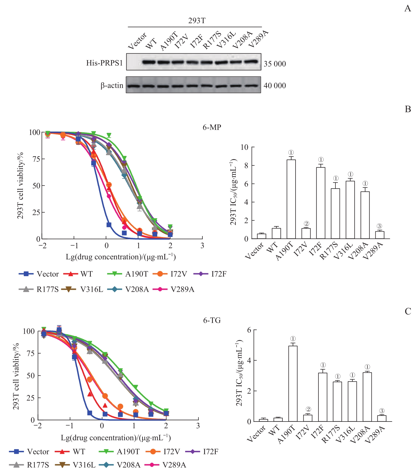
目的·研究磷酸核糖焦磷酸合成酶1(phosphoribosyl pyrophosphate synthetase 1,PRPS1)I72位点突变是否能使急性淋巴细胞白血病(acute lymphoblastic leukemia,ALL)细胞对巯嘌呤类化学治疗(化疗)药物6-巯基嘌呤(6-mercaptopurine,6-MP)、6-硫代鸟嘌呤(6-thioguanine,6-TG)产生耐药性,并解释其作用机制。方法·将临床上已发现的PRPS1基因的各突变(I72F、R177S、V316L)和2个ALL细胞系(KOPN72bi和RS4;11)中存在的PRPS1基因突变(V208A、V289A)分别插入融合了绿色荧光蛋白(green fluorescent protein,GFP)的载体pGV303中,用已证明的对巯嘌呤类化疗药耐药的PRPS1 A190T突变为阳性对照,对该类药不耐药的空载体pGV303(Vector)、PRPS1野生型(wild type,WT)及PRPS1 I72V突变为阴性对照。将上述构建好的载体瞬时转染入HEK-293T细胞(简称293T细胞)中,并采用蛋白质印迹法(Western blotting)检测PRPS1各突变体在293T细胞中的蛋白表达情况。采用药物敏感性实验检测并计算6-MP或6-TG对上述瞬转PRPS1各突变体的293T细胞的半数抑制浓度(half maximal inhibitory concentration,IC50)。随后,除PRPS1 I72F和I72V之外,将第72位异亮氨酸(isoleucine,I)变成其他氨基酸的多个突变即I72M、I72L、I72N、I72S、I72T分别插入载体pGV303中,瞬时转染入293T细胞后采用Western blotting、药物敏感性实验检测各突变体在293T细胞中的蛋白表达情况以及6-MP或6-TG的IC50。采用慢病毒感染法将PRPS1 WT、I72F、I72V、A190T及载体pGV303感染REH细胞系,通过流式细胞术分选GFP阳性细胞以获得PRPS1各突变体稳定表达的细胞,并采用Western blotting及药物敏感性实验检测各突变体在REH细胞中的蛋白表达情况以及6-MP或6-TG的IC50,用以验证293T细胞获得的药物敏感性实验结果。采用Annexin Ⅴ/DAPI双染法评估各REH细胞系的凋亡情况,并通过Western blotting检测各REH细胞系的DNA损伤相关蛋白[S139位点磷酸化的组蛋白H2AX(phosphorylated H2AX-S139,γ-H2AX)、磷酸化的细胞周期检验点激酶2(phosphorylated check point kinase 2,pCHK2)]和细胞凋亡相关蛋白聚(腺苷二磷酸核糖)聚合酶剪切体[cleaved poly(ADP-ribose)polymerase,cleaved PARP]的表达水平。使用PDB数据库(Protein Data Bank)中编号为2HCR(PDB code 2HCR)的PRPS1晶体结构图,通过三维成像及PyMOL软件预测并绘制I72位点、I72V和I72F的氨基酸残基及空间构象图。结果·Western blotting结果显示,瞬时转染的外源性PRPS1各突变蛋白在293T细胞中成功表达;药物敏感性实验结果显示,表达PRPS1 I72F、R177S、V316L、V208A与阳性对照A190T的293T细胞对6-MP或6-TG的IC50均远高于表达V289A及阴性对照Vector、PRPS1 WT、PRPS1 I72V的细胞(均P=0.000)。将第72位异亮氨酸变成其他氨基酸后,Western blotting结果显示瞬时转染的外源性PRPS1 I72位点的各突变蛋白在293T细胞中成功表达;药物敏感性实验结果显示,表达PRPS1 I72M、I72F、I72L、I72N、I72S、I72T与A190T的293T细胞对6-MP或6-TG的IC50均远高于表达Vector、PRPS1 WT、PRPS1 I72V的细胞(均P=0.000)。慢病毒感染REH细胞后,Western blotting结果显示在已构建的稳定细胞系中PRPS1 WT、A190T、I72F、I72V的蛋白高表达且表达量相似;药物敏感性实验结果显示,表达PRPS1 I72F、A190T的REH细胞对6-MP或6-TG的IC50均远高于表达Vector、PRPS1 WT、PRPS1 I72V的细胞(均P=0.000),与293T细胞中瞬时转染得到的药物敏感性结果一致。Annexin Ⅴ/DAPI双染法、DNA损伤和凋亡相关蛋白的Western blotting检测的结果均显示,经6-MP处理后,表达PRPS1 A190T、I72F的REH细胞系的DNA损伤和凋亡率明显低于表达Vector、PRPS1 WT、PRPS1 I72V的细胞(均P=0.000)。蛋白结构分析结果显示,PRPS1 I72F会使PRPS1的空间构象发生改变。结论·PRPS1 I72F、R177S、V316L、V208A、I72M、I72L、I72N、I72S、I72T突变可使细胞获得对巯嘌呤类化疗药的耐药性,PRPS1 V289A、I72V突变不影响细胞对巯嘌呤类化疗药的敏感性。293T中的药物敏感性实验结果和REH中的药物敏感性实验结果一致,证明293T细胞可以作为检测PRPS1突变对巯嘌呤类化疗药物耐药性的快速研究模型。PRPS1 I72位点突变对巯嘌呤类化疗药耐药性的影响可能与PRPS1结构发生改变有关。

关键词: 急性淋巴细胞白血病, 耐药复发, 磷酸核糖焦磷酸合成酶1, 巯嘌呤

Abstract:

Objective ·To study whether mutations at the I72 site of phosphoribosyl pyrophosphate synthetase 1 (PRPS1) can induce resistance in acute lymphoblastic leukemia (ALL) cells to thiopurine chemotherapy drugs 6-mercaptopurine (6-MP) and 6-thioguanine (6-TG), and explain their mechanisms of action. Methods ·The PRPS1 gene mutations (I72F, R177S and V316L) found in clinical practice and PRPS1 gene mutations (V208A and V289A) present in two ALL cell lines (KOPN72bi and RS4;11) were constructed into the vector pGV303 fused with green fluorescent protein (GFP), respectively. The PRPS1 A190T mutation that has been proven to be resistant to thiopurine chemotherapy drugs was used as the positive control, and the empty vector pGV303 (Vector), PRPS1 wild-type (WT) and PRPS1 I72V were used as the negative controls. The various mutants of PRPS1 were transiently transfected into HEK-293T cells (referred to as 293T cells), and the expression of these exogenous PRPS1 was detected by Western blotting. The half maximal inhibitory concentration (IC50) of 6-MP or 6-TG on the above 293T cell lines transiently transfected with PRPS1 mutants was detected and calculated by drug sensitivity experiments. Subsequently, in addition to PRPS1 I72F and I72V, multiple mutations I72M, I72L, I72N, I72S and I72T were constructed into the vector pGV303, respectively, by changing the isoleucine (I) at position 72 into other amino acids. The various mutants were transiently transfected into 293T cells, respectively, and the protein expression of each mutant and IC50 values of 6-MP or 6-TG were detected by Western blotting and drug sensitivity experiments. PRPS1 WT, I72F, I72V, A190T and pGV303 vectors were transfected into REH cell lines by lentivirus infection, and GFP-positive cells were sorted by flow cytometry to obtain cells with stable expression of PRPS1 mutants. The protein expression of each mutant in REH cells and IC50 values of 6-MP or 6-TG were detected by Western blotting and drug sensitivity experiments to verify the results of drug sensitivity experiments obtained by 293T cells. Annexin V/DAPI double staining was used to evaluate the apoptosis of each REH cell line, and Western blotting was used to detect the levels of DNA damage-related proteins [phosphorylation at S139 of histone H2AX (phosphorylated H2AX-S139, γ-H2AX), phosphorylated check point kinase 2 (pCHK2)], and apoptosis-related protein cleaved poly (ADP-ribose) polymerase (cleaved PARP) in each REH cell line. The diagrams of amino acid residues and spatial conformations of I72 locus, I72V and I72F were predicted and drawn through three-dimensional imaging and PyMOL software by using the crystal structure data of PRPS1-numbered 2HCR (PDB code 2HCR) in the PDB (Protein Data Bank) database. Results ·Western blotting results showed that the transiently transfected exogenous PRPS1-mutated proteins were successfully expressed in 293T cells. The drug sensitivity experiment results showed that the IC50 values of 6-MP or 6-TG in 293T cells expressing PRPS1 I72F, R177S, V316L, V208A and the positive control A190T were much higher than those in cells expressing V289A and the negative control Vector, PRPS1 WT and I72V (all P =0.000). After mutating the isoleucine (I) at position 72 with other amino acids, Western blotting results showed successful expression of exogenous PRPS1-mutated proteins at position I72 after transient transfection in 293T cells. Drug sensitivity experiments revealed the IC50 values of 6-MP or 6-TG in 293T cells expressing PRPS1 I72M, I72F, I72L, I72N, I72S, I72T and positive control A190T were much higher than those in cells expressing negative control Vector, PRPS1 WT and I72V (all P=0.000). Western blotting results showed that the protein expression levels of PRPS1 WT, A190T, I72F and I72V in the REH stable cell lines constructed by lentivirus were high and similar. The drug sensitivity experiment results showed that the IC50 values of 6-MP or 6-TG in REH cells expressing PRPS1 I72F and positive control A190T were much higher than those in cells expressing negative control Vector, PRPS1 WT and I72V (all P=0.000), which was consistent with the drug sensitivity results obtained by transient transfection in 293T cells. The results of Annexin V/DAPI double staining method and the detection of DNA damage and apoptosis-related proteins by Western blotting showed that after 6-MP treatment, the DNA damage and apoptosis rates of REH cell lines expressing PRPS1 A190T and I72F were significantly lower than those of cells expressing negative control Vector, PRPS1 WT and I72V (all P=0.000). The protein structure analysis results showed that PRPS1 I72F could change the conformation of PRPS1. Conclusion ·The PRPS1 I72F, R177S, V316L, V208A, I72M, I72L, I72N, I72S and I72T mutations can confer drug resistance to the thiopurine chemotherapy drugs in cells, while the PRPS1 V289A and I72V mutations do not affect cell sensitivity to the thiopurine chemotherapy drugs. The drug sensitivity experiment results in 293T cells are consistent with those in REH cells, demonstrating that 293T cells can serve as a rapid research model for detecting the resistance of PRPS1 mutations to thiopurine chemotherapy drugs. The effects of the PRPS1 I72 mutations on the resistance of the thiopurine chemotherapy drugs may be related to changes in the structure of PRPS1.

Key words: acute lymphoblastic leukemia (ALL), drug resistance and relapse, phosphoribosyl pyrophosphate synthetase 1 (PRPS1), thiopurine

中图分类号: