
上海交通大学学报(医学版) ›› 2026, Vol. 46 ›› Issue (10): 1339-1354.doi: 10.3969/j.issn.1674-8115.2026.10.004
• 论著 · 基础研究 • 上一篇
邓睿1,2, 方后顺1,2, 周吉全1,2, 陈尧1,2( ), 李慧1,2()
收稿日期:2026-04-15
接受日期:2026-06-20
出版日期:2026-10-15
发布日期:2026-10-15
通讯作者:
李 慧,研究员,博士;电子信箱:lihui@scmc.com.cn。基金资助:
Deng Rui1,2, Fang Houshun1,2, Zhou Jiquan1,2, Chen Yao1,2(), Li Hui1,2()
Received:2026-04-15
Accepted:2026-06-20
Online:2026-10-15
Published:2026-10-15
Contact:
Li Hui, E-mail: lihui@scmc.com.cn.Supported by:摘要:
目的·探讨泛素特异性蛋白酶37(ubiquitin-specific protease 37,USP37)在肝母细胞瘤(hepatoblastoma,HB)发生发展中的生物学功能,并探索其通过调控骨髓细胞瘤病毒癌基因同源物(myelocytomatosis viral oncogene homolog,c-MYC)蛋白稳定性影响肿瘤进展的分子机制。方法·检索PubMed数据库截至2026年3月31日发表的相关文献,结合HB转录组数据及临床病理特征,系统筛选可能参与c-MYC蛋白稳定性调控的USP家族成员。采用实时定量荧光反转录聚合酶链式反应(real time fluorescent quantitative PCR,qPCR)和受试者工作特征(receiver operating characteristic,ROC)曲线分析验证USP37的表达特征及诊断效能,并利用Kaplan-Meier Plotter数据库肝细胞癌队列进行探索性生存分析。在HuH6和HepG2细胞中构建USP37稳定敲低模型,通过细胞增殖及克隆形成实验评估其生物学功能,并采用shRNA抵抗型USP37表达载体进行回补验证。结合qPCR、蛋白质印迹法(Western blotting)、放线菌酮(cycloheximide,CHX)追踪实验、蛋白酶体抑制实验、免疫共沉淀(co-immunoprecipitation,Co-IP)及去泛素化实验,解析USP37对c-MYC表达及蛋白稳定性的调控机制。结果·通过文献证据、转录组差异表达及临床病理特征的交叉筛选,确定USP37为HB中潜在调控分子。临床样本及公共数据库分析显示,USP37在HB组织中显著上调。基于8对HB及配对癌旁组织的qPCR结果进行ROC曲线分析,曲线下面积(area under the curve,AUC)为0.942 7,提示USP37具有区分HB组织与癌旁组织的能力。33例HB患者临床资料分析显示,USP37高表达与肿瘤转移(P=0.028)及肿瘤体积增大(P=0.031)显著相关。体外功能实验表明,敲低USP37显著抑制HuH6和HepG2细胞增殖及克隆形成能力,而回补USP37可有效逆转细胞增殖抑制表型。机制研究显示,USP37过表达可剂量依赖性提高c-MYC蛋白水平,但不显著影响其mRNA表达;敲低USP37则显著降低c-MYC蛋白水平,而其mRNA表达无明显变化。CHX追踪实验显示,USP37过表达使293T细胞中c-MYC蛋白半衰期由2.357 h延长至4.796 h(P=0.006);敲低USP37使HuH6和HepG2细胞中c-MYC蛋白半衰期分别由1.504 h和1.585 h缩短至0.986 h和0.792 h(P=0.006、P=0.005)。蛋白酶体抑制剂MG132处理可恢复USP37敲低细胞中的c-MYC蛋白水平。正反向Co-IP实验进一步证实USP37与c-MYC存在相互作用;去泛素化实验显示,USP37过表达可降低c-MYC泛素化水平。结论·USP37作为去泛素化酶,通过稳定c-MYC蛋白促进HB细胞增殖及肿瘤进展,提示靶向USP37-c-MYC轴有望成为潜在干预靶点。
中图分类号:
邓睿, 方后顺, 周吉全, 陈尧, 李慧. 泛素特异性蛋白酶37通过去泛素化稳定骨髓细胞瘤病毒癌基因同源物蛋白促进肝母细胞瘤进展的机制研究[J]. 上海交通大学学报(医学版), 2026, 46(10): 1339-1354.
Deng Rui, Fang Houshun, Zhou Jiquan, Chen Yao, Li Hui. USP37-mediated deubiquitination maintains c-MYC protein homeostasis and contributes to hepatoblastoma progression[J]. Journal of Shanghai Jiao Tong University (Medical Science), 2026,(10): 1339-1354.
| shRNA name | shRNA sequence (5'→3') | |
|---|---|---|
| USP37 shRNA-1 | Forward | CCGGTCCGGGTAGAGGATCGATTAACTCGAGTTAATCGATCCTCTACCCGGATTTTTG |
| Reverse | AATTCAAAAATCCGGGTAGAGGATCGATTAACTCGAGTTAATCGATCCTCTACCCGGA | |
| USP37 shRNA-2 | Forward | CCGGCCGGATTTGCAGAAGATGATACTCGAGTATCATCTTCTGCAAATCCGGTTTTTG |
| Reverse | AATTCAAAAACCGGATTTGCAGAAGATGATACTCGAGTATCATCTTCTGCAAATCCGG |
表1 shRNA序列
Tab 1 shRNA sequences
| shRNA name | shRNA sequence (5'→3') | |
|---|---|---|
| USP37 shRNA-1 | Forward | CCGGTCCGGGTAGAGGATCGATTAACTCGAGTTAATCGATCCTCTACCCGGATTTTTG |
| Reverse | AATTCAAAAATCCGGGTAGAGGATCGATTAACTCGAGTTAATCGATCCTCTACCCGGA | |
| USP37 shRNA-2 | Forward | CCGGCCGGATTTGCAGAAGATGATACTCGAGTATCATCTTCTGCAAATCCGGTTTTTG |
| Reverse | AATTCAAAAACCGGATTTGCAGAAGATGATACTCGAGTATCATCTTCTGCAAATCCGG |
| Primer | Application | Forward | Reverse |
|---|---|---|---|
| USP37-Flag | Plasmid construction | CTAGAGGATCCCCGGGTACCGGTCGCCACCATGTCTCCTCTGAAGATACATGGT | CCGTCATGGTCTTTGTAGTCGCTAGCCAAGGCCTGACGGGTAGTCTT |
| USP37-HA | Plasmid construction | CTAGAGGATCCCCGGGTACCGGTCGCCACCATGTCTCCTCTGAAGATACATGGT | TCTGGGACATCATAAGGGTAGCTAGCCAAGGCCTGACGGGTAGTCTT |
| USP37-shRNA-resistant | Plasmid construction | CCCTGGCCGTGGCTCAATCAAGACTGTAGCAGGAAGTGGAATAGCTCGGA | GATTGAGCCACGGCCAGGGTTACCAAGAACTTTTCGAAATGG |
| USP37 | qPCR | GGAACACTCTTCTGGTGGCA | GTGAGAGGAAAGGGGCACTC |
| c-MYC | qPCR | GTGGTCTTCCCCTACCCTCT | TGTTCCTCCTCAGAGTCGCT |
| β-actin | qPCR | AACTGGGACGACATGGAGAAAA | GGATAGCACAGCCTGGATAGCA |
表2 引物序列及用途 (5′→3′)
Tab 2 Primer sequences and applications (5′→3′)
| Primer | Application | Forward | Reverse |
|---|---|---|---|
| USP37-Flag | Plasmid construction | CTAGAGGATCCCCGGGTACCGGTCGCCACCATGTCTCCTCTGAAGATACATGGT | CCGTCATGGTCTTTGTAGTCGCTAGCCAAGGCCTGACGGGTAGTCTT |
| USP37-HA | Plasmid construction | CTAGAGGATCCCCGGGTACCGGTCGCCACCATGTCTCCTCTGAAGATACATGGT | TCTGGGACATCATAAGGGTAGCTAGCCAAGGCCTGACGGGTAGTCTT |
| USP37-shRNA-resistant | Plasmid construction | CCCTGGCCGTGGCTCAATCAAGACTGTAGCAGGAAGTGGAATAGCTCGGA | GATTGAGCCACGGCCAGGGTTACCAAGAACTTTTCGAAATGG |
| USP37 | qPCR | GGAACACTCTTCTGGTGGCA | GTGAGAGGAAAGGGGCACTC |
| c-MYC | qPCR | GTGGTCTTCCCCTACCCTCT | TGTTCCTCCTCAGAGTCGCT |
| β-actin | qPCR | AACTGGGACGACATGGAGAAAA | GGATAGCACAGCCTGGATAGCA |
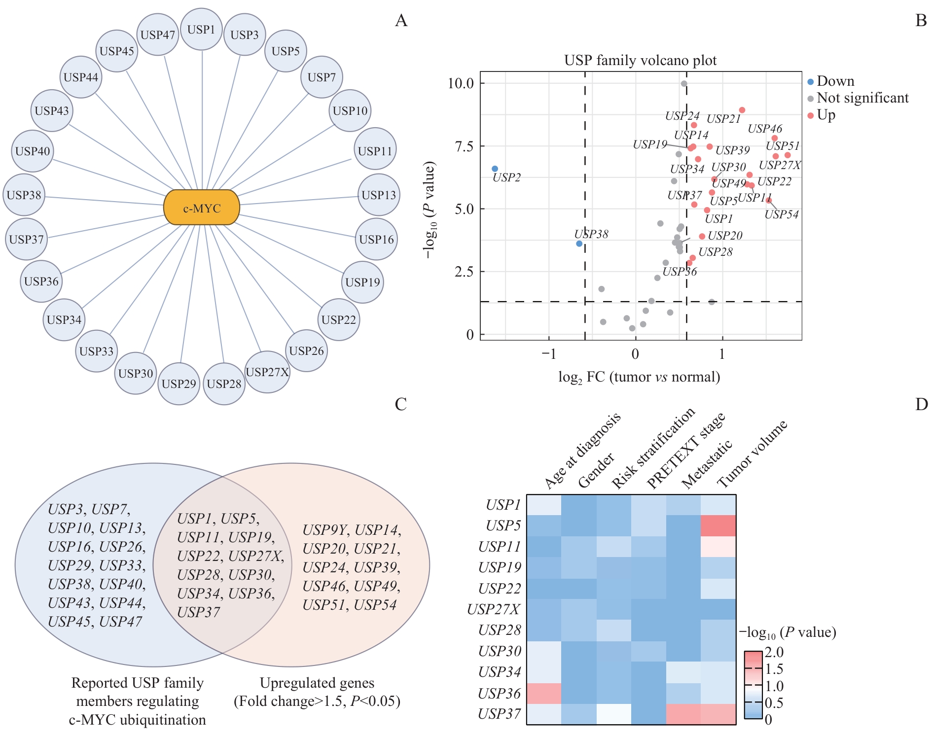
图1 HB中c-MYC相关USP家族成员的系统性筛选Note: A. Summary of USP family members reported to be involved in the regulation of c-MYC ubiquitination and stability, based on published studies retrieved from PubMed. B. Differential expression analysis of USP family members in the GSE104766 dataset. Genes with FPKM > 1 were included in the analysis, and the significance thresholds were set at P<0.05 and fold change > 1.5. C. Venn diagram showing the overlap between reported c-MYC-associated deubiquitinases and significantly upregulated USP family members in HB, identifying candidate USP regulators. D. Clinical correlation analysis of the candidate USP genes in 33 HB samples with available clinical information. P values were calculated using Fisher's exact test. Color intensity represents -log10(P value).
Fig 1 Systematic screening of c-MYC-associated USP family members in HB
| USP | Cell model | Reference |
|---|---|---|
| USP1 | Bladder cancer cell lines (T24, UM-UC-3) | [ |
| USP3 | Clear cell renal cell carcinoma cell lines (786-O, Caki-2) | [ |
| USP5 | Hepatocellular carcinoma cell lines (Huh-7, HepG2) | [ |
| USP7 | Pancreatic ductal adenocarcinoma cell lines (PANC1, CFPAC-1, AsPC1) | [ |
| USP10 | Osteosarcoma cell lines (143B, HOS) | [ |
| USP11 | Prostate cancer cell line (22Rv1) | [ |
| USP13 | Glioblastoma stem cells | [ |
| USP16 | Prostate cancer cell lines (PC3, DU145) | [ |
| USP19 | Hepatocellular carcinoma cell lines (Hep3b, Huh-7) | [ |
| USP22 | Breast cancer cell lines (MCF7, T47D) | [ |
| USP26 | Gastric cancer cell lines (SNU-216, HGC-27) | [ |
| USP27X | Breast cancer cell lines (MCF-7, MDA-MB-231); mouse embryonic fibroblasts | [ |
| USP28 | Osteosarcoma cell line (U2OS), cervical cancer cell line (HeLa); mouse intestinal model, MEFs, colorectal cancer model | [ |
| USP29 | Neuroblastoma cell line (SK-N-BE2), burkitt lymphoma cell line (Ramos), non-small cell lung cancer cell line (A549) | [ |
| USP30 | Oral squamous cell carcinoma cell lines (SCC4, HSC4) | [ |
| USP33 | Prostate cancer cell lines (22Rv1, DU145, PC3); ovarian cancer cell lines (SKOV3, ES2) | [ |
| USP34 | Hepatocellular carcinoma cell lines (Huh-7, HepG2, HCCLM3, MHCC97H) | [ |
| USP36 | Cervical cancer cell line (HeLa), non-small cell lung cancer cell line (H1299), breast cancer cell line (MDA-MB-231), non-tumorigenic breast epithelial cell line (MCF10A) | [ |
| USP37 | Non-small cell lung cancer cell lines (H1299) | [ |
| USP38 | Non-small cell lung cancer cell lines (A549), cervical cancer cell lines (HeLa) | [ |
| USP40 | Acute myeloid leukemia cell lines (HL-60, Kasumi-1) | [ |
| USP43 | Bladder cancer cell lines (T24, 5637, UM-UC-3) | [ |
| USP44 | Meningioma cell lines (IOMM-Lee, CH157) | [ |
| USP45 | Cervical cancer cell lines (HeLa, SiHa) | [ |
| USP47 | Non-small cell lung cancer cell lines (A549, Calu-1, H358) | [ |
表3 c-MYC泛素化与稳定性调控相关USP家族成员
Tab 3 USP family members involved in the regulation of c-MYC ubiquitination and stability
| USP | Cell model | Reference |
|---|---|---|
| USP1 | Bladder cancer cell lines (T24, UM-UC-3) | [ |
| USP3 | Clear cell renal cell carcinoma cell lines (786-O, Caki-2) | [ |
| USP5 | Hepatocellular carcinoma cell lines (Huh-7, HepG2) | [ |
| USP7 | Pancreatic ductal adenocarcinoma cell lines (PANC1, CFPAC-1, AsPC1) | [ |
| USP10 | Osteosarcoma cell lines (143B, HOS) | [ |
| USP11 | Prostate cancer cell line (22Rv1) | [ |
| USP13 | Glioblastoma stem cells | [ |
| USP16 | Prostate cancer cell lines (PC3, DU145) | [ |
| USP19 | Hepatocellular carcinoma cell lines (Hep3b, Huh-7) | [ |
| USP22 | Breast cancer cell lines (MCF7, T47D) | [ |
| USP26 | Gastric cancer cell lines (SNU-216, HGC-27) | [ |
| USP27X | Breast cancer cell lines (MCF-7, MDA-MB-231); mouse embryonic fibroblasts | [ |
| USP28 | Osteosarcoma cell line (U2OS), cervical cancer cell line (HeLa); mouse intestinal model, MEFs, colorectal cancer model | [ |
| USP29 | Neuroblastoma cell line (SK-N-BE2), burkitt lymphoma cell line (Ramos), non-small cell lung cancer cell line (A549) | [ |
| USP30 | Oral squamous cell carcinoma cell lines (SCC4, HSC4) | [ |
| USP33 | Prostate cancer cell lines (22Rv1, DU145, PC3); ovarian cancer cell lines (SKOV3, ES2) | [ |
| USP34 | Hepatocellular carcinoma cell lines (Huh-7, HepG2, HCCLM3, MHCC97H) | [ |
| USP36 | Cervical cancer cell line (HeLa), non-small cell lung cancer cell line (H1299), breast cancer cell line (MDA-MB-231), non-tumorigenic breast epithelial cell line (MCF10A) | [ |
| USP37 | Non-small cell lung cancer cell lines (H1299) | [ |
| USP38 | Non-small cell lung cancer cell lines (A549), cervical cancer cell lines (HeLa) | [ |
| USP40 | Acute myeloid leukemia cell lines (HL-60, Kasumi-1) | [ |
| USP43 | Bladder cancer cell lines (T24, 5637, UM-UC-3) | [ |
| USP44 | Meningioma cell lines (IOMM-Lee, CH157) | [ |
| USP45 | Cervical cancer cell lines (HeLa, SiHa) | [ |
| USP47 | Non-small cell lung cancer cell lines (A549, Calu-1, H358) | [ |
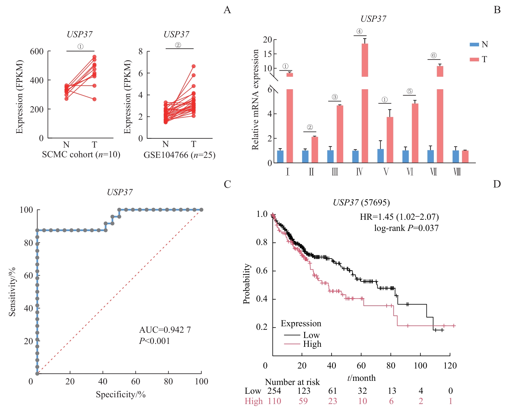
图2 USP37 在HB中的表达分析及其临床相关性Note: A. Transcriptomic analysis of USP37 expression in HB (T) and adjacent non-tumorous liver tissues (N) from the SCMC cohort (n=10) and the GSE104766 dataset (n=25). Each dot represents an individual sample. Statistical significance was assessed using a paired t test; ①P=0.003,②P<0.001. B. qPCR analysis of USP37 mRNA expression in HB patient tissue samples. Data are presented as x±s. Each sample was analyzed in triplicate. Roman numerals (Ⅰ‒Ⅷ) indicate individual paired patient samples. Statistical significance was determined using paired t test, ①P<0.001, ②P=0.003, ③P=0.002, ④P=0.004, ⑤P=0.002, and ⑥P=0.001. C. Receiver operating characteristic (ROC) curve analysis evaluating the diagnostic performance of USP37 expression in distinguishing HB tissues from non-tumorous tissues. The area under the curve (AUC) is shown. D. Kaplan-Meier survival analysis of the hepatocellular carcinoma (HCC) cohort stratified by USP37 expression levels. Overall survival was compared between the high- and low-expression groups using the log-rank test. The hazard ratio (HR) and 95% confidence interval (CI) are indicated.
Fig 2 Expression analysis and prognostic evaluation of USP37 in HB
| Characteristic | USP37 Expression | P value | |
|---|---|---|---|
| Low (n=17) | High (n=16) | ||
| Age at diagnosis | 0.176 | ||
| ≤24 months | 11 (33.33) | 6 (18.18) | |
| >24 months | 6 (18.18) | 10 (30.30) | |
| Gender | 0.514 | ||
| Male | 9 (27.27) | 11 (33.33) | |
| Female | 8 (24.24) | 5 (15.15) | |
| Risk stratification | 0.142 | ||
| Low/intermediate-risk | 14 (42.42) | 9 (27.27) | |
| High-risk | 3 (9.09) | 7 (21.21) | |
| PRETEXT stage | 1.000 | ||
| Ⅰ/Ⅱ | 7 (21.21) | 7 (21.21) | |
| Ⅲ/Ⅳ | 10 (30.30) | 9 (27.27) | |
| Metastatic | 0.028 | ||
| Non-metastatic | 16 (48.48) | 8 (24.24) | |
| Metastatic | 1 (3.03) | 6 (18.18) | |
| NA | 0 | 2 (6.06) | |
| Tumor volume | 0.031 | ||
| ≤500 cm3 | 11 (33.33) | 3 (9.09) | |
| >500 cm3 | 2 (6.06) | 7 (21.21) | |
| NA | 4 (12.12) | 6 (18.18) | |
表4 USP37 表达水平与HB临床特征的关系[n(%)]
Tab 4 Association between USP37 expression and clinical features in HB [n(%)]
| Characteristic | USP37 Expression | P value | |
|---|---|---|---|
| Low (n=17) | High (n=16) | ||
| Age at diagnosis | 0.176 | ||
| ≤24 months | 11 (33.33) | 6 (18.18) | |
| >24 months | 6 (18.18) | 10 (30.30) | |
| Gender | 0.514 | ||
| Male | 9 (27.27) | 11 (33.33) | |
| Female | 8 (24.24) | 5 (15.15) | |
| Risk stratification | 0.142 | ||
| Low/intermediate-risk | 14 (42.42) | 9 (27.27) | |
| High-risk | 3 (9.09) | 7 (21.21) | |
| PRETEXT stage | 1.000 | ||
| Ⅰ/Ⅱ | 7 (21.21) | 7 (21.21) | |
| Ⅲ/Ⅳ | 10 (30.30) | 9 (27.27) | |
| Metastatic | 0.028 | ||
| Non-metastatic | 16 (48.48) | 8 (24.24) | |
| Metastatic | 1 (3.03) | 6 (18.18) | |
| NA | 0 | 2 (6.06) | |
| Tumor volume | 0.031 | ||
| ≤500 cm3 | 11 (33.33) | 3 (9.09) | |
| >500 cm3 | 2 (6.06) | 7 (21.21) | |
| NA | 4 (12.12) | 6 (18.18) | |
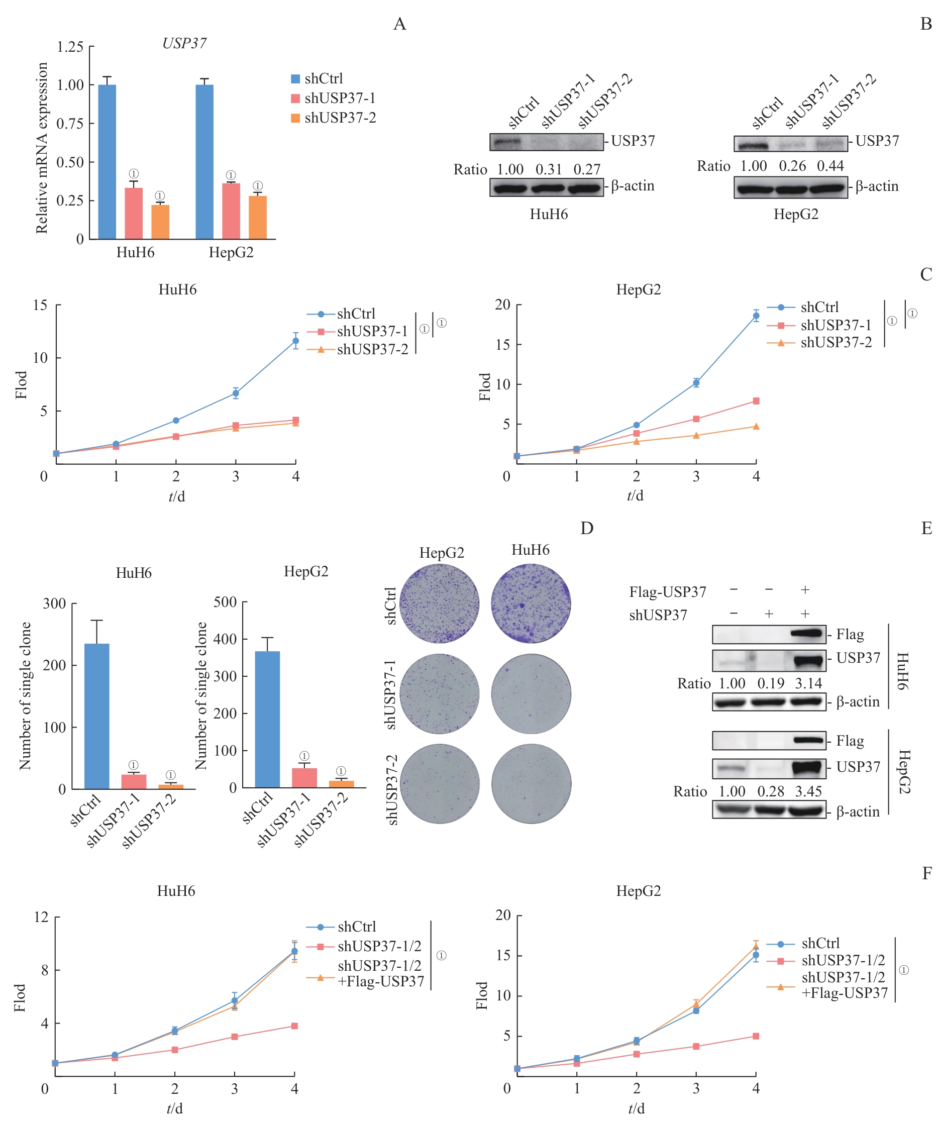
图3 USP37 敲低对HB细胞增殖的影响Note: A. qPCR analysis of USP37 mRNA levels in HuH6 and HepG2 cells transduced with control shRNA (shCtrl) or two independent USP37-targeting shRNAs (shUSP37-1 and shUSP37-2). Data are presented as x±s from n=3 independent experiments. Statistical significance was assessed using Student's t test; ①P<0.001. B. Western blotting analysis showing USP37 protein levels in HuH6 and HepG2 cells expressing shCtrl, shUSP37-1, or shUSP37-2. β-actin was used as a loading control. Relative protein levels were quantified by densitometric analysis and normalized to β-actin. C. Cell proliferation assays of HuH6 and HepG2 cells expressing shCtrl, shUSP37-1, or shUSP37-2. Data are shown as x±s from n=6 independent experiments. Statistical significance was determined using student's t test; ①P<0.001. D. Colony formation assays performed in HuH6 and HepG2 cells following USP37 knockdown. Representative images and quantification of colony numbers are shown. Data are presented as x±s from n=3 independent experiments. Statistical significance was assessed using Student's t test; ①P<0.001. E. Western blotting analysis showing USP37 and Flag-USP37 protein levels in HuH6 and HepG2 cells expressing shCtrl, shUSP37-1/2, or shUSP37-1/2+Flag-USP37. β-actin was used as a loading control. Relative protein levels were quantified by densitometric analysis and normalized to β-actin. F. Cell proliferation assays of HuH6 and HepG2 cells expressing shCtrl, shUSP37-1/2, or shUSP37-1/2+Flag-USP37. Data are shown as x±s from n=6 independent experiments. Statistical significance was determined using one-way ANOVA; ①P<0.001.
Fig 3 Effects of USP37 knockdown on HB cell proliferation
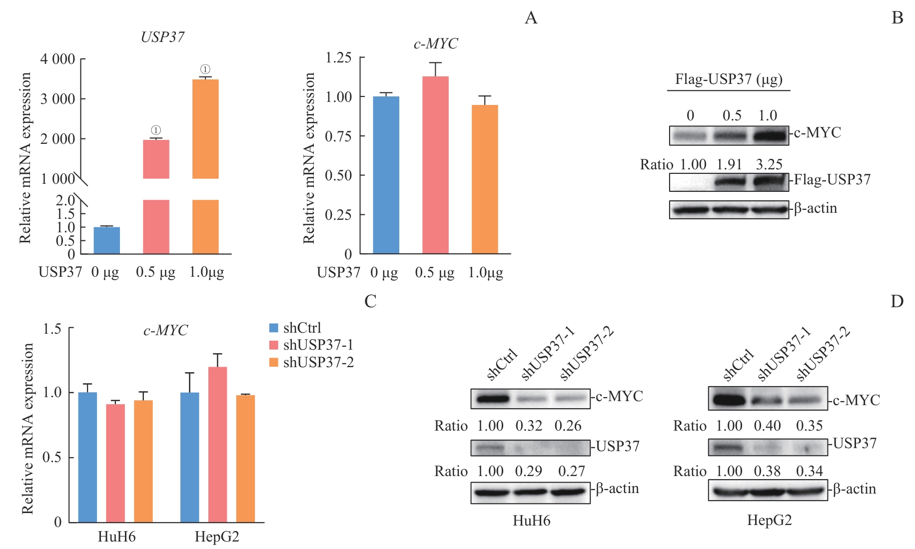
图4 USP37 表达水平变化对 c-MYC mRNA及蛋白水平的影响Note: A. qPCR analysis of USP37 and c-MYC mRNA levels in 293T cells transfected with increasing amounts of Flag-USP37 plasmid (0, 0.5, and 1 μg). mRNA levels were calculated using the 2-ΔΔCt method, with the 0 μg group set as 1. Data are presented as x±s from n=3 independent experiments. Statistical significance was assessed using Student's t test: for USP37 mRNA. B.Western blotting analysis of c-MYC and Flag-USP37 protein levels in 293T cells transfected with increasing amounts of Flag-USP37 plasmid (0, 0.5, and 1 μg). β-actin was used as the loading control. c-MYC protein levels were quantified by densitometry, normalized to β-actin, and expressed relative to the 0 μg group (1, 1.91, and 3.25 for 0, 0.5, and 1 μg, respectively). C. qPCR analysis of c-MYC mRNA levels in HuH6 and HepG2 cells expressing shCtrl, shUSP37-1, or shUSP37-2. mRNA levels were calculated using the 2-ΔΔCt method, with the shCtrl group set as 1. Data are presented as x̄±s from n=3 independent experiments. Statistical significance was determined using Student's t test: in HuH6 cells. D. Western blotting analysis of c-MYC and USP37 protein levels in HuH6 and HepG2 cells expressing shCtrl, shUSP37-1, or shUSP37-2. β-actin was used as the loading control. c-MYC protein levels were quantified by densitometry, normalized to β-actin, and expressed relative to the shCtrl group: HuH6, 1/0.32/0.26 (shCtrl/shUSP37-1/shUSP37-2); HepG2, 1/0.40/0.35.
Fig 4 Effects of USP37 expression modulation on c-MYC mRNA and protein levels
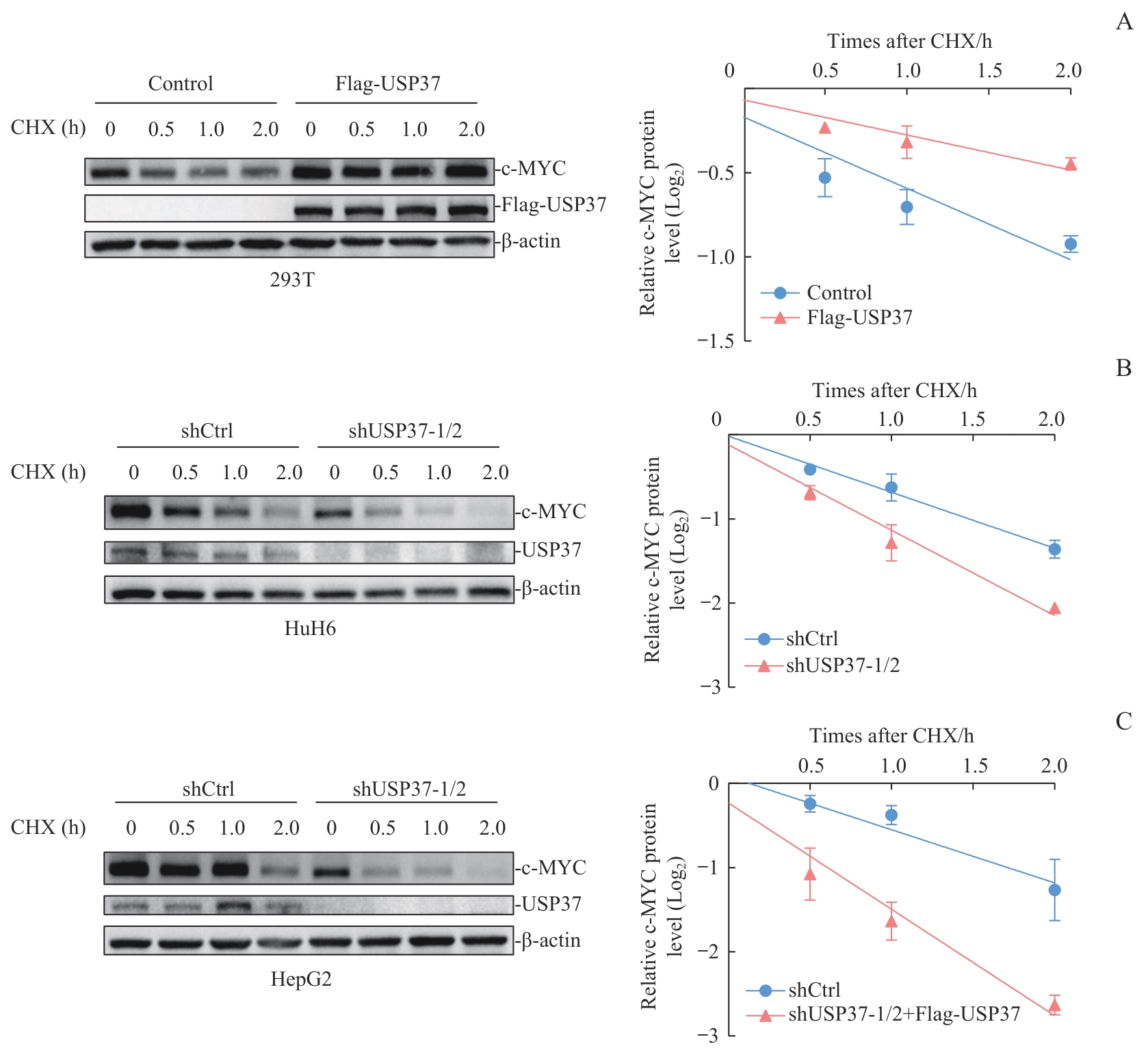
图5 USP37 表达水平变化对c-MYC蛋白稳定性的影响分析Note: A. CHX chase assay in 293T cells transfected with control vector or Flag-USP37. Cells were treated with CHX (50 µg/mL) and harvested at 0, 0.5, 1, and 2 h. Left: representative immunoblots of c-MYC, Flag-USP37, and β-actin. Right: quantification of c-MYC protein levels. c-MYC signals were normalized to β-actin, further normalized to the 0 h time point within each group, and log₂-transformed. Data are presented as x±s from n=2 independent experiments. Statistical significance between the two degradation curves was assessed by two-way ANOVA (P=0.006). The estimated half-life of c-MYC was 2.357 h in the control group and 4.796 h in the USP37-overexpression group. B. CHX chase assay in HuH6 cells expressing shCtrl or shUSP37-1/2. Cells were treated with CHX (50 µg/mL) and harvested at 0, 0.5, 1, and 2 h. Left: representative immunoblots of c-MYC, USP37, and β-actin. Right: quantification of c-MYC protein levels, normalized to β-actin, then to the 0 h time point within each group, and log₂-transformed. Data are presented as x̄±s from n=2 independent experiments. Statistical significance between the two degradation curves was assessed by two-way ANOVA (P=0.006). The estimated half-life of c-MYC was 1.504 h in shCtrl cells and 0.986 h in shUSP37 cells. C. CHX chase assay in HepG2 cells expressing shCtrl or shUSP37-1/2. Cells were treated with CHX (50 µg/mL) and harvested at 0, 0.5, 1, and 2 h. Left: representative immunoblots of c-MYC, USP37, and β-actin. Right: quantification of c-MYC protein levels, normalized to β-actin, then to the 0 h time point within each group, and log2-transformed. Data are presented as x̄±s from n=2 independent experiments. Statistical significance between the two degradation curves was assessed by two-way ANOVA (P=0.005). The estimated half-life of c-MYC was 1.585 h in shCtrl cells and 0.792 h in shUSP37 cells.
Fig 5 Analysis of c-MYC protein stability following modulation of USP37 expression
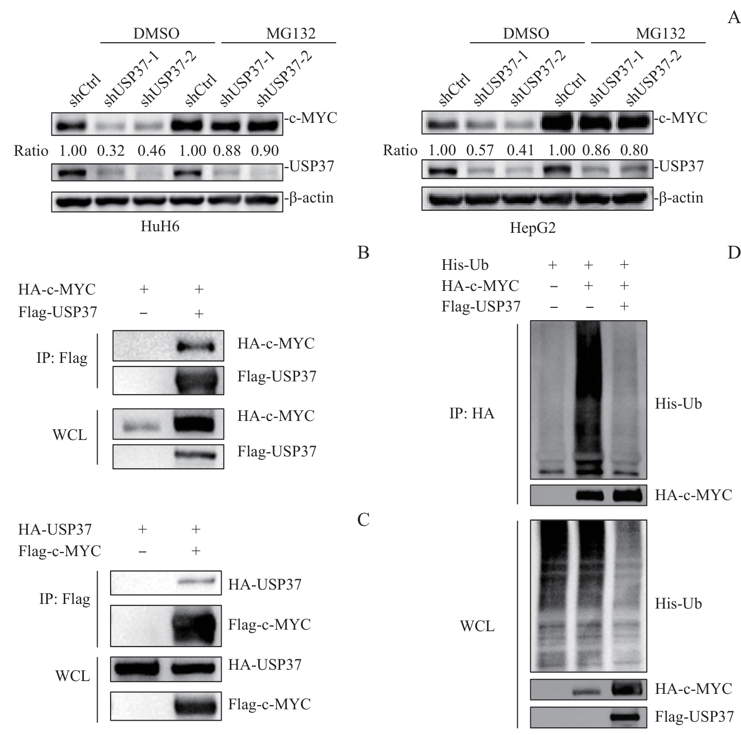
图6 USP37 通过去泛素化调控c-MYC蛋白稳定性的机制分析Note: A. HuH6 and HepG2 cells expressing shCtrl or shUSP37 were treated with DMSO or the proteasome inhibitor MG132 (20 μmol/L for 6 h). Protein levels of c-MYC and USP37 were analyzed by Western blotting. β-actin was used as a loading control. Relative protein levels were quantified by densitometric analysis and normalized to β-actin. B. Co-immunoprecipitation (Co-IP) analysis of the interaction between USP37 and c-MYC. 293T cells were transfected with HA-c-MYC with or without Flag-USP37, as indicated. Cell lysates were immunoprecipitated with anti-Flag beads, and the precipitated proteins were immunoblotted with anti-HA and anti-Flag antibodies. Whole-cell lysates (WCL) were included as input controls to confirm protein expression. C. Reverse Co-IP analysis of the interaction between USP37 and c-MYC. 293T cells were transfected with HA-USP37 with or without Flag-c-MYC, as indicated. Cell lysates were immunoprecipitated with anti-Flag beads, and the precipitated proteins were immunoblotted with anti-HA and anti-Flag antibodies. WCL were included as input controls. D. In vivo deubiquitination assay. 293T cells were transfected with His-Ub and HA-c-MYC, with or without Flag-USP37, as indicated. At 24-36 h after transfection, cells were treated with MG132 (20 μmol/L) for 6 h before harvest. HA-c-MYC was immunoprecipitated with anti-HA beads, and the ubiquitinated c-MYC was detected by immunoblotting with an anti-His antibody. The immunoprecipitated HA-c-MYC levels are shown below as a loading control for the IP. WCL was analyzed to confirm the expression of His-Ub, HA-c-MYC, and Flag-USP37.
Fig 6 Mechanistic analysis of USP37-mediated deubiquitination and stabilization of c-MYC
| [1] | Hager J, Sergi C M. Liver Cancer[M]. Brisbane (AU): Exon Publications, 2021:145-164. |
| [2] | Pio L, O'Neill A F, Woodley H, et al. Hepatoblastoma[J]. Nat Rev Dis Primers, 2025, 11: 36. |
| [3] | Tang M J, Ma X L, He X L, et al. A multicenter prospective study on the management of hepatoblastoma in children: a report from the Chinese Children's Cancer Group[J]. World J Pediatr, 2024, 20(6): 590-601. |
| [4] | Zhou S M, Malvar J, Chi Y Y, et al. Independent assessment of the children's hepatic tumors international collaboration risk stratification for hepatoblastoma and the association of tumor histological characteristics with prognosis[J]. JAMA Netw Open, 2022, 5(2): e2148013. |
| [5] | Stine Z E, Walton Z E, Altman B J, et al. MYC, metabolism, and cancer[J]. Cancer Discov, 2015, 5(10): 1024-1039. |
| [6] | Dang C V. MYC on the path to cancer[J]. Cell, 2012, 149(1): 22-35. |
| [7] | Venkatraman S, Balasubramanian B, Thuwajit C, et al. Targeting MYC at the intersection between cancer metabolism and oncoimmunology[J]. Front Immunol, 2024, 15: 1324045. |
| [8] | Cairo S, Armengol C, de Reyniès A, et al. Hepatic stem-like phenotype and interplay of Wnt/beta-catenin and Myc signaling in aggressive childhood liver cancer[J]. Cancer Cell, 2008, 14(6): 471-484. |
| [9] | Wang D L, Tian J M, Yan Z Y, et al. Mitochondrial fragmentation is crucial for c-Myc-driven hepatoblastoma-like liver tumors[J]. Mol Ther, 2022, 30(4): 1645-1660. |
| [10] | Farrell A S, Sears R C. MYC degradation[J]. Cold Spring Harb Perspect Med, 2014, 4(3): a014365. |
| [11] | Lee J M, Hammarén H M, Savitski M M, et al. Control of protein stability by post-translational modifications[J]. Nat Commun, 2023, 14(1): 201. |
| [12] | Wang C, Zhang J W, Yin J, et al. Alternative approaches to target Myc for cancer treatment[J]. Signal Transduct Target Ther, 2021, 6(1): 117. |
| [13] | Nguyen L H, Robinton D A, Seligson M T, et al. Lin28b is sufficient to drive liver cancer and necessary for its maintenance in murine models[J]. Cancer Cell, 2014, 26(2): 248-261. |
| [14] | Sumazin P, Chen Y D, Treviño L R, et al. Genomic analysis of hepatoblastoma identifies distinct molecular and prognostic subgroups[J]. Hepatology, 2017, 65(1): 104-121. |
| [15] | Kwon Y T, Ciechanover A. The ubiquitin code in the ubiquitin-proteasome system and autophagy[J]. Trends Biochem Sci, 2017, 42(11): 873-886. |
| [16] | Mevissen T E T, Komander D. Mechanisms of deubiquitinase specificity and regulation[J]. Annu Rev Biochem, 2017, 86: 159-192. |
| [17] | Liu F F, Chen J Y, Li K, et al. Ubiquitination and deubiquitination in cancer: from mechanisms to novel therapeutic approaches[J]. Mol Cancer, 2024, 23(1): 148. |
| [18] | Sun T S, Liu Z N, Yang Q. The role of ubiquitination and deubiquitination in cancer metabolism[J]. Mol Cancer, 2020, 19(1): 146. |
| [19] | Hong K, Hu L X, Liu X J, et al. USP37 promotes deubiquitination of HIF2α in kidney cancer[J]. Proc Natl Acad Sci U S A, 2020, 117(23): 13023-13032. |
| [20] | Yang W C, Shih H M. The deubiquitinating enzyme USP37 regulates the oncogenic fusion protein PLZF/RARA stability[J]. Oncogene, 2013, 32(43): 5167-5175. |
| [21] | Pan J, Deng Q, Jiang C, et al. USP37 directly deubiquitinates and stabilizes c-Myc in lung cancer[J]. Oncogene, 2015, 34(30): 3957-3967. |
| [22] | Zhang X, Peng P, Bao L W, et al. Ubiquitin-specific protease 1 promotes bladder cancer progression by stabilizing c-MYC[J]. Cells, 2024, 13(21): 1798. |
| [23] | Xiao Z L, Wang Y, Pan D H, et al. USP3 promotes clear cell renal cell carcinoma progression by stabilizing MYC and enhancing glycolysis[J]. Biochim Biophys Acta Gen Subj, 2025, 1869(6): 130801. |
| [24] | Xia P, Zhang H, Lu H F, et al. METTL5 stabilizes c-Myc by facilitating USP5 translation to reprogram glucose metabolism and promote hepatocellular carcinoma progression[J]. Cancer Commun (Lond), 2023, 43(3): 338-364. |
| [25] | Gu J C, Xiao X, Zou C F, et al. Ubiquitin-specific protease 7 maintains c-Myc stability to support pancreatic cancer glycolysis and tumor growth[J]. J Transl Med, 2024, 22(1): 1135. |
| [26] | Li A N, Wang S J, Li F, et al. PCMTD1-127aa suppresses osteosarcoma progression by competitively binding to USP10 to promote c-MYC degradation[J]. iScience, 2026, 29(3): 114765. |
| [27] | Pornour M, Jeon H Y, Ryu H, et al. USP11 promotes prostate cancer progression by up-regulating AR and c-Myc activity[J]. Proc Natl Acad Sci U S A, 2024, 121(31): e2403331121. |
| [28] | Fang X G, Zhou W C, Wu Q L, et al. Deubiquitinase USP13 maintains glioblastoma stem cells by antagonizing FBXL14-mediated Myc ubiquitination[J]. J Exp Med, 2017, 214(1): 245-267. |
| [29] | Ge J C, Yu W D, Li J H, et al. USP16 regulates castration-resistant prostate cancer cell proliferation by deubiquitinating and stablizing c-Myc[J]. J Exp Clin Cancer Res, 2021, 40(1): 59. |
| [30] | Zhang D, Zhang X W, Chang S, et al. E2F1 activates USP19 to affect the stability of c-Myc to facilitate the progression of hepatocellular carcinoma[J]. Mutat Res, 2025, 830: 111902. |
| [31] | Kim D, Hong A, Park H I, et al. Deubiquitinating enzyme USP22 positively regulates c-Myc stability and tumorigenic activity in mammalian and breast cancer cells[J]. J Cell Physiol, 2017, 232(12): 3664-3676. |
| [32] | Ren D D, Cai Z Q, Zhang R, et al. USP26 promotes cell proliferation of gastric cancer by stabilizing c-myc[J]. DNA Cell Biol, 2025, 44(12): 662-670. |
| [33] | Han X, Ren C E, Lu C, et al. Phosphorylation of USP27X by PIM2 promotes glycolysis and breast cancer progression via deubiquitylation of MYC[J]. Oncogene, 2024, 43(33): 2493-2503. |
| [34] | Popov N, Wanzel M, Madiredjo M, et al. The ubiquitin-specific protease USP28 is required for MYC stability[J]. Nat Cell Biol, 2007, 9(7): 765-774. |
| [35] | Diefenbacher M E, Chakraborty A, Blake S M, et al. Usp28 counteracts Fbw7 in intestinal homeostasis and cancer[J]. Cancer Res, 2015, 75(7): 1181-1186. |
| [36] | Tu R F, Kang W Q, Yang M J, et al. USP29 coordinates MYC and HIF1α stabilization to promote tumor metabolism and progression[J]. Oncogene, 2021, 40(46): 6417-6429. |
| [37] | Zhang X Y, Han Y, Liu S L, et al. MF-094 nanodelivery inhibits oral squamous cell carcinoma by targeting USP30[J]. Cell Mol Biol Lett, 2022, 27(1): 107. |
| [38] | Ding T, Zhu Y J, Jin H M, et al. Circular RNA circ_0057558 controls prostate cancer cell proliferation through regulating miR-206/USP33/c-Myc axis[J]. Front Cell Dev Biol, 2021, 9: 644397. |
| [39] | Chen D J, Zhao Y, Zhang X B, et al. USP33-mediated stabilization of c-Myc drives glycolytic reprogramming and promotes ovarian cancer progression[J]. Biochim Biophys Acta Gen Subj, 2025, 1869(9): 130830. |
| [40] | Liu H L, Wei X L, Nie Y, et al. The knockdown of USP34 inhibits the progression of hepatocellular carcinoma by accelerating c-Myc degradation[J]. Turk J Gastroenterol, 2025, 36(9): 573-582. |
| [41] | Sun X X, He X, Yin L, et al. The nucleolar ubiquitin-specific protease USP36 deubiquitinates and stabilizes c-Myc[J]. Proc Natl Acad Sci U S A, 2015, 112(12): 3734-3739. |
| [42] | Xu Z J, Hu H, Fang D B, et al. The deubiquitinase USP38 promotes cell proliferation through stabilizing c-Myc[J]. Int J Biochem Cell Biol, 2021, 137: 106023. |
| [43] | Fu W, Wu Q L, Yao Y R, et al. Ubiquitin-specific peptidase 40 promotes cellular proliferation and inhibits apoptosis through deubiquitination-mediated stabilization of c-MYC in acute myeloid leukemia[J]. Biochim Biophys Acta Mol Basis Dis, 2026, 1872(3): 168138. |
| [44] | Li M X, Yu J T, Ju L G, et al. USP43 stabilizes c-Myc to promote glycolysis and metastasis in bladder cancer[J]. Cell Death Dis, 2024, 15(1): 44. |
| [45] | Cheng H X, Hua L Y, Tang H L, et al. CBX7 reprograms metabolic flux to protect against meningioma progression by modulating the USP44/c-MYC/LDHA axis[J]. J Mol Cell Biol, 2024, 15(10): mjad057. |
| [46] | Tu X, Li C C, Sun W, et al. Suppression of cancer cell stemness and drug resistance via MYC destabilization by deubiquitinase USP45 inhibition with a natural small molecule[J]. Cancers, 2023, 15(3): 930. |
| [47] | Shin H, Hwang S, Jeong J H, et al. Targeting USP47 enhances the efficacy of KRAS inhibitor in KRASG12C mutated non-small cell lung cancer by controlling deubiquitination of c-Myc[J]. Pharmacol Res, 2025, 215: 107722. |
| [48] | Ye M J, He J J, Zhang J J, et al. USP7 promotes hepatoblastoma progression through activation of PI3K/AKT signaling pathway[J]. Cancer Biomark, 2021, 31(2): 107-117. |
| [49] | Wu L, Zhao N, Zhou Z L, et al. PLAGL2 promotes the proliferation and migration of gastric cancer cells via USP37-mediated deubiquitination of Snail1[J]. Theranostics, 2021, 11(2): 700-714. |
| [50] | Das C M, Taylor P, Gireud M, et al. The deubiquitylase USP37 links REST to the control of p27 stability and cell proliferation[J]. Oncogene, 2013, 32(13): 1691-1701. |
| [51] | Dobson T H W, Hatcher R J, Swaminathan J, et al. Regulation of USP37 expression by REST-associated G9a-dependent histone methylation[J]. Mol Cancer Res, 2017, 15(8): 1073-1084. |
| [1] | 陈赟琪, 那迪娜·帕尔哈提, 张鹏善, 黄陈. 去泛素化酶USP38通过稳定RBM14促进胃癌进展的机制研究[J]. 上海交通大学学报(医学版), 2026, 46(8): 1037-1052. |
| [2] | 翟元晖, 王莹莹, 王宇轩, 吴文萱, 朱楚娇, 张幼萍, 白雯会, 雷虎, 徐含章, 赵亚雪, 吴英理. 甲基巴多索隆作为新型泛素特异性蛋白酶48抑制剂抑制结直肠癌的作用及机制研究[J]. 上海交通大学学报(医学版), 2026, 46(8): 1026-1036. |
| [3] | 那迪娜·帕尔哈提, 张鹏善, 徐亦天, 陈赟琪, 黄陈. 人去泛素化酶圆柱瘤蛋白截短体质粒的构建及其对胃癌细胞表型的调控研究[J]. 上海交通大学学报(医学版), 2025, 45(9): 1149-1160. |
| [4] | 童桐, 秦星, 谢非, 蒋英英, 石剑波, 张建军. USP47通过促进抗凋亡蛋白表达介导头颈鳞癌细胞顺铂耐药[J]. 上海交通大学学报(医学版), 2021, 41(4): 434-441. |
| [5] | 朱之星1, 2,纪 伟3,顾坚磊1, 4,吕 晖1, 2, 4,田国力3. 葡萄糖 -6- 磷酸脱氢酶基因4种突变及蛋白结构分析[J]. 上海交通大学学报(医学版), 2020, 40(12): 1571-1578. |
| [6] | 屈国君 1,陆元凤 2,李宇 1. PDGFRα下游新底物 CCT2对肿瘤细胞生长的影响及其机制[J]. 上海交通大学学报(医学版), 2019, 39(1): 28-. |
| [7] | 李则,任尊,涂鸿鹄,简政,余健秀,邓蓉. BAP1及其抑癌作用研究新进展[J]. 上海交通大学学报(医学版), 2017, 37(5): 689-. |
| [8] | 谢文娟,秦东军,张 健,等. 紫菀酮对去泛素化酶2活性的抑制作用[J]. 上海交通大学学报(医学版), 2014, 34(11): 1563-. |
| [9] | 盛 琦, 袁晓军, 张 勤, 等. 12例儿童肝母细胞瘤的诊断与治疗[J]. , 2011, 31(4): 517-. |
| 阅读次数 | ||||||
|
全文 |
|
|||||
|
摘要 |
|
|||||
