JOURNAL OF SHANGHAI JIAOTONG UNIVERSITY (MEDICAL SCIENCE) ›› 2021, Vol. 41 ›› Issue (6): 732-740.doi: 10.3969/j.issn.1674-8115.2021.06.005
• Basic research • Previous Articles Next Articles
Qing WANG( ), Wei WANG, Da-jun JIANG, Wei-tao JIA()
Online:2021-06-28
Published:2021-06-29
Contact:
Wei-tao JIA
E-mail:paidaqing@outlook.com;jiaweitao@shsmu.edu.cn
Supported by:CLC Number:
Qing WANG, Wei WANG, Da-jun JIANG, Wei-tao JIA. Evaluation of JDBM porous scaffold coated with DCPD in promoting angiogenesis and repairing bone defects[J]. JOURNAL OF SHANGHAI JIAOTONG UNIVERSITY (MEDICAL SCIENCE), 2021, 41(6): 732-740.
Add to citation manager EndNote|Ris|BibTeX
URL: https://xuebao.shsmu.edu.cn/EN/10.3969/j.issn.1674-8115.2021.06.005
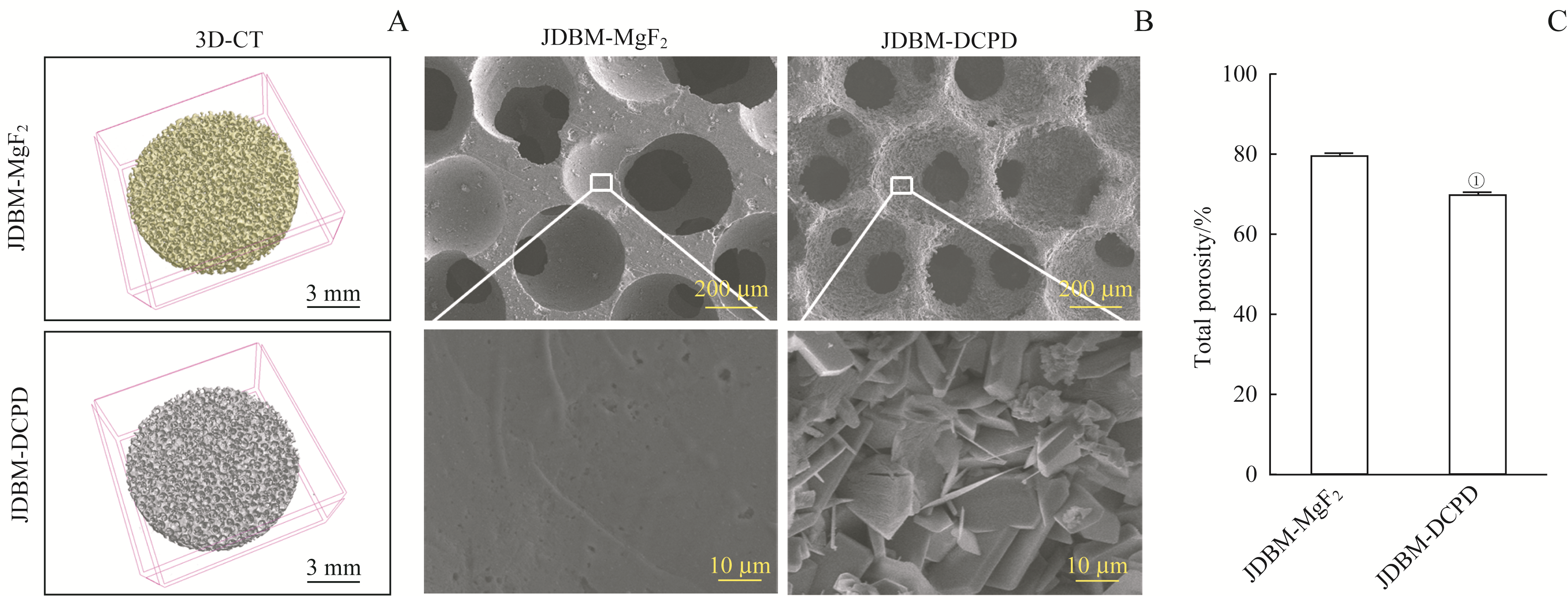
Fig 1 Comparison of JDBM-DCPD and JDBM-MgF2 porous scaffolds in characteristics
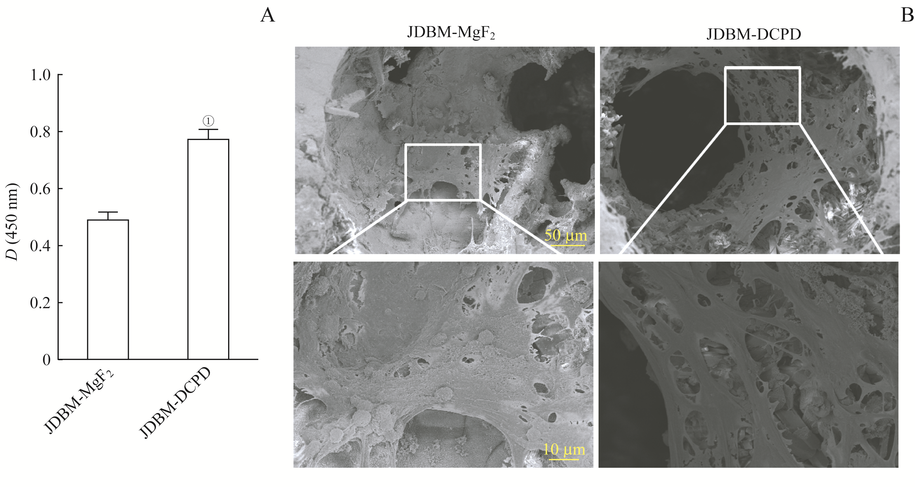
Fig 2 Comparison of JDBM-DCPD and JDBM-MgF2 in biocompatibility
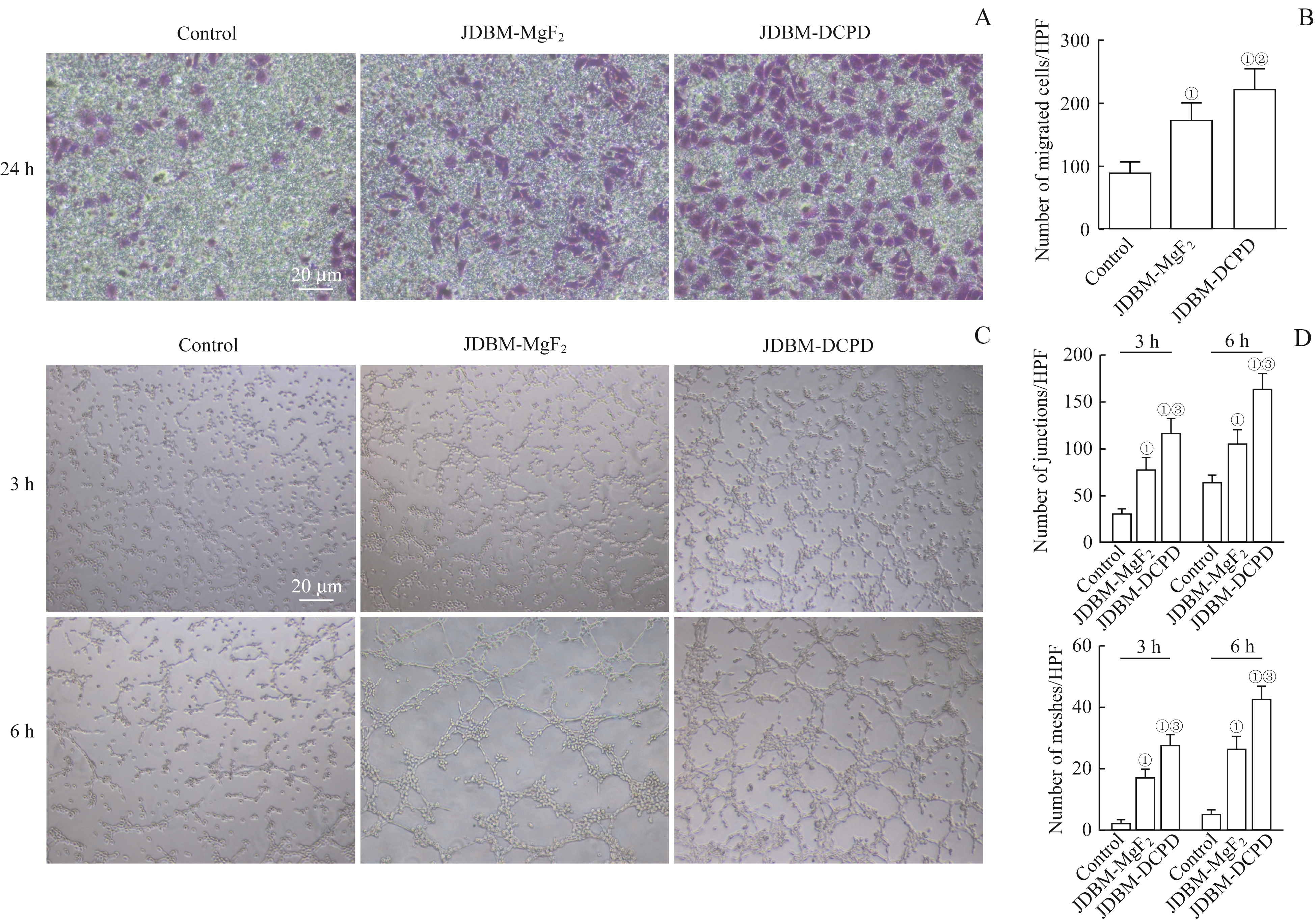
Fig 3 Comparison of the effects of JDBM-DCPD and JDBM-MgF2 scaffold extracts on Ea.hy926 cell migration and tube formation
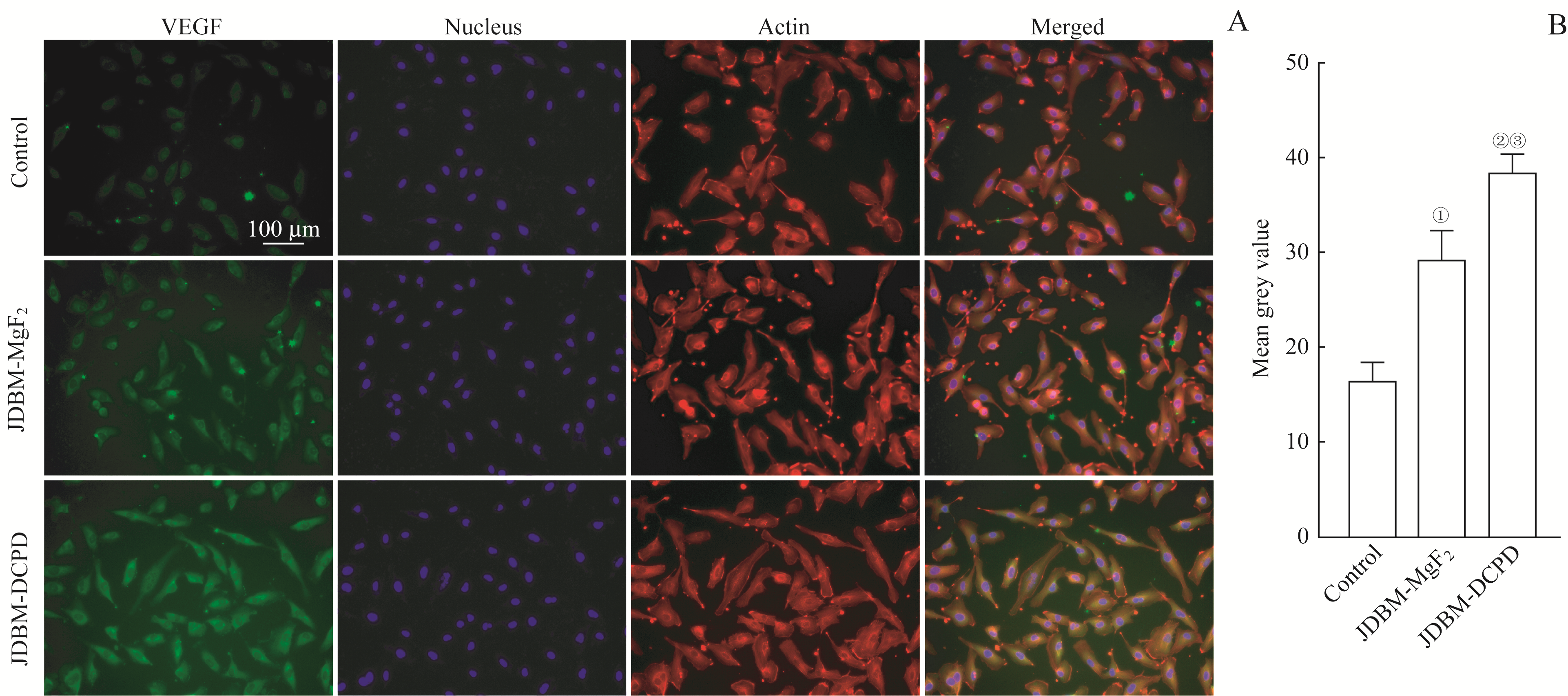
Fig 4 Comparison of the effects of JDBM-DCPD and JDBM-MgF2 scaffold extracts on the expression of VEGF in Ea.hy926 cells

Fig 5 Comparison of effects of JDBM-DCPD and JDBM-MgF2 scaffold extracts on osteogenic differentiation of BMSCs
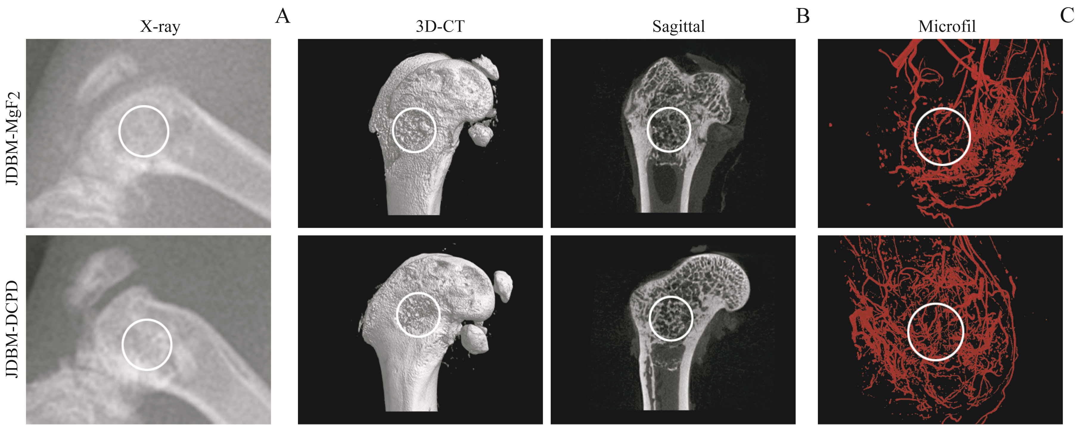
Fig 6 Osteogenesis and angiogenesis of JDBM-DCPD and JDBM-MgF2 scaffolds in vivo detected by radiography 8 weeks after implantation
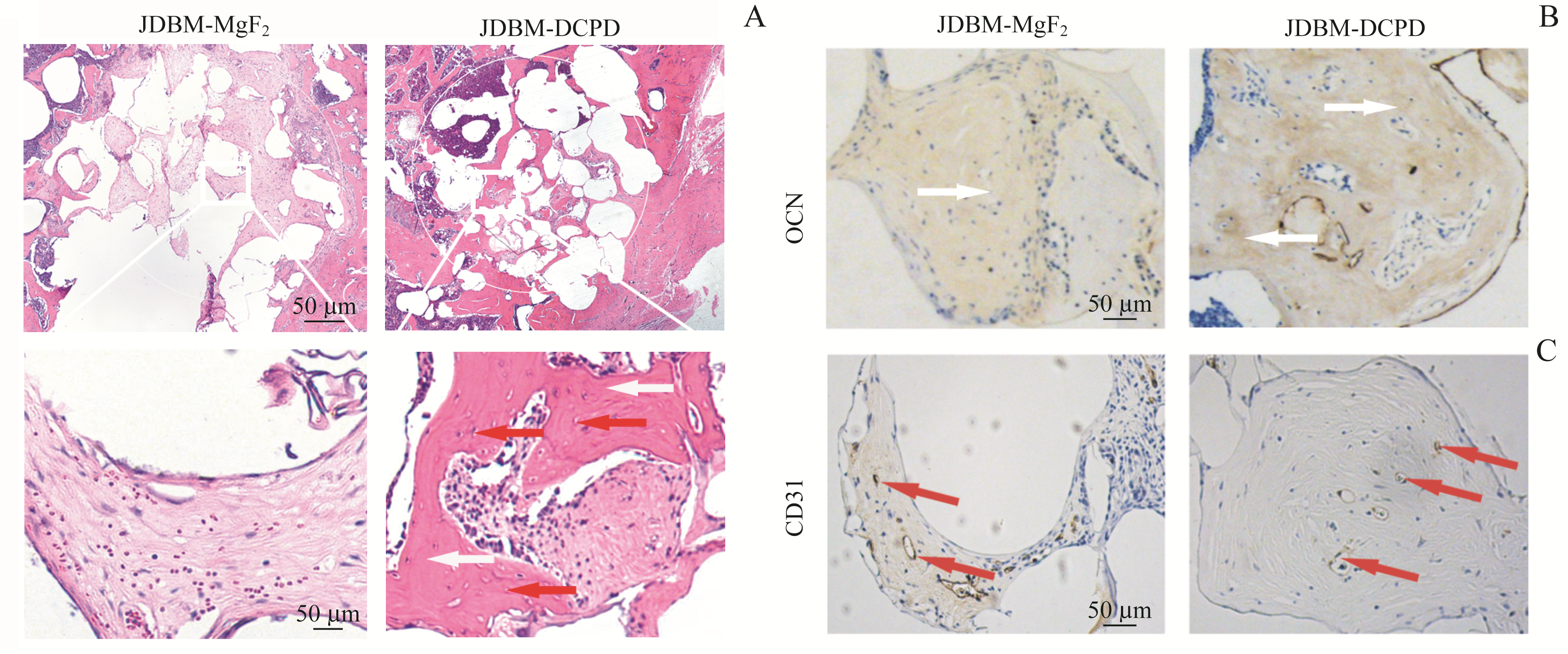
Fig 7 Histological observation of the bone defect areas in JDBM-DCPD group and JDBM-MgF2 group rats 8 weeks after implantation
| 1 | Giannoudis PV, Dinopoulos H, Tsiridis E. Bone substitutes: an update[J]. Injury, 2005, 36(): S20-S27. |
| 2 | Moore WR, Graves SE, Bain GI. Synthetic bone graft substitutes[J]. ANZ J Surg, 2001, 71(6): 354-361. |
| 3 | Samartzis D, Shen FH, Goldberg EJ, et al. Is autograft the gold standard in achieving radiographic fusion in one-level anterior cervical discectomy and fusion with rigid anterior plate fixation?[J]. Spine, 2005, 30(15): 1756-1761. |
| 4 | Jin L, Li P, Wang YC, et al. Studies of superb microvascular imaging and contrast-enhanced ultrasonography in the evaluation of vascularization in early bone regeneration[J]. J Ultrasound Med, 2019, 38(11): 2963-2971. |
| 5 | Brandi ML, Collin-Osdoby P. Vascular biology and the skeleton[J]. J Bone Miner Res, 2006, 21(2): 183-192. |
| 6 | Parfitt AM. The mechanism of coupling: a role for the vasculature[J]. Bone, 2000, 26(4): 319-323. |
| 7 | Novosel EC, Kleinhans C, Kluger PJ. Vascularization is the key challenge in tissue engineering[J]. Adv Drug Deliv Rev, 2011, 63(4/5): 300-311. |
| 8 | Rouwkema J, Rivron NC, van Blitterswijk CA. Vascularization in tissue engineering[J]. Trends Biotechnol, 2008, 26(8): 434-441. |
| 9 | Kanczler JM, Oreffo RO. Osteogenesis and angiogenesis: the potential for engineering bone[J]. Eur Cell Mater, 2008, 15: 100-114. |
| 10 | Thevenot P, Nair A, Dey J, et al. Method to analyze three-dimensional cell distribution and infiltration in degradable scaffolds[J]. Tissue Eng Part C Methods, 2008, 14(4): 319-331. |
| 11 | Staiger MP, Pietak AM, Huadmai J, et al. Magnesium and its alloys as orthopedic biomaterials: a review[J]. Biomaterials, 2006, 27(9): 1728-1734. |
| 12 | Velasco MA, Narváez-Tovar CA, Garzón-Alvarado DA. Design, materials, and mechanobiology of biodegradable scaffolds for bone tissue engineering[J]. Biomed Res Int, 2015, 2015: 729076. |
| 13 | Kunjukunju S, Roy A, Ramanathan M, et al. A layer-by-layer approach to natural polymer-derived bioactive coatings on magnesium alloys[J]. Acta Biomater, 2013, 9(10): 8690-8703. |
| 14 | Zhang J, Ma X, Lin D, et al. Magnesium modification of a calcium phosphate cement alters bone marrow stromal cell behavior via an integrin-mediated mechanism[J]. Biomaterials, 2015, 53: 251-264. |
| 15 | Qin H, Zhao Y, An Z, et al. Enhanced antibacterial properties, biocompatibility, and corrosion resistance of degradable Mg-Nd-Zn-Zr alloy[J]. Biomaterials, 2015, 53: 211-220. |
| 16 | Kong X, Wang L, Li G, et al. Mg-based bone implants show promising osteoinductivity and controllable degradation: a long-term study in a goat femoral condyle fracture model[J]. Mater Sci Eng C Mater Biol Appl, 2018, 86: 42-47. |
| 17 | Guan X, Xiong M, Zeng F, et al. Enhancement of osteogenesis and biodegradation control by brushite coating on Mg-Nd-Zn-Zr alloy for mandibular bone repair[J]. ACS Appl Mater Interfaces, 2014, 6(23): 21525-21533. |
| 18 | Tamimi F, Sheikh Z, Barralet J. Dicalcium phosphate cements: brushite and monetite[J]. Acta Biomater, 2012, 8(2): 474-487. |
| 19 | Apelt D, Theiss F, El-Warrak AO, et al. In vivo behavior of three different injectable hydraulic calcium phosphate cements[J]. Biomaterials, 2004, 25(7): 1439-1451. |
| 20 | Malhotra A, Habibovic P. Calcium phosphates and angiogenesis: implications and advances for bone regeneration[J]. Trends Biotechnol, 2016, 34(12): 983-992. |
| 21 | Wang W, Jia G, Wang Q, et al. The in vitro and in vivo biological effects and osteogenic activity of novel biodegradable porous Mg alloy scaffolds[J]. Mater Des, 2020, 189: 108514. |
| 22 | Pijuan J, Barceló C, Moreno DF, et al. In vitro cell migration, invasion, and adhesion assays: from cell imaging to data analysis[J]. Front Cell Dev Biol, 2019, 7: 107. |
| 23 | Hu H, Chen Y, Zou Z, et al. Panax notoginseng saponins prevent bone loss by promoting angiogenesis in an osteoporotic mouse model[J]. Biomed Res Int, 2020, 2020: 8412468. |
| 24 | Dai C, Guo H, Lu J, et al. Osteogenic evaluation of calcium/magnesium-doped mesoporous silica scaffold with incorporation of rhBMP-2 by synchrotron radiation-based μCT[J]. Biomaterials, 2011, 32(33): 8506-8517. |
| 25 | Griffith LG. Emerging design principles in biomaterials and scaffolds for tissue engineering[J]. Ann N Y Acad Sci, 2002, 961: 83-95. |
| 26 | Karageorgiou V, Kaplan D. Porosity of 3D biomaterial scaffolds and osteogenesis[J]. Biomaterials, 2005, 26(27): 5474-5491. |
| 27 | Chiesa R, Sandrini E, Santin M, et al. Osteointegration of titanium and its alloys by anodic spark deposition and other electrochemical techniques: a review[J]. J Appl Biomater Biomech, 2003, 1(2): 91-107. |
| 28 | Liang C, Wang H, Yang J, et al. Femtosecond laser-induced micropattern and Ca/P deposition on Ti implant surface and its acceleration on early osseointegration[J]. ACS Appl Mater Interfaces, 2013, 5(16): 8179-8186. |
| 29 | Ma H, Luo J, Sun Z, et al. 3D printing of biomaterials with mussel-inspired nanostructures for tumor therapy and tissue regeneration[J]. Biomaterials, 2016, 111: 138-148. |
| 30 | Xu F, Ding H, Song F, et al. Effects of preparation methods on the bone formation potential of apatite-coated chitosan microspheres[J]. J Biomater Sci Polym Ed, 2014, 25(18): 2080-2093. |
| 31 | Yu W, Zhao H, Ding Z, et al. In vitro and in vivo evaluation of MgF2 coated AZ31 magnesium alloy porous scaffolds for bone regeneration[J]. Colloids Surf B Biointerfaces, 2017, 149: 330-340. |
| 32 | Lafage-Proust MH, Prisby R, Roche B, et al. Bone vascularization and remodeling[J]. Joint Bone Spine, 2010, 77(6): 521-524. |
| 33 | Kusumbe AP, Ramasamy SK, Adams RH. Coupling of angiogenesis and osteogenesis by a specific vessel subtype in bone[J]. Nature, 2014, 507(7492): 323-328. |
| 34 | Dhandapani R, Krishnan PD, Zennifer A, et al. Additive manufacturing of biodegradable porous orthopaedic screw[J]. Bioact Mater, 2020, 5(3): 458-467. |
| 35 | Xie H, Cui Z, Wang L, et al. PDGF-BB secreted by preosteoclasts induces angiogenesis during coupling with osteogenesis[J]. Nat Med, 2014, 20(11): 1270-1278. |
| 36 | Liu Q, Zhou YF, Li ZB. PDGF‑BB promotes the differentiation and proliferation of MC3T3‑E1 cells through the Src/JAK2 signaling pathway[J]. Mol Med Rep, 2018, 18(4): 3719-3726. |
| 37 | Liu W, Guo S, Tang Z, et al. Magnesium promotes bone formation and angiogenesis by enhancing MC3T3-E1 secretion of PDGF-BB[J]. Biochem Biophys Res Commun, 2020, 528(4): 664-670. |
| 38 | Saghiri MA, Asatourian A, Orangi J, et al. Functional role of inorganic trace elements in angiogenesis—part Ⅰ: N, Fe, Se, P, Au, and Ca[J]. Crit Rev Oncol Hematol, 2015, 96(1): 129-142. |
| 39 | Song GL, Song SZ. A possible biodegradable magnesium implant material[J]. Adv Eng Mater, 2007, 9(4): 298-302. |
| 40 | Gu XN, Zheng YF, Chen LJ. Influence of artificial biological fluid composition on the biocorrosion of potential orthopedic Mg-Ca, Az31, Az91 alloys[J]. Biomed Mater, 2009, 4(6): 065011. |
| 41 | Fischer J, Prosenc MH, Wolff M, et al. Interference of magnesium corrosion with tetrazolium-based cytotoxicity assays[J]. Acta Biomater, 2010, 6(5): 1813-1823. |
| Viewed | ||||||
|
Full text |
|
|||||
|
Abstract |
|
|||||