Journal of Shanghai Jiao Tong University (Medical Science) ›› 2023, Vol. 43 ›› Issue (2): 180-187.doi: 10.3969/j.issn.1674-8115.2023.02.006
• Clinical research • Previous Articles
WU Bing1( ), LI Xiaomin1, LIU Siyu1, ZHAO Lulu1, WU Wen2, HAO Yongqiang2, AI Songtao1()
Received:2022-09-22
Accepted:2023-01-11
Online:2023-02-28
Published:2023-02-28
Contact:
AI Songtao
E-mail:wubing-wb@sjtu.edu.cn;ai.songtao@qq.com
Supported by:CLC Number:
WU Bing, LI Xiaomin, LIU Siyu, ZHAO Lulu, WU Wen, HAO Yongqiang, AI Songtao. Preliminary application of improved 3D printed pathological section box to assisting stitching pathological images of bone tumor[J]. Journal of Shanghai Jiao Tong University (Medical Science), 2023, 43(2): 180-187.
Add to citation manager EndNote|Ris|BibTeX
URL: https://xuebao.shsmu.edu.cn/EN/10.3969/j.issn.1674-8115.2023.02.006
| Sequence | TR/ms | TE/ms | FA/( °) | ST/mm | SG/mm | FOV/cm | b value/(s·mm -2) |
|---|---|---|---|---|---|---|---|
| T1 | 615 (axial) | 18 | 120 | 5 | 6 | 360 | N/A |
| T2 FS | 5 000 (axial) 5 000 (coronal) | 85 (axial) 80 (coronal) | 130 | 5 | 6 | 360 | N/A |
| DWI | 6 100 | 74 | 90 | 3 | 3 | 360 | 1 000 |
Tab 1 MRI scanning parameters
| Sequence | TR/ms | TE/ms | FA/( °) | ST/mm | SG/mm | FOV/cm | b value/(s·mm -2) |
|---|---|---|---|---|---|---|---|
| T1 | 615 (axial) | 18 | 120 | 5 | 6 | 360 | N/A |
| T2 FS | 5 000 (axial) 5 000 (coronal) | 85 (axial) 80 (coronal) | 130 | 5 | 6 | 360 | N/A |
| DWI | 6 100 | 74 | 90 | 3 | 3 | 360 | 1 000 |
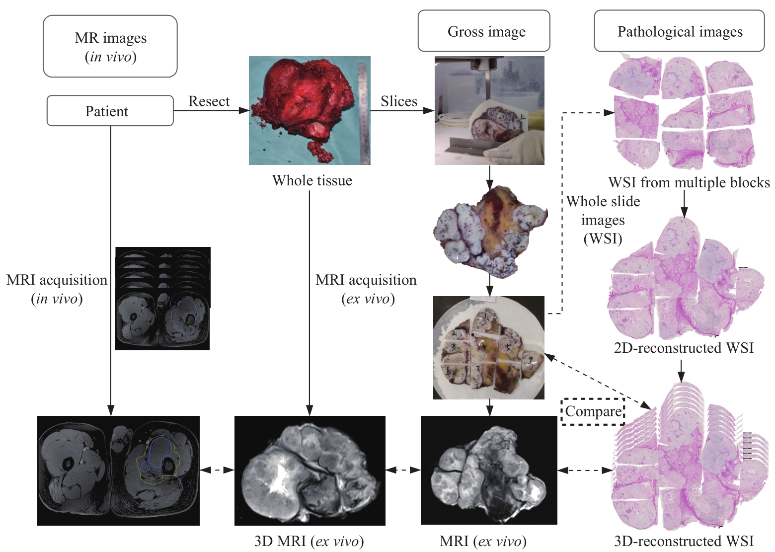
Fig 1 Image-pathology control flow chart
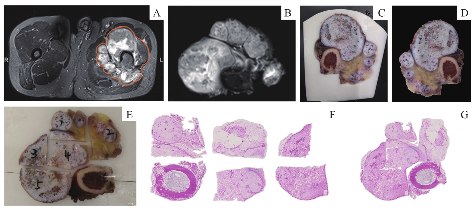
Fig 2 Flow chart of stitching pathological images
| Case No. | Gender | Age/year | Diagnosis | Tumor location | Tumor maximum diameter/cm | Surgery performed |
|---|---|---|---|---|---|---|
| 1 | Male | 25 | Osteochondroma | Femur | 6.1 | Excision + EPR |
| 2 | Male | 62 | Renal cell carcinoma bone metastasis | Ilium | 13.9 | Excision + EPR |
| 3 | Female | 12 | Osteosarcoma | Femur | 13.8 | Excision + EPR |
| 4 | Male | 62 | Papillary thyroid carcinoma bone metastases | Femur | 14.6 | Excision + EPR |
Tab 2 Clinical information of the patients
| Case No. | Gender | Age/year | Diagnosis | Tumor location | Tumor maximum diameter/cm | Surgery performed |
|---|---|---|---|---|---|---|
| 1 | Male | 25 | Osteochondroma | Femur | 6.1 | Excision + EPR |
| 2 | Male | 62 | Renal cell carcinoma bone metastasis | Ilium | 13.9 | Excision + EPR |
| 3 | Female | 12 | Osteosarcoma | Femur | 13.8 | Excision + EPR |
| 4 | Male | 62 | Papillary thyroid carcinoma bone metastases | Femur | 14.6 | Excision + EPR |
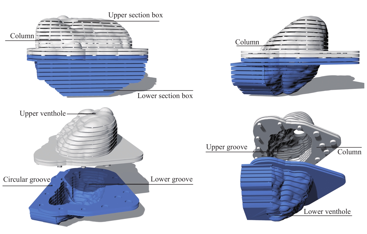
Fig 3 Schematic diagram of the structure of the 3D printed pathological section box
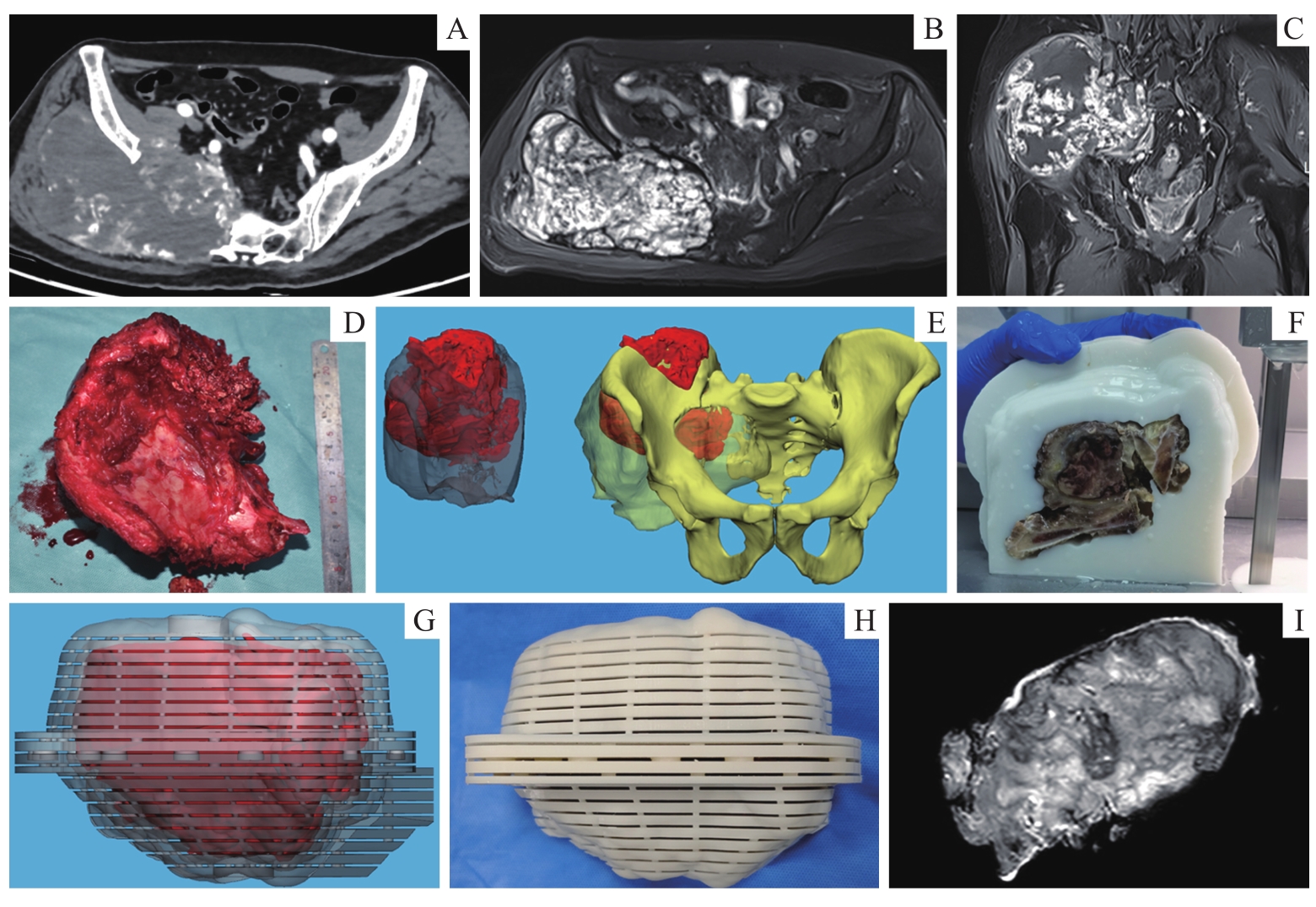
Fig 4 Application of improved 3D printed pathological slide box to bone tumor

Fig 5 CT images of fitting degree of 3D printing pathological section box and bone tumor specimen
| 1 | ANDERSON W J, DOYLE L A. Updates from the 2020 World Health Organization classification of soft tissue and bone tumours[J]. Histopathology, 2021, 78(5): 644-657. |
| 2 | JIN T, DENG Z P, LIU W F, et al. Magnetic resonance imaging for the assessment of long bone tumors[J]. Chin Med J (Engl), 2017, 130(21): 2547-2550. |
| 3 | MIN J H, LEE M W, PARK H S, et al. Interobserver variability and diagnostic performance of gadoxetic acid-enhanced MRI for predicting microvascular invasion in hepatocellular carcinoma[J]. Radiology, 2020, 297(3): 573-581. |
| 4 | YUAN Y, ZENG D W, LIU Y J, et al. DWI and IVIM are predictors of Ki67 proliferation index: direct comparison of MRI images and pathological slices in a murine model of rhabdomyosarcoma[J]. Eur Radiol, 2020, 30(3): 1334-1341. |
| 5 | ZHANG T, YU J M, WANG Y Q, et al. WHO grade Ⅰ meningioma subtypes: MRI features and pathological analysis[J]. Life Sci, 2018, 213: 50-56. |
| 6 | ABSINTA M, NAIR G, FILIPPI M, et al. Postmortem magnetic resonance imaging to guide the pathologic cut: individualized, 3-dimensionally printed cutting boxes for fixed brains[J]. J Neuropathol Exp Neurol, 2014, 73(8): 780-788. |
| 7 | DWIVEDI D K, CHATZINOFF Y, ZHANG Y, et al. Development of a patient-specific tumor mold using magnetic resonance imaging and 3-dimensional printing technology for targeted tissue procurement and radiomics analysis of renal masses[J]. Urology, 2018, 112: 209-214. |
| 8 | GUY J R, SATI P, LEIBOVITCH E, et al. Custom fit 3D-printed brain holders for comparison of histology with MRI in marmosets[J]. J Neurosci Methods, 2016, 257: 55-63. |
| 9 | 李小敏, 曲扬, 武文, 等. 3D打印切片盒在骨盆肿瘤边界三维定位中的应用初探[J]. 上海交通大学学报(医学版), 2020, 40(10): 1408-1413. |
| LI X M, QU Y, WU W, et al. Preliminary application of MR imaging-pathology co-localization by 3D printing box in pelvic tumor assessment[J]. Journal of Shanghai Jiao Tong University (Medical Science), 2020, 40(10): 1408-1413. | |
| 10 | CHAPPELOW J, TOMASZEWSKI J E, FELDMAN M, et al. HistoStitcher©: an interactive program for accurate and rapid reconstruction of digitized whole histological sections from tissue fragments[J]. Comput Med Imaging Graph, 2011, 35(7/8): 557-567. |
| 11 | SOOD R R, SHAO W, KUNDER C, et al. 3D Registration of pre-surgical prostate MRI and histopathology images via super-resolution volume reconstruction[J]. Med Image Anal, 2021, 69: 101957. |
| 12 | 曲扬, 艾松涛, 杨飞, 等. CT和MRI图像配准融合联合3D打印技术在难治性骨盆肿瘤术前规划中的应用[J]. 上海交通大学学报(医学版), 2017, 37(9): 1239-1244, 1238. |
| QU Y, AI S T, YANG F, et al. Application of CT/MRI image registration and fusion combined with 3D printing technique in pre-surgical planning of refractory pelvic tumors[J]. Journal of Shanghai Jiao Tong University (Medical Science), 2017, 37(9): 1239-1244, 1238. | |
| 13 | QU Y, LI X M, YAN Z N, et al. Surgical planning of pelvic tumor using multi-view CNN with relation-context representation learning[J]. Med Image Anal, 2021, 69: 101954. |
| 14 | 翁丹枫, 傅晓丹, 陈淑惠, 等. EDTA脱钙在骨组织大切片制片中的应用效果[J]. 临床与实验病理学杂志, 2021, 37(3): 350-352. |
| WENG D F, FU X D, CHEN X H, et al. Application effect of EDTA decalcification in the preparation of large sections of bone tissue[J]. Chinese Journal of Clinical and Experimental Pathology, 2021, 37(3): 350-352. | |
| 15 | PRIESTER A, WU H, KHOSHNOODI P, et al. Registration accuracy of patient-specific, three-dimensional-printed prostate molds for correlating pathology with magnetic resonance imaging[J]. IEEE Trans Biomed Eng, 2019, 66(1): 14-22. |
| 16 | WU H H, PRIESTER A, KHOSHNOODI P, et al. A system using patient-specific 3D-printed molds to spatially align in vivo MRI with ex vivo MRI and whole-mount histopathology for prostate cancer research[J]. J Magn Reson Imaging, 2019, 49(1): 270-279. |
| 17 | LI X M, WU B, ZOU Y X, et al. Development of a 3D-printed pelvic CT phantom combined with fresh pathological tissues of bone tumor[J]. Quant Imaging Med Surg, 2022, 12(9): 4647-4657. |
| 18 | COSTA D N, CHATZINOFF Y, PASSONI N M, et al. Improved magnetic resonance imaging-pathology correlation with imaging-derived, 3D-printed, patient-specific whole-mount molds of the prostate[J]. Invest Radiol, 2017, 52(9): 507-513. |
| 19 | HUANG L, XIA W, ZHANG B, et al. MSFCN-multiple supervised fully convolutional networks for the osteosarcoma segmentation of CT images[J]. Comput Methods Programs Biomed, 2017, 143: 67-74. |
| 20 | ZHANG R, HUANG L, XIA W, et al. Multiple supervised residual network for osteosarcoma segmentation in CT images[J]. Comput Med Imaging Graph, 2018, 63: 1-8. |
| 21 | RAUSCHECKER A M, RUDIE J D, XIE L, et al. Artificial intelligence system approaching neuroradiologist-level differential diagnosis accuracy at brain MRI[J]. Radiology, 2020, 295(3): 626-637. |
| 22 | JIANG Y L, EDWARDS A V, NEWSTEAD G M. Artificial intelligence applied to breast MRI for improved diagnosis[J]. Radiology, 2021, 298(1): 38-46. |
| [1] | LIU Siyu, WU Bing, LI Xiaomin, ZHAO Lulu, CHEN Jun, AI Songtao. Preliminary exploration of diffusion-weighted imaging in pre-surgical planning of dermatofibrosarcoma protuberans [J]. Journal of Shanghai Jiao Tong University (Medical Science), 2022, 42(8): 1095-1102. |
| [2] | CHEN Liqi, XUE Zhuowei, WU Qingkai. Review of MRI-based three-dimensional digital model reconstruction of female pelvic floor organs [J]. Journal of Shanghai Jiao Tong University (Medical Science), 2022, 42(3): 381-386. |
| [3] | Xuehong WANG, Xuzhuo CHEN, Yi MAO, Da SHEN, Shanyong ZHANG. Difference in recurrence rates after temporomandibular joint disc repositioning surgery with miniscrew anchor at different developmental stages in adolescents [J]. JOURNAL OF SHANGHAI JIAOTONG UNIVERSITY (MEDICAL SCIENCE), 2022, 42(2): 173-177. |
| [4] | Yihuan WANG, Ruokun LI, Huanhuan CHONG, Fuhua YAN. Research progress of Gd-EOB-DTPA-enhanced magnetic resonance imaging in the evaluation of biological behavior of hepatocellular carcinoma [J]. JOURNAL OF SHANGHAI JIAOTONG UNIVERSITY (MEDICAL SCIENCE), 2022, 42(1): 130-134. |
| [5] | Cui CHEN, Ye JIN, Lin WANG, Hongli LI, Caifeng WAN, Lixin JIANG. Comparative analysis of 30 cases of metaplastic carcinoma of the breast [J]. JOURNAL OF SHANGHAI JIAOTONG UNIVERSITY (MEDICAL SCIENCE), 2022, 42(1): 70-76. |
| [6] | Yan-qing LU, Xing ZHOU, Jiao LI, Jian-ping PENG, Chuan-dong WANG, Xiao-ling ZHANG. Promotive effect of antitumor drug etoposide on osteogenic differentiation of mesenchymal stem cells [J]. JOURNAL OF SHANGHAI JIAOTONG UNIVERSITY (MEDICAL SCIENCE), 2021, 41(7): 849-857. |
| [7] | Zi-hao LIU, Wen-fang TANG, Hui WANG. Research progress in amino acid PET imaging of pediatric brain tumors [J]. JOURNAL OF SHANGHAI JIAOTONG UNIVERSITY (MEDICAL SCIENCE), 2021, 41(7): 949-952. |
| [8] | Zhu-sheng ZHANG, Qi LIU, Qi-yuan BAO, Yu-hui SHEN, Wei-bin ZHANG, Rong WAN. Surgical approach and prognosis evaluation of primary benign and aggressive bone tumors involving the acetabulum [J]. JOURNAL OF SHANGHAI JIAOTONG UNIVERSITY (MEDICAL SCIENCE), 2021, 41(11): 1470-1477. |
| [9] | JI Ying-ying, XUE Bin, HUANG Yue, ZHANG Jian-wei. Efficacy and safety of oral midazolam in combination with intranasal dexmedetomidine for paediatric magnetic resonance imaging sedation [J]. JOURNAL OF SHANGHAI JIAOTONG UNIVERSITY (MEDICAL SCIENCE), 2020, 40(8): 1098-1102. |
| [10] | XIA Zhi-peng1, YUAN Ying2, YANG Xi2, GU Hao2, LIN Xiao-xi 2#, TAO Xiao-feng1#. Value of dynamic contrast-enhanced MRI in selecting sclerosants for endovascular sclerosis of venous malformation [J]. JOURNAL OF SHANGHAI JIAOTONG UNIVERSITY (MEDICAL SCIENCE), 2020, 40(7): 873-880. |
| [11] | ZHOU Yue-ling, DING Wei, AI Hong-lan, LU Jian-xin, DING Feng, HU Chun. Cerebral structural abnormalities and cognitive function in end-stage renal disease patients undergoing maintenance hemodialysis [J]. JOURNAL OF SHANGHAI JIAOTONG UNIVERSITY (MEDICAL SCIENCE), 2020, 40(7): 962-967. |
| [12] | YANG Tao, CHEN Jun, FANG Yi-ru. Advances in magnetic resonance imaging study of bipolar Ⅰdisorder [J]. JOURNAL OF SHANGHAI JIAOTONG UNIVERSITY (MEDICAL SCIENCE), 2020, 40(12): 1660-1664. |
| [13] | ZHENG Ji-si1, JIAO Zi-xian1, BAI Guo1, ZHANG Shan-yong1, CHEN Min-jie1, JIANG Wen-bo2, YANG Chi1. Clinical application of custom-made temporomandibular joint-mandible combined prosthesis [J]. JOURNAL OF SHANGHAI JIAOTONG UNIVERSITY (MEDICAL SCIENCE), 2020, 40(10): 1376-1381. |
| [14] | LI Feng-shi, LIU Guang, LIU Xiao-bing#, LU Xin-wu#. Early outcomes of 3D printing-assisted fenestrated/branched endovascular repair in treatment of aortic diseases involving visceral branches [J]. JOURNAL OF SHANGHAI JIAOTONG UNIVERSITY (MEDICAL SCIENCE), 2020, 40(10): 1388-1392. |
| [15] | YUE Xiu-hui, KONG Wei-dan, REN Ji-liang, YUAN Ying#, TAO Xiao-feng#. Value of 3.0-T MR diffusion-weighted imaging combined with dynamic contrast-enhanced imaging in differentiating benign and malignant thyroid nodules [J]. JOURNAL OF SHANGHAI JIAOTONG UNIVERSITY (MEDICAL SCIENCE), 2020, 40(10): 1393-1397. |
| Viewed | ||||||
|
Full text |
|
|||||
|
Abstract |
|
|||||