Journal of Shanghai Jiao Tong University (Medical Science) ›› 2023, Vol. 43 ›› Issue (4): 406-416.doi: 10.3969/j.issn.1674-8115.2023.04.002
• Basic research • Previous Articles
LI Xuran1,2,3( ), TAO Shicong1,2,3(), GUO Shangchun1,2,3()
Received:2022-12-23
Accepted:2023-03-27
Online:2023-04-28
Published:2023-04-28
Contact:
TAO Shicong,GUO Shangchun
E-mail:15737905921@163.com;sctao@shsmu.edu.cn;scguo@shsmu.edu.cn
Supported by:CLC Number:
LI Xuran, TAO Shicong, GUO Shangchun. Ameliorative effects on osteoporosis of small extracellular vesicles derived from bone marrow mesenchymal stem cells[J]. Journal of Shanghai Jiao Tong University (Medical Science), 2023, 43(4): 406-416.
Add to citation manager EndNote|Ris|BibTeX
URL: https://xuebao.shsmu.edu.cn/EN/10.3969/j.issn.1674-8115.2023.04.002
| Primer | Sequence |
|---|---|
| β-actin forward | 5'-CCTCTATGCCAACACAGT-3' |
| β-actin reverse | 5'-AGCCACCAATCCACACAG-3' |
| CREB forward | 5'-CCTTGCTTTCCGAATCCTC-3' |
| CREB reverse | 5'-CACTTTGGCTGGACATCTTG-3' |
| c-Jun forward | 5'-AGCAACTTTCCTGACCCAGAG-3' |
| c-Jun reverse | 5'-TCTTTACAGTCTCGGTGGCAG-3' |
| CTSK forward | 5'-CCAGAATCTTGTGGACTGTGT-3' |
| CTSK reverse | 5'-CATCTTCAGAGTCAATGCCTC-3' |
Tab 1 Primer sequences for qPCR
| Primer | Sequence |
|---|---|
| β-actin forward | 5'-CCTCTATGCCAACACAGT-3' |
| β-actin reverse | 5'-AGCCACCAATCCACACAG-3' |
| CREB forward | 5'-CCTTGCTTTCCGAATCCTC-3' |
| CREB reverse | 5'-CACTTTGGCTGGACATCTTG-3' |
| c-Jun forward | 5'-AGCAACTTTCCTGACCCAGAG-3' |
| c-Jun reverse | 5'-TCTTTACAGTCTCGGTGGCAG-3' |
| CTSK forward | 5'-CCAGAATCTTGTGGACTGTGT-3' |
| CTSK reverse | 5'-CATCTTCAGAGTCAATGCCTC-3' |
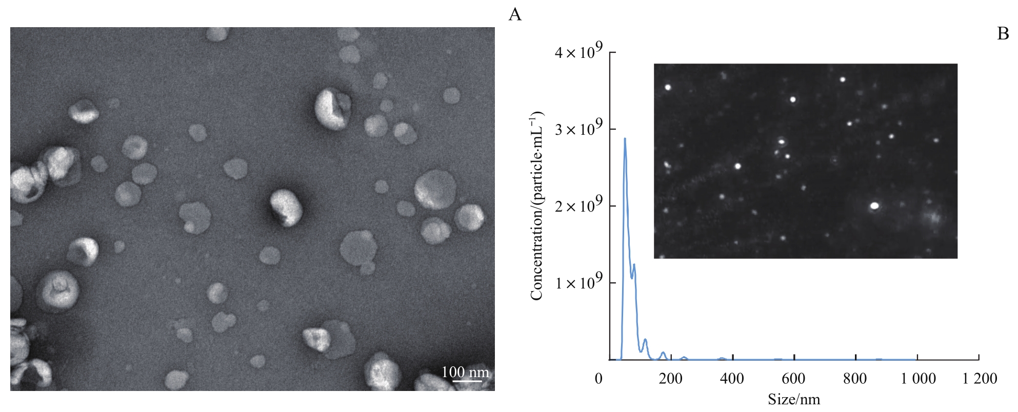
Fig 1 Identification of sEVs from BMSCs by TEM and NTA
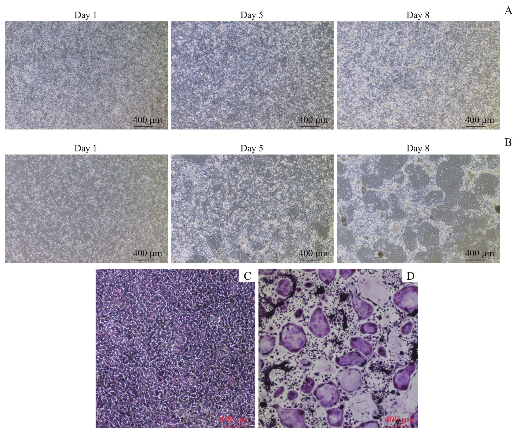
Fig 2 Formation of osteoclasts and the results of TRAP staining
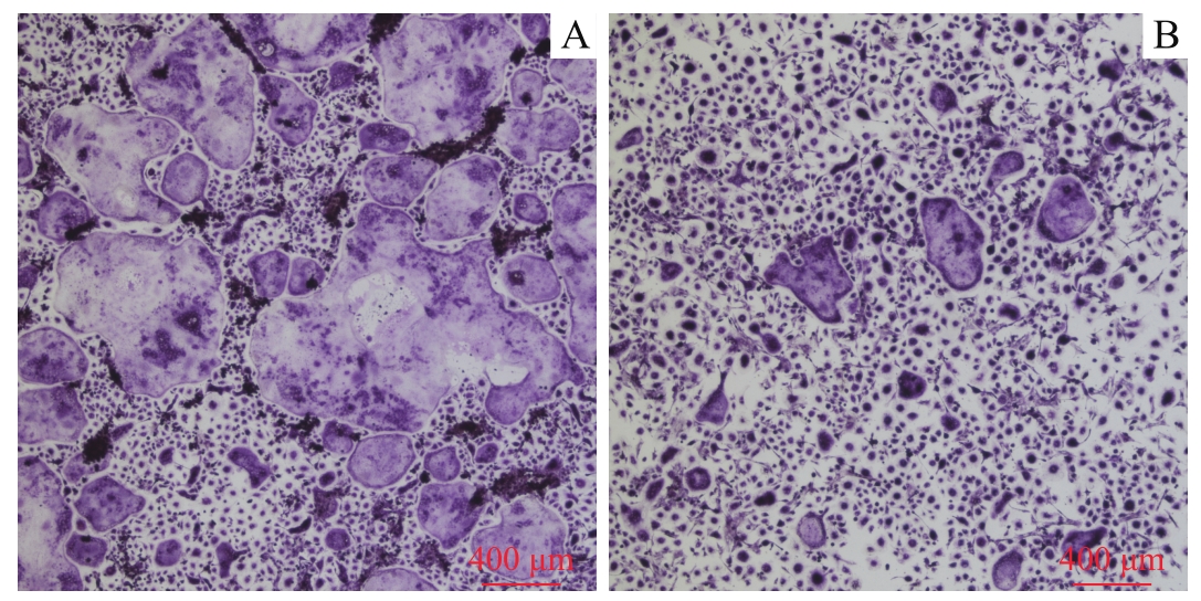
Fig 3 TRAP staining results of two groups of cells differentiated into osteoclasts (×40)
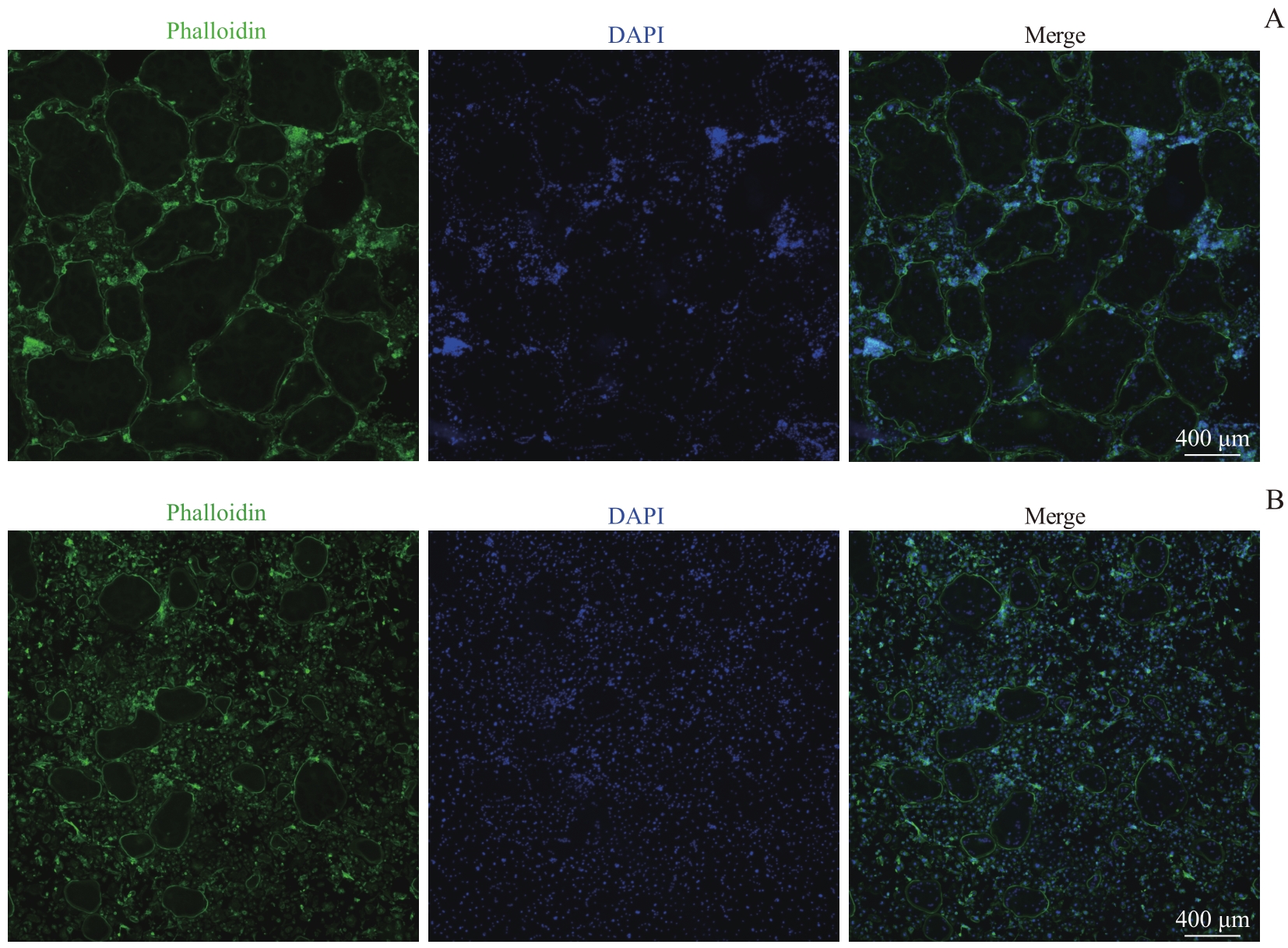
Fig 4 Phalloidin staining results of two groups of cells differentiated into osteoclasts (×40)
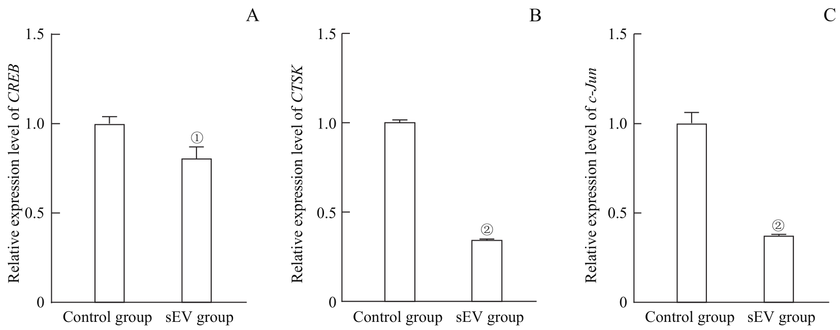
Fig 5 mRNA levels of CREB (A), CTSK (B) and c-Jun (C) detected by qPCR after the cells differentiated into osteoclasts in the two groups
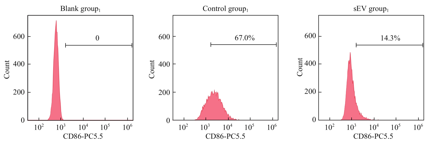
Fig 6 Expression level of M1 macrophage marker (CD86) detected by flow cytometry
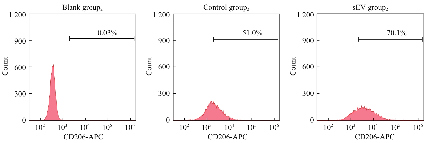
Fig 7 Expression level of M2 macrophage marker (CD206) detected by flow cytometry
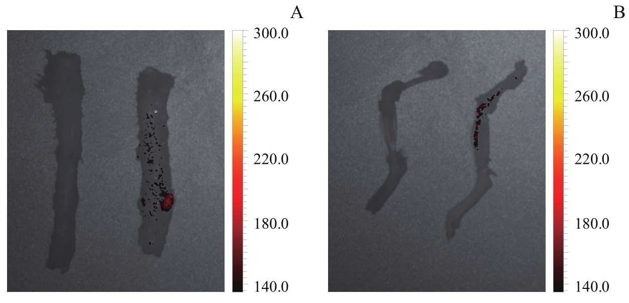
Fig 8 Observation of sEVs distribution in mouse bones by living imaging
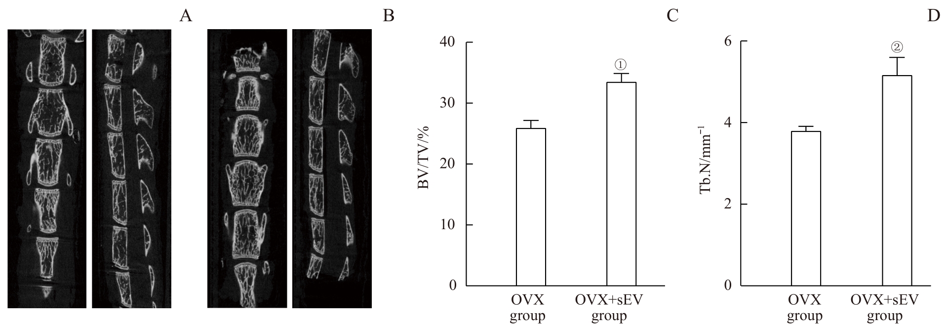
Fig 9 Micro-CT imaging analysis and bone parameters of the osteoporosis mice and the sEV-intervened mice
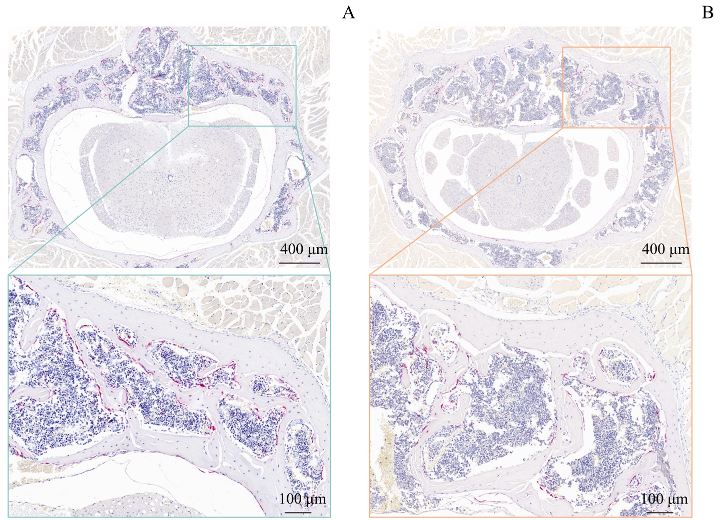
Fig 10 TRAP staining images of the osteoporosis mice and the sEV-intervened mice
| 1 | CHEATHAM S W, HANNEY W, KOLBER M, et al. Osteoporosis: exercise programming insight for the sports medicine professional[J]. Strength Cond J, 2017, 39: 2-13. |
| 2 | SI L, WINZENBERG T M, JIANG Q, et al. Projection of osteoporosis-related fractures and costs in China: 2010‒2050[J]. Osteoporos Int, 2015, 26(7): 1929-1937. |
| 3 | YU F, XIA W B. The epidemiology of osteoporosis, associated fragility fractures, and management gap in China[J]. Arch Osteoporos, 2019, 14(1): 32. |
| 4 | LEE C W, LIN H C, WANG B Y, et al. Ginkgolide B monotherapy reverses osteoporosis by regulating oxidative stress-mediated bone homeostasis[J]. Free Radic Biol Med, 2021, 168: 234-246. |
| 5 | LIU P, LEE S, KNOLL J, et al. Loss of menin in osteoblast lineage affects osteocyte-osteoclast crosstalk causing osteoporosis[J]. Cell Death Differ, 2017, 24(4): 672-682. |
| 6 | LEE K M, LEE C Y, ZHANG G, et al. Methylglyoxal activates osteoclasts through JNK pathway leading to osteoporosis[J]. Chem Biol Interact, 2019, 308: 147-154. |
| 7 | BARSONY J, XU Q, VERBALIS J G. Hyponatremia elicits gene expression changes driving osteoclast differentiation and functions[J]. Mol Cell Endocrinol, 2022, 554: 111724. |
| 8 | JACOME-GALARZA C E, PERCIN G I, MULLER J T, et al. Developmental origin, functional maintenance and genetic rescue of osteoclasts[J]. Nature, 2019, 568(7753): 541-545. |
| 9 | PESCE VIGLIETTI A I, GIAMBARTOLOMEI G H, DELPINO M V. Endocrine modulation of Brucella abortus-infected osteocytes function and osteoclastogenesis via modulation of RANKL/OPG[J]. Microbes Infect, 2019, 21(7): 287-295. |
| 10 | KIM J M, LIN C J, STAVRE Z, et al. Osteoblast-osteoclast communication and bone homeostasis[J]. Cells, 2020, 9(9): 2073. |
| 11 | QIAN J, HE Y, ZHAO J, et al. IL4/IL4R signaling promotes the osteolysis in metastatic bone of CRC through regulating the proliferation of osteoclast precursors[J]. Mol Med, 2021, 27(1): 152. |
| 12 | YAO Z, GETTING S J, LOCKE I C. Regulation of TNF-induced osteoclast differentiation[J]. Cells, 2021, 11(1): 132. |
| 13 | KANG M Y, HUANG C C, LU Y, et al. Bone regeneration is mediated by macrophage extracellular vesicles[J]. Bone, 2020, 141: 115627. |
| 14 | CAI F Y, LIU S L, LEI Y X, et al. Epigallocatechin-3 gallate regulates macrophage subtypes and immunometabolism to ameliorate experimental autoimmune encephalomyelitis[J]. Cell Immunol, 2021, 368: 104421. |
| 15 | ZHANG Z G, ZHANG C Y, ZHANG S R. Irisin activates M1 macrophage and suppresses Th2-type immune response in rats with pelvic inflammatory disease[J]. Evid Based Complement Alternat Med, 2022, 2022: 5215915. |
| 16 | EOM J, YOO J, KIM J J, et al. Viperin deficiency promotes polarization of macrophages and secretion of M1 and M2 cytokines[J]. Immune Netw, 2018, 18(4): e32. |
| 17 | ZHANG W J, GUAN N, ZHANG X M, et al. Study on the imbalance of M1/M2 macrophage polarization in severe chronic periodontitis[J]. Technol Health Care, 2023, 31(1): 117-124. |
| 18 | WANG W H, LIU H, LIU T, et al. Insights into the role of macrophage polarization in the pathogenesis of osteoporosis[J]. Oxid Med Cell Longev, 2022, 2022: 2485959. |
| 19 | YU L, HU M, CUI X, et al. M1 macrophage-derived exosomes aggravate bone loss in postmenopausal osteoporosis via a microRNA-98/DUSP1/JNK axis[J]. Cell Biol Int, 2021, 45(12): 2452-2463. |
| 20 | LU Y P, LIU S S, YANG P P, et al. Exendin-4 and eldecalcitol synergistically promote osteogenic differentiation of bone marrow mesenchymal stem cells through M2 macrophages polarization via PI3K/AKT pathway[J]. Stem Cell Res Ther, 2022, 13(1): 113. |
| 21 | CHEN M, LIN W M, YE R, et al. PPARβ/δ agonist alleviates diabetic osteoporosis via regulating M1/M2 macrophage polarization[J]. Front Cell Dev Biol, 2021, 9: 753194. |
| 22 | WEI H, CHEN Q, LIN L, et al. Regulation of exosome production and cargo sorting[J]. Int J Biol Sci, 2021, 17(1): 163-177. |
| 23 | LI M D, JIA J, LI S S, et al. Exosomes derived from tendon stem cells promote cell proliferation and migration through the TGF β signal pathway[J]. Biochem Biophys Res Commun, 2021, 536: 88-94. |
| 24 | WANG S W, JU T Y, WANG J J, et al. Migration of BEAS-2B cells enhanced by H1299 cell derived-exosomes[J]. Micron, 2021, 143: 103001. |
| 25 | SHARIATI NAJAFABADI S, AMIRPOUR N, AMINI S, et al. Human adipose derived stem cell exosomes enhance the neural differentiation of PC12 cells[J]. Mol Biol Rep, 2021, 48(6): 5033-5043. |
| 26 | YANG S D, GUO S, TONG S, et al. Promoting osteogenic differentiation of human adipose-derived stem cells by altering the expression of exosomal miRNA[J]. Stem Cells Int, 2019, 2019: 1351860. |
| 27 | ZHANG B B, ZHAXI D W, LI C, et al. M2 macrophagy-derived exosomal miRNA-26a-5p induces osteogenic differentiation of bone mesenchymal stem cells[J]. J Orthop Surg Res, 2022, 17(1): 137. |
| 28 | WEN X, HU G, XIAO X, et al. FGF2 positively regulates osteoclastogenesis via activating the ERK-CREB pathway[J]. Arch Biochem Biophys, 2022, 727: 109348. |
| 29 | ZHU G C, CHEN W, TANG C Y, et al. Knockout and double knockout of cathepsin K and Mmp9 reveals a novel function of cathepsin K as a regulator of osteoclast gene expression and bone homeostasis[J]. Int J Biol Sci, 2022, 18(14): 5522-5538. |
| 30 | HE F T, LUO S H, LIU S J, et al. Zanthoxylum bungeanum seed oil inhibits RANKL-induced osteoclastogenesis by suppressing ERK/c-JUN/NFATc1 pathway and regulating cell cycle arrest in RAW264.7 cells[J]. J Ethnopharmacol, 2022, 289: 115094. |
| 31 | KUMAR A, HUGHES T M, CRAFT S, et al. A novel approach to isolate brain-cell-derived exosomes from plasma to better understand pathogenesis of Alzheimer's disease[J]. Alzheimer's Dement, 2020, 16(Suppl 4): e044894. |
| 32 | LI K, WONG D K, HONG K Y, et al. Cushioned-density gradient ultracentrifugation (C-DGUC): a refined and high performance method for the isolation, characterization, and use of exosomes[J]. Methods Mol Biol, 2018, 1740: 69-83. |
| 33 | HELWA I, CAI J W, DREWRY M D, et al. A comparative study of serum exosome isolation using differential ultracentrifugation and three commercial reagents[J]. PLoS One, 2017, 12(1): e0170628. |
| 34 | DING M, WANG C, LU X L, et al. Comparison of commercial exosome isolation kits for circulating exosomal microRNA profiling[J]. Anal Bioanal Chem, 2018, 410(16): 3805-3814. |
| 35 | LIANG B, BURLEY G, LIN S, et al. Osteoporosis pathogenesis and treatment: existing and emerging avenues[J]. Cell Mol Biol Lett, 2022, 27(1): 72. |
| 36 | LI K, XIU C M, ZHOU Q, et al. A dual role of cholesterol in osteogenic differentiation of bone marrow stromal cells[J]. J Cell Physiol, 2019, 234(3): 2058-2066. |
| 37 | CHE Y T, YANG J Z, TANG F, et al. New function of cholesterol oxidation products involved in osteoporosis pathogenesis[J]. Int J Mol Sci, 2022, 23(4): 2020. |
| 38 | LI K Q, CHEN S H, CAI P Y, et al. MiRNA-483-5p is involved in the pathogenesis of osteoporosis by promoting osteoclast differentiation[J]. Mol Cell Probes, 2020, 49: 101479. |
| 39 | PARK E, LEE C G, LIM E, et al. Osteoprotective effects of loganic acid on osteoblastic and osteoclastic cells and osteoporosis-induced mice[J]. Int J Mol Sci, 2020, 22(1): 233. |
| 40 | LAI G H, ZHAO R L, ZHUANG W D, et al. BMSC-derived exosomal miR-27a-3p and miR-196b-5p regulate bone remodeling in ovariectomized rats[J]. PeerJ, 2022, 10: e13744. |
| 41 | SONG H Y, LI X Q, ZHAO Z C, et al. Reversal of osteoporotic activity by endothelial cell-secreted bone targeting and biocompatible exosomes[J]. Nano Lett, 2019, 19(5): 3040-3048. |
| 42 | CHEN X T, WAN Z, YANG L, et al. Exosomes derived from reparative M2-like macrophages prevent bone loss in murine periodontitis models via IL-10 mRNA[J]. J Nanobiotechnology, 2022, 20(1): 110. |
| 43 | ZHU L F, LI L, WANG X Q, et al. M1 macrophages regulate TLR4/AP1 via paracrine to promote alveolar bone destruction in periodontitis[J]. Oral Dis, 2019, 25(8): 1972-1982. |
| 44 | LIANG B L, WANG H C, WU D, et al. Macrophage M1/M2 polarization dynamically adapts to changes in microenvironment and modulates alveolar bone remodeling after dental implantation[J]. J Leukoc Biol, 2021, 110(3): 433-447. |
| 45 | SHI M S, WANG C, WANG Y L, et al. Deproteinized bovine bone matrix induces osteoblast differentiation via macrophage polarization[J]. J Biomed Mater Res A, 2018, 106(5): 1236-1246. |
| 46 | SHI C, YUAN F, LI Z L, et al. MSN@IL-4 sustainingly mediates macrophagocyte M2 polarization and relieves osteoblast damage via NF-κB pathway-associated apoptosis[J]. Biomed Res Int, 2022, 2022: 2898729. |
| 47 | Horibe K, Hara M, Nakamura H. M2-like macrophage infiltration and transforming growth factor-β secretion during socket healing process in mice[J]. Arch Oral Biol, 2021, 123: 105042. |
| 48 | WANG X Y, JI Q B, HU W H, et al. Isobavachalcone prevents osteoporosis by suppressing activation of ERK and NF-κB pathways and M1 polarization of macrophages[J]. Int Immunopharmacol, 2021, 94: 107370. |
| 49 | LI Z K, ZHU X D, XU R J, et al. Deacylcynaropicrin inhibits RANKL-induced osteoclastogenesis by inhibiting NF-κB and MAPK and promoting M2 polarization of macrophages[J]. Front Pharmacol, 2019, 10: 599. |
| 50 | YAO M Y, CUI B, ZHANG W H, et al. Exosomal miR-21 secreted by IL-1β-primed-mesenchymal stem cells induces macrophage M2 polarization and ameliorates sepsis[J]. Life Sci, 2021, 264: 118658. |
| 51 | MA J, CHEN L, ZHU X, et al. Mesenchymal stem cell-derived exosomal miR-21a-5p promotes M2 macrophage polarization and reduces macrophage infiltration to attenuate atherosclerosis[J]. Acta Biochim Biophys Sin (Shanghai), 2021, 53(9): 1227-1236. |
| 52 | LI R, LI D Z, WANG H N, et al. Exosomes from adipose-derived stem cells regulate M1/M2 macrophage phenotypic polarization to promote bone healing via miR-451a/MIF[J]. Stem Cell Res Ther, 2022, 13(1): 149. |
| 53 | LI R, ZHAO K C, RUAN Q, et al. Bone marrow mesenchymal stem cell-derived exosomal microRNA-124-3p attenuates neurological damage in spinal cord ischemia-reperfusion injury by downregulating Ern1 and promoting M2 macrophage polarization[J]. Arthritis Res Ther, 2020, 22(1): 75. |
| [1] | LIU Chenjun, YIN Bohao, SUN Hui, ZHANG Wei. Application of non-invasive methods of radiology to the osteoporosis [J]. Journal of Shanghai Jiao Tong University (Medical Science), 2023, 43(3): 385-390. |
| [2] | Hong-yan XUAN, Li-hua WANG, Hua-fang LI. Review of the factors influencing bone metabolism in schizophrenia [J]. JOURNAL OF SHANGHAI JIAOTONG UNIVERSITY (MEDICAL SCIENCE), 2021, 41(7): 972-976. |
| [3] | Miao-miao CAI, Yan-hong GAO. Research progress of osteosarcopenia [J]. JOURNAL OF SHANGHAI JIAOTONG UNIVERSITY (MEDICAL SCIENCE), 2021, 41(5): 678-683. |
| [4] | LUO Hong1, 2, WU Hong-yan1, 3, TAN Xi1, 3, DAI Hong-wei1, 2, 3, HUANG Lan1, 2, 3. Study on the difference of orthodontic tooth movement rate and pressure side bone remodeling between obese and obesity-resistant rats [J]. JOURNAL OF SHANGHAI JIAOTONG UNIVERSITY (MEDICAL SCIENCE), 2020, 40(8): 1055-1062. |
| [5] | WANG Feng-wei, SHEN Qiu-ming, SHI Yue, ZHANG Shu-xian, WANG Hu-wen, CHANG Rui-jie, YANG Ying-hua, WAN He-ping, SHEN Tian, CAI Yong. Analysis on the prevention of osteoporosis in middle-aged and elderly residents of Shanghai community [J]. , 2020, 40(4): 525-. |
| [6] | CUI Ya-qi, BAI Yu-bing, XU Yi-chen, TAN Xin-chen, LI Meng-ying, JIA Hao. Research progress of adipose-derived mesenchymal stem cell and its extracellular vesicles in osteogenesis [J]. JOURNAL OF SHANGHAI JIAOTONG UNIVERSITY (MEDICAL SCIENCE), 2020, 40(12): 1672-1676. |
| [7] | YANG Yi-qi, TANG Ting-ting. SIRT1 signaling pathway in bone metabolism [J]. , 2019, 39(11): 1335-. |
| [8] | LI Zi-lin1, GU Wen-qing2, SHEN Tian1. Advances in research on relationship between osteoporosis and intestinal microbe [J]. , 2019, 39(10): 1214-. |
| [9] | ZHAO Jing-yu, HUANG Ming-jian, ZHANG Xiao-ling. Effects of estrogen on proliferation, apoptosis and differentiation of bone marrow macrophage [J]. , 2018, 38(7): 745-. |
| [10] | SHI Yue1, WANG Ze-zhou1, SHEN Qiu-ming1, Lhakpa Tsamlag1, WAN He-ping2, YANG Ying-hua3, SHEN Tian1, CAI Yong1. Validity and reliability of osteoporosis prevention and control behavior scale for health care workers in community [J]. , 2018, 38(4): 439-. |
| [11] | WANG Jun1*, LIANG Jing1*, HE Yi-feng2, 3. A preliminary study on the impact of Notch1 on RANKL/RANK system in osteoclast via RAW264.7 cells [J]. , 2018, 38(12): 1440-. |
| [12] | YANG Qian-hao, ZHU Dao-yu, CHEN Yi-xuan, GAO You-shui, ZHANG Chang-qing. Research progress of roles of mammalian target of rapamycin signaling in bone homeostasis and associated diseases [J]. , 2018, 38(11): 1391-. |
| [13] | KONG Xiang-xin, LIU Hui-fang, CHEN Feng-ling. CaMKKβ promotes mouse macrophage M2 polarization by activating AMPK/JAK2/STAT3 signaling [J]. , 2017, 37(7): 914-. |
| [14] | XU Zi-jun, CUI Ying-chao, Lü Cheng, WU Ying-yan, CAI Yong, SHEN Tian. Health behavior intervention models and theories for osteoporosis patients [J]. , 2017, 37(1): 118-. |
| [15] | HU Yan, GAO Yan-hong. Research progress of bone-targeting estrogen-like drugs in treatment of osteoporosis [J]. , 2016, 36(3): 437-. |
| Viewed | ||||||
|
Full text |
|
|||||
|
Abstract |
|
|||||