Journal of Shanghai Jiao Tong University (Medical Science) ›› 2024, Vol. 44 ›› Issue (5): 552-559.doi: 10.3969/j.issn.1674-8115.2024.05.002
• High-risk?pregnancy column • Previous Articles
ZHOU Tianfan1( ), SHAO Feixue1, WAN Sheng1, ZHOU Chenchen1, ZHOU Sijin2, HUA Xiaolin1()
Received:2023-12-21
Accepted:2024-05-08
Online:2024-05-28
Published:2024-05-28
Contact:
HUA Xiaolin
E-mail:zhoutf9789@126.com;xiaolin_hua@tongji.edu.cn
Supported by:CLC Number:
ZHOU Tianfan, SHAO Feixue, WAN Sheng, ZHOU Chenchen, ZHOU Sijin, HUA Xiaolin. Feasibility study on quantifying retinal vascular features for predicting preeclampsia based on artificial intelligence models[J]. Journal of Shanghai Jiao Tong University (Medical Science), 2024, 44(5): 552-559.
Add to citation manager EndNote|Ris|BibTeX
URL: https://xuebao.shsmu.edu.cn/EN/10.3969/j.issn.1674-8115.2024.05.002
| Characteristic | Unaffected group (n=685) | HDP group (n=104) | Pc value | |||
|---|---|---|---|---|---|---|
| GH group (n=36) | Pa value | PE group (n=68) | Pb value | |||
| Age/year | 31.00 (29.00, 34.00) | 32.00 (30.00, 34.00) | 0.307 | 32.00 (29.00, 35.00) | 0.153 | 0.234 |
| Pre-pregnancy BMI/(kg·m-2) | 22.43 (20.32, 25.15) | 22.72 (21.04, 27.29) | 0.194 | 23.87 (20.77, 26.66) | 0.013 | 0.025 |
| Previous PE history/n(%) | 5 (0.73) | 1 (2.78) | 0.265 | 0 (0) | 1.000 | 0.317 |
| Primiparity/n(%) | 493 (71.97) | 29 (80.56) | 0.339 | 54 (79.41) | 0.203 | 0.270 |
| ART/n(%) | 51 (7.45) | 2 (5.56) | 1.000 | 12 (17.65) | 0.009 | 0.021 |
| CH/n(%) | 14 (2.04) | 1 (2.78) | 0.540 | 8 (11.76) | 0.000 | 0.000 |
| Renal disease/n(%) | 3 (0.44) | 0 (0) | 1.000 | 2 (2.94) | 0.067 | 0.080 |
| AD/n(%) | 29 (4.23) | 2 (5.56) | 0.664 | 5 (7.35) | 0.222 | 0.318 |
| PGDM/n(%) | 4 (0.58) | 1 (2.78) | 0.227 | 3 (4.41) | 0.019 | 0.016 |
| GDM/n(%) | 76 (11.09) | 7 (19.44) | 0.173 | 9 (13.24) | 0.550 | 0.252 |
| PCOS/n(%) | 10 (1.46) | 1 (2.78) | 0.443 | 0 (0) | 0.612 | 0.436 |
| Second-trimester MAP/mmHg | 82.67 (76.33, 88.33) | 88.17 (85.67, 96.33) | 0.000 | 91.33 (84.67, 95.33) | 0.000 | 0.000 |
| Second-trimester EFW/g | 1 445.89 (1 320.37, 1 566.41) | 1 443.56 (1 341.89, 1 583.57) | 0.604 | 1 347.73 (1 218.09, 1 511.01) | 0.003 | 0.009 |
| Delivery method/n(%) | 0.167 | 0.000 | 0.000 | |||
| Spontaneous delivery | 393 (57.37) | 16 (44.44) | 20 (29.41) | |||
| Caesarean section | 292 (42.63) | 20 (55.56) | 48 (70.59) | |||
| Gestational weeks/week | 39.43 (38.57, 40.14) | 39.00 (38.29, 39.71) | 0.028 | 37.64 (36.04, 39.00) | 0.000 | 0.000 |
| PPH/n(%) | 19 (2.77) | 1 (2.78) | 1.000 | 4 (5.88) | 0.146 | 0.283 |
| Postpartum hospitalization days/n(%) | 3.00 (3.00, 4.00) | 4.00 (3.00, 5.00) | 0.001 | 6.00 (5.00, 7.00) | 0.000 | 0.000 |
| Fetal birth weight/g | 3 380.00 (3 115.00, 3 670.00) | 3 407.50 (3 135.00, 3 655.00) | 0.725 | 3 005.00 (2 446.25, 3 292.50) | 0.000 | 0.000 |
| 1 min Apgar score/score | 9.00 (9.00, 10.00) | 9.00 (9.00, 10.00) | 0.964 | 9.00 (9.00, 9.00) | 0.000 | 0.000 |
| 5 min Apgar score/score | 10.00 (10.00, 10.00) | 10.0 (10.00, 10.00) | 0.029 | 10.00 (9.00, 10.00) | 0.000 | 0.000 |
Tab 1 Comparison of clinical data of pregnant women and neonatal outcome data
| Characteristic | Unaffected group (n=685) | HDP group (n=104) | Pc value | |||
|---|---|---|---|---|---|---|
| GH group (n=36) | Pa value | PE group (n=68) | Pb value | |||
| Age/year | 31.00 (29.00, 34.00) | 32.00 (30.00, 34.00) | 0.307 | 32.00 (29.00, 35.00) | 0.153 | 0.234 |
| Pre-pregnancy BMI/(kg·m-2) | 22.43 (20.32, 25.15) | 22.72 (21.04, 27.29) | 0.194 | 23.87 (20.77, 26.66) | 0.013 | 0.025 |
| Previous PE history/n(%) | 5 (0.73) | 1 (2.78) | 0.265 | 0 (0) | 1.000 | 0.317 |
| Primiparity/n(%) | 493 (71.97) | 29 (80.56) | 0.339 | 54 (79.41) | 0.203 | 0.270 |
| ART/n(%) | 51 (7.45) | 2 (5.56) | 1.000 | 12 (17.65) | 0.009 | 0.021 |
| CH/n(%) | 14 (2.04) | 1 (2.78) | 0.540 | 8 (11.76) | 0.000 | 0.000 |
| Renal disease/n(%) | 3 (0.44) | 0 (0) | 1.000 | 2 (2.94) | 0.067 | 0.080 |
| AD/n(%) | 29 (4.23) | 2 (5.56) | 0.664 | 5 (7.35) | 0.222 | 0.318 |
| PGDM/n(%) | 4 (0.58) | 1 (2.78) | 0.227 | 3 (4.41) | 0.019 | 0.016 |
| GDM/n(%) | 76 (11.09) | 7 (19.44) | 0.173 | 9 (13.24) | 0.550 | 0.252 |
| PCOS/n(%) | 10 (1.46) | 1 (2.78) | 0.443 | 0 (0) | 0.612 | 0.436 |
| Second-trimester MAP/mmHg | 82.67 (76.33, 88.33) | 88.17 (85.67, 96.33) | 0.000 | 91.33 (84.67, 95.33) | 0.000 | 0.000 |
| Second-trimester EFW/g | 1 445.89 (1 320.37, 1 566.41) | 1 443.56 (1 341.89, 1 583.57) | 0.604 | 1 347.73 (1 218.09, 1 511.01) | 0.003 | 0.009 |
| Delivery method/n(%) | 0.167 | 0.000 | 0.000 | |||
| Spontaneous delivery | 393 (57.37) | 16 (44.44) | 20 (29.41) | |||
| Caesarean section | 292 (42.63) | 20 (55.56) | 48 (70.59) | |||
| Gestational weeks/week | 39.43 (38.57, 40.14) | 39.00 (38.29, 39.71) | 0.028 | 37.64 (36.04, 39.00) | 0.000 | 0.000 |
| PPH/n(%) | 19 (2.77) | 1 (2.78) | 1.000 | 4 (5.88) | 0.146 | 0.283 |
| Postpartum hospitalization days/n(%) | 3.00 (3.00, 4.00) | 4.00 (3.00, 5.00) | 0.001 | 6.00 (5.00, 7.00) | 0.000 | 0.000 |
| Fetal birth weight/g | 3 380.00 (3 115.00, 3 670.00) | 3 407.50 (3 135.00, 3 655.00) | 0.725 | 3 005.00 (2 446.25, 3 292.50) | 0.000 | 0.000 |
| 1 min Apgar score/score | 9.00 (9.00, 10.00) | 9.00 (9.00, 10.00) | 0.964 | 9.00 (9.00, 9.00) | 0.000 | 0.000 |
| 5 min Apgar score/score | 10.00 (10.00, 10.00) | 10.0 (10.00, 10.00) | 0.029 | 10.00 (9.00, 10.00) | 0.000 | 0.000 |
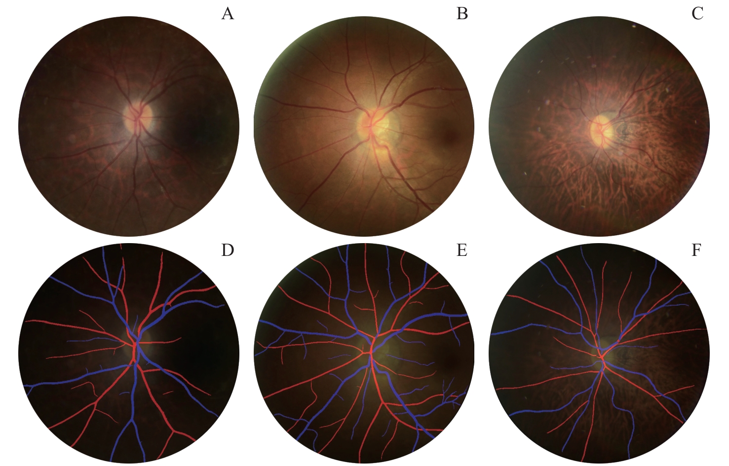
Fig 1 Typical fundus images and schematic diagrams of segmentation of retinal blood vessels of the three groups
| Characteristic | Unaffected group (n=685) | HDP group (n=104) | Pc value | |||
|---|---|---|---|---|---|---|
| GH group (n=36) | Pa value | PE group (n=68) | Pb value | |||
| Decreased elasticity of retinal arteries/n(%) | 196 (28.61) | 12 (33.33) | 0.572 | 23 (33.82) | 0.401 | 0.532 |
| Leopard pattern change/n(%) | 456 (66.57) | 23 (63.89) | 0.721 | 48 (70.59) | 0.589 | 0.746 |
| Arteriosclerosis/n(%) | 4 (0.58) | 1 (2.78) | 0.227 | 1 (1.47) | 0.378 | 0.181 |
| Vitreous warts/n(%) | 42 (6.13) | 2 (5.56) | 1.000 | 4 (5.88) | 1.000 | 1.000 |
| Retinal sporadic bleeding/n(%) | 7 (1.02) | 1 (2.78) | 0.338 | 1 (1.47) | 0.533 | 0.337 |
| CRAE | 94.00 (87.00, 99.00) | 89.00 (86.00, 95.00) | 0.150 | 87.00 (80.00, 94.00) | 0.000 | 0.000 |
| CRVE | 122.00 (116.00, 129.00) | 120.00 (116.00, 122.50) | 0.101 | 120.00 (111.00, 126.50) | 0.017 | 0.019 |
| AVR | 0.75 (0.71, 0.81) | 0.74 (0.70, 0.79) | 0.432 | 0.72 (0.67, 0.77) | 0.002 | 0.006 |
| Retinal artery tortuosity | 0.05 (0.04, 0.07) | 0.04 (0.04, 0.07) | 0.567 | 0.04 (0.03, 0.06) | 0.004 | 0.015 |
| Retinal vein tortuosity | 0.09 (0.07, 0.11) | 0.07 (0.06, 0.11) | 0.186 | 0.08 (0.06, 0.10) | 0.120 | 0.141 |
| Retinal artery fractal dimension | 1.48 (1.41, 1.54) | 1.47 (1.41, 1.51) | 0.245 | 1.45 (1.38, 1.51) | 0.003 | 0.007 |
| Retinal vein fractal dimension | 1.49 (1.42, 1.56) | 1.51 (1.45, 1.55) | 0.569 | 1.48 (1.44, 1.55) | 0.990 | 0.848 |
| VCDR | 0.28 (0.22, 0.34) | 0.29 (0.20, 0.33) | 0.688 | 0.27 (0.22, 0.34) | 0.522 | 0.764 |
| HCDR | 0.39 (0.31, 0.46) | 0.40 (0.31, 0.45) | 0.967 | 0.39 (0.31, 0.45) | 0.740 | 0.941 |
Tab 2 Comparison of fundus characteristics and retinal vascular characteristic parameters among the three groups of pregnant women
| Characteristic | Unaffected group (n=685) | HDP group (n=104) | Pc value | |||
|---|---|---|---|---|---|---|
| GH group (n=36) | Pa value | PE group (n=68) | Pb value | |||
| Decreased elasticity of retinal arteries/n(%) | 196 (28.61) | 12 (33.33) | 0.572 | 23 (33.82) | 0.401 | 0.532 |
| Leopard pattern change/n(%) | 456 (66.57) | 23 (63.89) | 0.721 | 48 (70.59) | 0.589 | 0.746 |
| Arteriosclerosis/n(%) | 4 (0.58) | 1 (2.78) | 0.227 | 1 (1.47) | 0.378 | 0.181 |
| Vitreous warts/n(%) | 42 (6.13) | 2 (5.56) | 1.000 | 4 (5.88) | 1.000 | 1.000 |
| Retinal sporadic bleeding/n(%) | 7 (1.02) | 1 (2.78) | 0.338 | 1 (1.47) | 0.533 | 0.337 |
| CRAE | 94.00 (87.00, 99.00) | 89.00 (86.00, 95.00) | 0.150 | 87.00 (80.00, 94.00) | 0.000 | 0.000 |
| CRVE | 122.00 (116.00, 129.00) | 120.00 (116.00, 122.50) | 0.101 | 120.00 (111.00, 126.50) | 0.017 | 0.019 |
| AVR | 0.75 (0.71, 0.81) | 0.74 (0.70, 0.79) | 0.432 | 0.72 (0.67, 0.77) | 0.002 | 0.006 |
| Retinal artery tortuosity | 0.05 (0.04, 0.07) | 0.04 (0.04, 0.07) | 0.567 | 0.04 (0.03, 0.06) | 0.004 | 0.015 |
| Retinal vein tortuosity | 0.09 (0.07, 0.11) | 0.07 (0.06, 0.11) | 0.186 | 0.08 (0.06, 0.10) | 0.120 | 0.141 |
| Retinal artery fractal dimension | 1.48 (1.41, 1.54) | 1.47 (1.41, 1.51) | 0.245 | 1.45 (1.38, 1.51) | 0.003 | 0.007 |
| Retinal vein fractal dimension | 1.49 (1.42, 1.56) | 1.51 (1.45, 1.55) | 0.569 | 1.48 (1.44, 1.55) | 0.990 | 0.848 |
| VCDR | 0.28 (0.22, 0.34) | 0.29 (0.20, 0.33) | 0.688 | 0.27 (0.22, 0.34) | 0.522 | 0.764 |
| HCDR | 0.39 (0.31, 0.46) | 0.40 (0.31, 0.45) | 0.967 | 0.39 (0.31, 0.45) | 0.740 | 0.941 |
| Characteristic | Univariate Logistic analysis | Multivariate Logistic analysis | ||||
|---|---|---|---|---|---|---|
| OR | 95% CI | P value | aOR | 95% CI | P value | |
| Second-trimester MAP | 1.110 | 1.076‒1.148 | 0.000 | 1.106 | 1.068‒1.147 | 0.000 |
| Second-trimester EFW | 0.702 | 0.571‒0.869 | 0.001 | 0.700 | 0.560‒0.870 | 0.000 |
| CRAE | 0.936 | 0.904‒0.965 | 0.000 | 0.940 | 0.910‒0.970 | 0.000 |
| CRVE | 0.972 | 0.952‒0.993 | 0.009 | 0.970 | 0.950‒0.990 | 0.010 |
| AVR | 0.010 | 0.000‒0.288 | 0.008 | 0.020 | 0.000‒0.500 | 0.020 |
| Retinal artery tortuosity | 0.000 | 0.000‒0.012 | 0.012 | 0.000 | 0.000‒0.050 | 0.020 |
| Retinal artery fractal dimension | 0.046 | 0.006‒0.393 | 0.004 | 0.070 | 0.010‒0.720 | 0.020 |
Tab 3 Univariate and multivariate Logistic regression analysis of the influencing factors of PE occurrence
| Characteristic | Univariate Logistic analysis | Multivariate Logistic analysis | ||||
|---|---|---|---|---|---|---|
| OR | 95% CI | P value | aOR | 95% CI | P value | |
| Second-trimester MAP | 1.110 | 1.076‒1.148 | 0.000 | 1.106 | 1.068‒1.147 | 0.000 |
| Second-trimester EFW | 0.702 | 0.571‒0.869 | 0.001 | 0.700 | 0.560‒0.870 | 0.000 |
| CRAE | 0.936 | 0.904‒0.965 | 0.000 | 0.940 | 0.910‒0.970 | 0.000 |
| CRVE | 0.972 | 0.952‒0.993 | 0.009 | 0.970 | 0.950‒0.990 | 0.010 |
| AVR | 0.010 | 0.000‒0.288 | 0.008 | 0.020 | 0.000‒0.500 | 0.020 |
| Retinal artery tortuosity | 0.000 | 0.000‒0.012 | 0.012 | 0.000 | 0.000‒0.050 | 0.020 |
| Retinal artery fractal dimension | 0.046 | 0.006‒0.393 | 0.004 | 0.070 | 0.010‒0.720 | 0.020 |
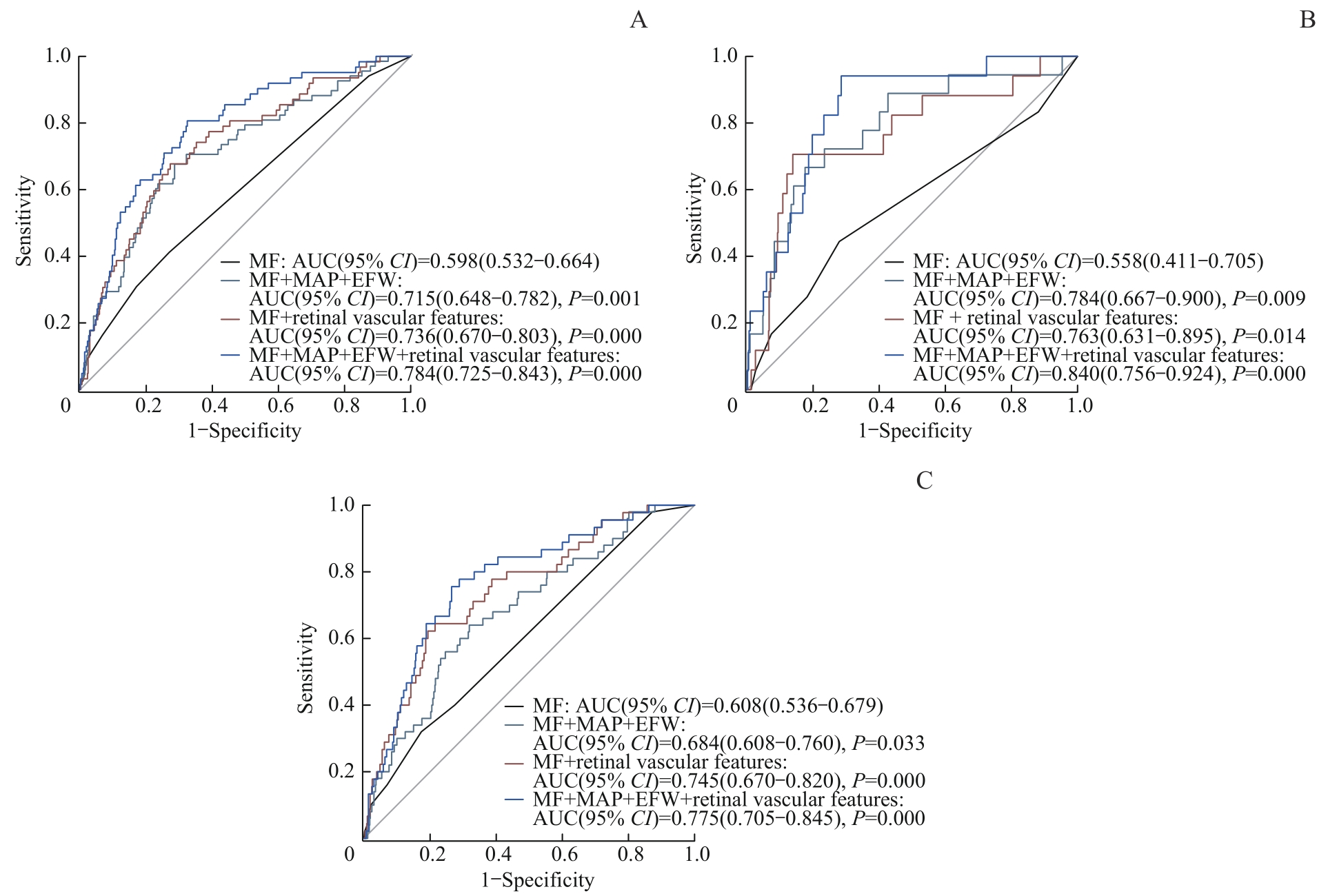
Fig 2 Analysis of ROC curves for predicting PE, early-onset PE and late-onset PE using different models
| 1 | RANA S, LEMOINE E, GRANGER J P, et al. Preeclampsia: pathophysiology, challenges, and perspectives[J]. Circ Res, 2019, 124(7): 1094-1112. |
| 2 | CHAPPELL L C, CLUVER C A, KINGDOM J, et al. Pre-eclampsia[J]. Lancet, 2021, 398(10297): 341-354. |
| 3 | Gestational Hypertension and Preeclampsia: ACOG Practice Bulletin, Number 222[J]. Obstet Gynecol, 2020, 135(6): e237-e260. |
| 4 | WRIGHT D, WRIGHT A, NICOLAIDES K H. The competing risk approach for prediction of preeclampsia[J]. Am J Obstet Gynecol, 2020, 223(1): 12-23. e7. |
| 5 | WRIGHT D, SYNGELAKI A, AKOLEKAR R, et al. Competing risks model in screening for preeclampsia by maternal characteristics and medical history[J]. Am J Obstet Gynecol, 2015, 213(1): 62.e1-62.e10. |
| 6 | TAN M Y, WRIGHT D, SYNGELAKI A, et al. Comparison of diagnostic accuracy of early screening for pre-eclampsia by NICE guidelines and a method combining maternal factors and biomarkers: results of SPREE[J]. Ultrasound Obstet Gynecol, 2020, 51(6): 743-750. |
| 7 | ZEISLER H, LLURBA E, CHANTRAINE F, et al. Predictive value of the sFlt-1: PLGF ratio in women with suspected preeclampsia[J]. N Engl J Med, 2016, 374(1): 13-22. |
| 8 | ACOG committee opinion No. 743: low-dose aspirin use during pregnancy[J]. Obstet Gynecol, 2018, 132(1): e44-e52. |
| 9 | ZHU Z T, CHEN Y F, WANG W, et al. Association of retinal age gap with arterial stiffness and incident cardiovascular disease[J]. Stroke, 2022, 53(11): 3320-3328. |
| 10 | WHELTON P K, CAREY R M, ARONOW W S, et al. 2017 ACC/AHA/AAPA/ABC/ACPM/AGS/APhA/ASH/ASPC/NMA/PCNA guideline for the prevention, detection, evaluation, and management of high blood pressure in adults: a report of the American College of Cardiology/American Heart Association Task Force on Clinical Practice Guidelines[J]. Hypertension, 2018, 71(6): 1269-1324. |
| 11 | ABU SAMRA K. The eye and visual system in the preeclampsia/eclampsia syndrome: what to expect?[J]. Saudi J Ophthalmol, 2013, 27(1): 51-53. |
| 12 | STERN E M, BLACE N. Ophthalmic pathology of preeclampsia[M]. Treasure Island (FL): StatPearls Publishing, 2022. |
| 13 | ZHOU L Q, WU X L, HUANG S Y, et al. Lymph node metastasis prediction from primary breast cancer US images using deep learning[J]. Radiology, 2020, 294(1): 19-28. |
| 14 | HADLOCK F P, HARRIST R B, SHARMAN R S, et al. Estimation of fetal weight with the use of head, body, and femur measurements: a prospective study[J]. Am J Obstet Gynecol, 1985, 151(3): 333-337. |
| 15 | 黄烨霖, 赵昕. 用于对视网膜眼底图像进行分析的方法及其相关产品: CN114359284B[P]. 2022-06-21. |
| HUANG Y L, ZHAO X. Method and related products for analyzing retinal fundus images: CN114359284B[P]. 2022-06-21. | |
| 16 | 黄烨霖, 史晓宇. 确定视网膜眼底血管的弯曲度的方法、设备和存储介质: CN115511883B[P]. 2023-04-18. |
| HUANG Y L, SHI X Y. Method, equipment, and storage medium for determining the curvature of retinal fundus blood vessels: CN115511883B[P]. 2023-04-18. | |
| 17 | 黄烨霖, 赵昕, 和超, 等. 一种训练眼底图像分割模型的方法以及动静脉分割方法: CN113920077A[P]. 2022-01-11. |
| HUANG Y L, ZHAO X, HE C, et al. A method for training fundus image segmentation models and arteriovenous segmentation method: CN113920077A [P]. 2022-01-11. | |
| 18 | 中华医学会妇产科学分会妊娠期高血压疾病学组. 妊娠期高血压疾病诊治指南(2020)[J]. 中华妇产科杂志, 2020, 55(4): 227-238. |
| Hypertensive Disorders in Pregnancy Subgroup, Chinese Society of Obstetrics and Gynecology, Chinese Medical Association. Diagnosis and treatment of hypertension and pre-eclampsia in pregnancy: a clinical practice guideline in China(2020)[J]. Chinese Journal of Obstetrics and Gynecology, 2020, 55(4): 227-238. | |
| 19 | STIRNEMANN J, VILLAR J, SALOMON L J, et al. International estimated fetal weight standards of the INTERGROWTH-21st Project[J]. Ultrasound Obstet Gynecol, 2017, 49(4): 478-486. |
| 20 | KALAFAT E, LAORETI A, KHALIL A, et al. Ophthalmic artery Doppler for prediction of pre-eclampsia: systematic review and meta-analysis[J]. Ultrasound Obstet Gynecol, 2018, 51(6): 731-737. |
| 21 | CILOGLU E, OKCU N T, DOGAN N Ç. Optical coherence tomography angiography findings in preeclampsia[J]. Eye, 2019, 33(12): 1946-1951. |
| 22 | CHAEMSAITHONG P, GIL M M, CHAIYASIT N, et al. Accuracy of placental growth factor alone or in combination with soluble fms-like tyrosine kinase-1 or maternal risk factors in detecting preeclampsia in asymptomatic women in the second and third trimesters: a systematic review and meta-analysis[J]. Am J Obstet Gynecol, 2023, 229(3): 222-247. |
| 23 | NICOLAIDES K H, SARNO M, WRIGHT A. Ophthalmic artery Doppler in the prediction of preeclampsia[J]. Am J Obstet Gynecol, 2022, 226(2S): S1098-S1101. |
| 24 | MCCARTHY F P, RYAN R M, CHAPPELL L C. Prospective biomarkers in preterm preeclampsia: a review[J]. Pregnancy Hypertens, 2018, 14: 72-78. |
| 25 | CHEUNG C Y, XU D J, CHENG C Y, et al. A deep-learning system for the assessment of cardiovascular disease risk via the measurement of retinal-vessel calibre[J]. Nat Biomed Eng, 2021, 5(6): 498-508. |
| [1] | MA Ruilin, LIU Yu, XU Guixiang, SHI Haoran, CUI Jianjian, YANG Zejun, MAO Yan, ZHAO Yin. Relationship between Doppler ultrasound parameters of uterine artery, umbilical artery, middle cerebral artery and placental vasculopathology and pregnancy outcome in preeclampsia rat model [J]. Journal of Shanghai Jiao Tong University (Medical Science), 2024, 44(5): 543-551. |
| [2] | MA Ben, ZHAO Cheng, SHU Yijun, DONG Ping. Application progress of CT radiomics in gastrointestinal stromal tumor [J]. Journal of Shanghai Jiao Tong University (Medical Science), 2023, 43(7): 923-930. |
| [3] | Xiaofeng WANG, Lu ZHOU, Leyu YAO, Fan HE, Haixia PENG, Daming YANG, Xiaolin HUANG. Effect of DCNN model-assisted colorectal polyp detection system on the detection of colorectal polyps by junior physicians [J]. JOURNAL OF SHANGHAI JIAOTONG UNIVERSITY (MEDICAL SCIENCE), 2022, 42(2): 205-210. |
| [4] | Fang ZHANG, Xiao-jin WANG, Jue MA, Yun-ting HE, Hao HE, Jing ZHAI, Yu-na GUO, Yan CHEN, Bing-shun WANG. Association between blood lipid profile in early pregnancy and risk of preeclampsia: a study based on real world data [J]. JOURNAL OF SHANGHAI JIAOTONG UNIVERSITY (MEDICAL SCIENCE), 2021, 41(4): 483-488. |
| [5] | RONG Wen-wen1, WANG Gang1, ZHU Qi-li2. Discussion on value of medical records-structured specialized disease database based on artificial intelligence in clinical research [J]. JOURNAL OF SHANGHAI JIAOTONG UNIVERSITY (MEDICAL SCIENCE), 2020, 40(7): 995-1000. |
| [6] | LU Yan-qiao, SHEN Lan, HE Ben. Application of artificial intelligence in assisted diagnosis and treatment of cardiovascular disease [J]. , 2020, 40(2): 259-. |
| Viewed | ||||||
|
Full text |
|
|||||
|
Abstract |
|
|||||