Journal of Shanghai Jiao Tong University (Medical Science) ›› 2026, Vol. 46 ›› Issue (1): 34-42.doi: 10.3969/j.issn.1674-8115.2026.01.004
• Basic research • Previous Articles Next Articles
Zhang Yue, Chen Qingjian( )
)
Received:2025-04-01
Accepted:2025-08-16
Online:2026-01-28
Published:2026-01-30
Contact:
Chen Qingjian
E-mail:chenqingjian2010@163.com
About author:First author contact:Zhang Yue was responsible for experimental design, experimental operations, and manuscript writing and revision. Chen Qingjian was responsible for project design, bioinformatics analysis, and manuscript revision. Both authors have read the last version of paper and consented to submission.
Supported by:CLC Number:
Zhang Yue, Chen Qingjian. Mass cytometry reveals prognostic immune microenvironment features in breast cancer[J]. Journal of Shanghai Jiao Tong University (Medical Science), 2026, 46(1): 34-42.
Add to citation manager EndNote|Ris|BibTeX
URL: https://xuebao.shsmu.edu.cn/EN/10.3969/j.issn.1674-8115.2026.01.004
| Metal label | Protein marker |
|---|---|
| 141Pr | SMA |
| 142Nd | CD14 |
| 144Nd | GAPDH |
| 145Nd | CD11C |
| 146Nd | CD16 |
| 147Sm | CD68 |
| 148Nd | CD15 |
| 149Sm | CD57 |
| 150Nd | CD25 |
| 152Sm | CD45 |
| 156Gd | PD1 |
| 158Gd | CDH1 |
| 159Tb | CD206 |
| 160Gd | CD24 |
| 162Dy | CD8a |
| 163Dy | CD83 |
| 164Dy | CD11A |
| 165Ho | CD4 |
| 166Er | HIF1A |
| 167Er | ALDH1 |
| 168Er | Ki67 |
| 169Tm | CD31 |
| 171Yb | CD27 |
| 172Yb | CD66b |
| 173Yb | CD62L |
| 176Yb | CD3 |
Tab 1 Panel for profiling breast cancer immune microenvironment
| Metal label | Protein marker |
|---|---|
| 141Pr | SMA |
| 142Nd | CD14 |
| 144Nd | GAPDH |
| 145Nd | CD11C |
| 146Nd | CD16 |
| 147Sm | CD68 |
| 148Nd | CD15 |
| 149Sm | CD57 |
| 150Nd | CD25 |
| 152Sm | CD45 |
| 156Gd | PD1 |
| 158Gd | CDH1 |
| 159Tb | CD206 |
| 160Gd | CD24 |
| 162Dy | CD8a |
| 163Dy | CD83 |
| 164Dy | CD11A |
| 165Ho | CD4 |
| 166Er | HIF1A |
| 167Er | ALDH1 |
| 168Er | Ki67 |
| 169Tm | CD31 |
| 171Yb | CD27 |
| 172Yb | CD66b |
| 173Yb | CD62L |
| 176Yb | CD3 |
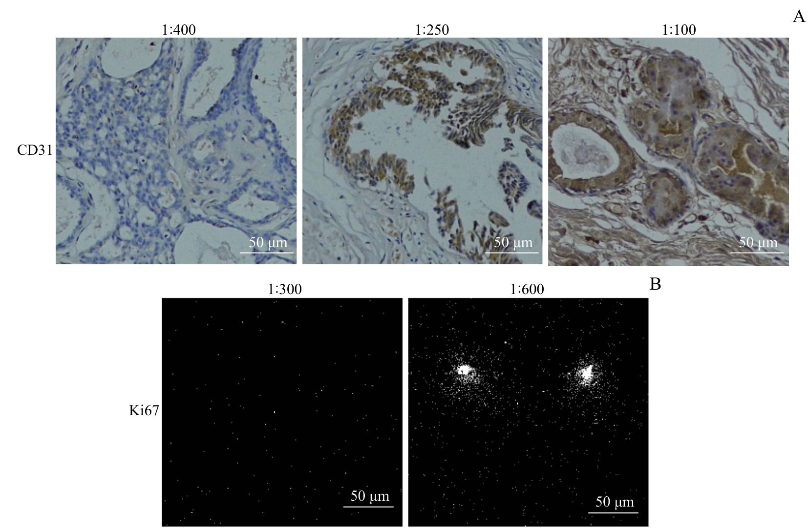
Fig 1 Comparison of pre-experimental effects of IHC and IMC before and after improving experimental conditions
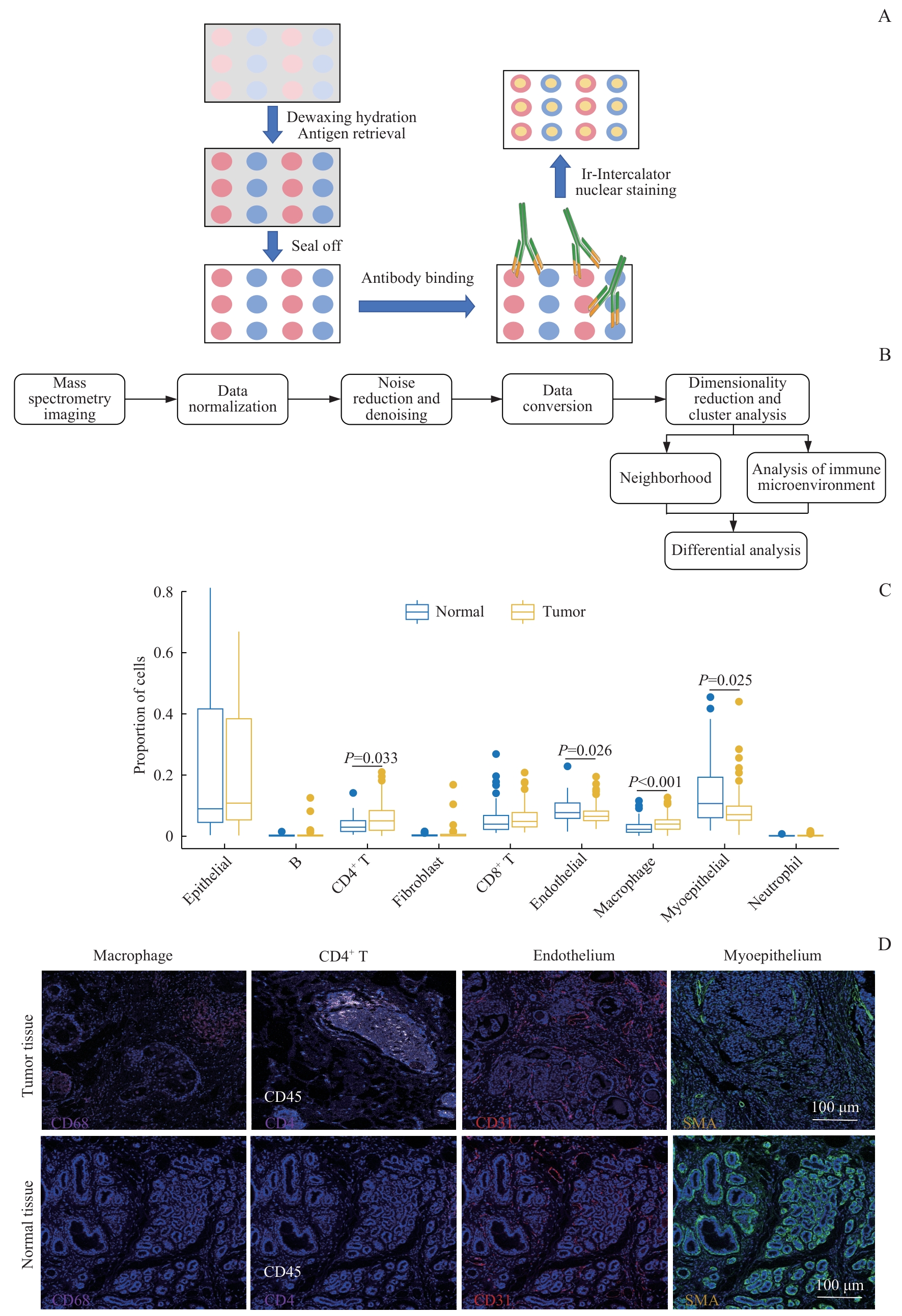
Fig 2 Analysis of composition of breast cancer immune microenvironment and spatial distribution of immune-related molecules
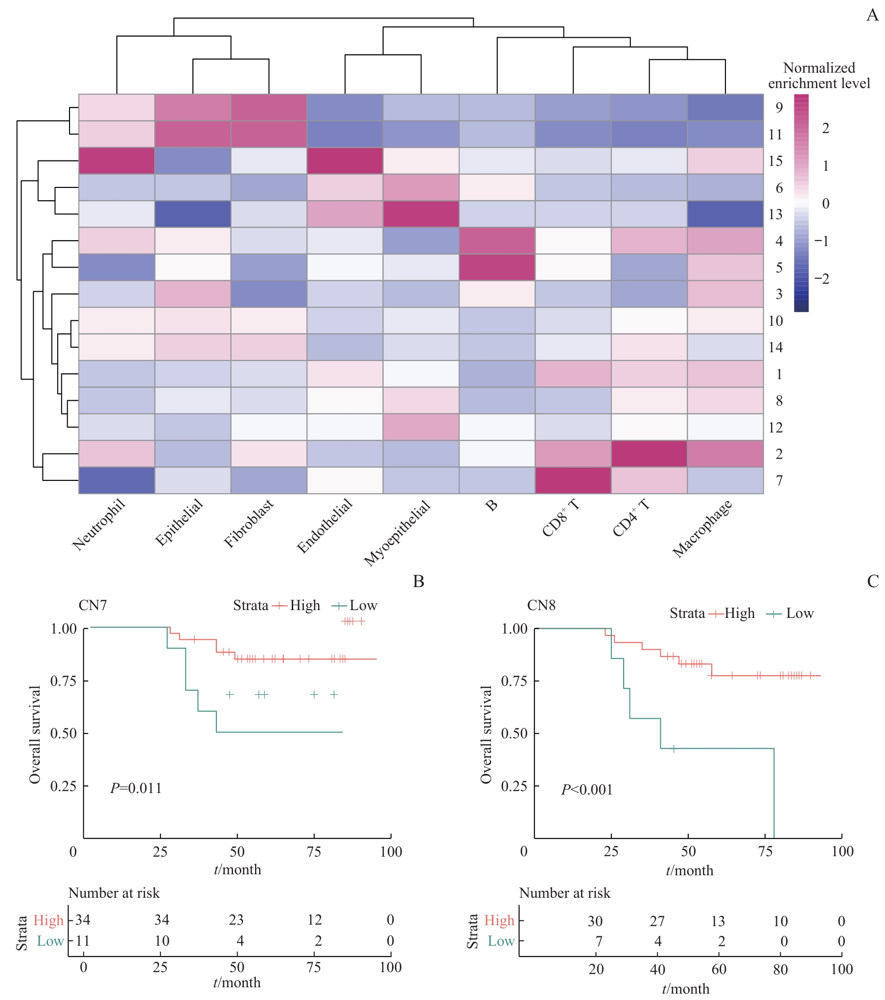
Fig 3 Functional analysis of the breast cancer immune microenvironment using the cell neighborhood method
| [1] | Dvir K, Giordano S, Leone J P. Immunotherapy in breast cancer[J]. Int J Mol Sci, 2024, 25(14): 7517. |
| [2] | Xiong X, Zheng L W, Ding Y, et al. Breast cancer: pathogenesis and treatments[J]. Signal Transduct Target Ther, 2025, 10(1): 49. |
| [3] | Vandereyken K, Sifrim A, Thienpont B, et al. Methods and applications for single-cell and spatial multi-omics[J]. Nat Rev Genet, 2023, 24(8): 494-515. |
| [4] | Pandey A, Bhutani N. Profiling joint tissues at single-cell resolution: advances and insights[J]. Nat Rev Rheumatol, 2024, 20(1): 7-20. |
| [5] | Rai M F, Wu C L, Capellini T D, et al. Single cell omics for musculoskeletal research[J]. Curr Osteoporos Rep, 2021, 19(2): 131-140. |
| [6] | Arnett L P, Rana R, Chung W W, et al. Reagents for mass cytometry[J]. Chem Rev, 2023, 123(3): 1166-1205. |
| [7] | Glasson Y, ChéPeaux L A, Dumé A S, et al. Single-cell high-dimensional imaging mass cytometry: one step beyond in oncology[J]. Semin Immunopathol, 2023, 45(1): 17-28. |
| [8] | Kante A, Chevalier M F, SèNe D, et al. Mass cytometry: exploring the immune landscape of systemic autoimmune and inflammatory diseases in the past fourteen years[J]. Front Immunol, 2024, 15: 1509782. |
| [9] | Le Rochais M, Hemon P, Pers J O, et al. Application of high-throughput imaging mass cytometry hyperion in cancer research[J]. Front Immunol, 2022, 13: 859414. |
| [10] | Naderi-Azad S, Croitoru D, Khalili S, et al. Research techniques made simple: experimental methodology for imaging mass cytometry[J]. J Invest Dermatol, 2021, 141(3): 467-473.e1. |
| [11] | Gray G K, Li C M, Rosenbluth J M, et al. A human breast atlas integrating single-cell proteomics and transcriptomics[J]. Dev Cell, 2022, 57(11): 1400-1420.e7. |
| [12] | Fan Y Y, Kao C Y, Yang F, et al. Integrated multi-omics analysis model to identify biomarkers associated with prognosis of breast cancer[J]. Front Oncol, 2022, 12: 899900. |
| [13] | Wang X, Chai Y Y, Quan Y, et al. NPM1 inhibits tumoral antigen presentation to promote immune evasion and tumor progression[J]. J Hematol Oncol, 2024, 17(1): 97. |
| [14] | Li Y, Jiang M L, Ling A Y, et al. UPP1 promotes lung adenocarcinoma progression through the induction of an immunosuppressive microenvironment[J]. Nat Commun, 2024, 15(1): 1200. |
| [15] | Liu H F, Zhao Q W, Tan L Y, et al. Neutralizing IL-8 potentiates immune checkpoint blockade efficacy for glioma[J]. Cancer Cell, 2023, 41(4): 693-710.e8. |
| [16] | Ma J L, Pang Y H, Shang Y F, et al. CyTOF analysis revealed platelet heterogeneity in breast cancer patients received T-DM1 treatment[J]. Clin Immunol, 2024, 263: 110227. |
| [17] | Lo Y C, Keyes T J, Jager A, et al. CytofIn enables integrated analysis of public mass cytometry datasets using generalized anchors[J]. Nat Commun, 2022, 13(1): 934. |
| [18] | Rybakowska P, Van Gassen S, Quintelier K, et al. Data processing workflow for large-scale immune monitoring studies by mass cytometry[J]. Comput Struct Biotechnol J, 2021, 19: 3160-3175. |
| [19] | Liu X, Song W C, Wong B Y, et al. A comparison framework and guideline of clustering methods for mass cytometry data[J]. Genome Biol, 2019, 20(1): 297. |
| [20] | Li X H, Zhang L, Liu C C, et al. Construction of mitochondrial quality regulation genes-related prognostic model based on bulk-RNA-seq analysis in multiple myeloma[J]. Biofactors, 2025, 51(1): e2135. |
| [21] | Artyomov M N, Van Den Bossche J. Immunometabolism in the single-cell era[J]. Cell Metab, 2020, 32(5): 710-725. |
| [22] | Peeters F, Cappuyns S, Piqué-Gili M, et al. Applications of single-cell multi-omics in liver cancer[J]. JHEP Rep, 2024, 6(7): 101094. |
| [23] | Rigamonti A, Viatore M, Polidori R, et al. Integrating AI-powered digital pathology and imaging mass cytometry identifies key classifiers of tumor cells, stroma, and immune cells in non-small cell lung cancer[J]. Cancer Res, 2024, 84(7): 1165-1177. |
| [24] | Xie P Y, Guo L, Yu Q, et al. ACE2 enhances sensitivity to PD-L1 blockade by inhibiting macrophage-induced immunosuppression and angiogenesis[J]. Cancer Res, 2025, 85(2): 299-313. |
| [25] | Brightman S E, Becker A, Thota R R, et al. Neoantigen-specific stem cell memory-like CD4+ T cells mediate CD8+ T cell-dependent immunotherapy of MHC class Ⅱ-negative solid tumors[J]. Nat Immunol, 2023, 24: 1345-1357. |
| [26] | Moradpoor R, Salimi M. Crosstalk between tumor cells and immune system leads to epithelial-mesenchymal transition induction and breast cancer progression[J]. Iran Biomed J, 2021, 25(1): 1-7. |
| [27] | Gulati G S, D'Silva J P, Liu Y H, et al. Profiling cell identity and tissue architecture with single-cell and spatial transcriptomics[J]. Nat Rev Mol Cell Biol, 2025, 26(1): 11-31. |
| [28] | Nolan E, Lindeman G J, Visvader J E. Deciphering breast cancer: from biology to the clinic[J]. Cell, 2023, 186(8): 1708-1728. |
| [29] | Barras D, Ghisoni E, Chiffelle J, et al. Response to tumor-infiltrating lymphocyte adoptive therapy is associated with preexisting CD8+ T-myeloid cell networks in melanoma[J]. Sci Immunol, 2024, 9(92): eadg7995. |
| [30] | Park J, Hsueh P C, Li Z Y, et al. Microenvironment-driven metabolic adaptations guiding CD8+ T cell anti-tumor immunity[J]. Immunity, 2023, 56(1): 32-42. |
| [31] | van Elsas M J, Middelburg J, Labrie C, et al. Immunotherapy-activated T cells recruit and skew late-stage activated M1-like macrophages that are critical for therapeutic efficacy[J]. Cancer Cell, 2024, 42(6): 1032-1050.e10. |
| [32] | Nasir I, Mcguinness C, Poh A R, et al. Tumor macrophage functional heterogeneity can inform the development of novel cancer therapies[J]. Trends Immunol, 2023, 44(12): 971-985. |
| [33] | Beck J D, Diken M, Suchan M, et al. Long-lasting mRNA-encoded interleukin-2 restores CD8+ T cell neoantigen immunity in MHC class Ⅰ-deficient cancers[J]. Cancer Cell, 2024, 42(4): 568-582.e11. |
| [34] | Kersten K, Hu K H, Combes A J, et al. Spatiotemporal co-dependency between macrophages and exhausted CD8+ T cells in cancer[J]. Cancer Cell, 2022, 40(6): 624-638.e9. |
| [35] | Wang K W, Yang Y Q, Wu F J, et al. Comparative analysis of dimension reduction methods for cytometry by time-of-flight data[J]. Nat Commun, 2023, 14(1): 1836. |
| [1] | WANG Jingyi, DENG Jiali, ZHU Yi, DING Xinyi, GUO Jiajing, WANG Zhongling. Experimental study on novel pH-responsive manganese-based nanoprobes for ferroptosis and magnetic resonance imaging in breast cancer [J]. Journal of Shanghai Jiao Tong University (Medical Science), 2025, 45(9): 1183-1193. |
| [2] | TANG Kairan, FENG Chengling, HAN Bangmin. Integrated single-cell and transcriptome sequencing to construct a prognostic model of M2 macrophage-related genes in prostate cancer [J]. Journal of Shanghai Jiao Tong University (Medical Science), 2025, 45(5): 549-561. |
| [3] | DENG Jiali, GUO Jiajing, WANG Jingyi, DING Xinyi, ZHU Yi, WANG Zhongling. Self -assembled drug -loaded nanoprobes for pyroptosis sensitization and chemical exchange saturation transfer imaging in breast cancer [J]. Journal of Shanghai Jiao Tong University (Medical Science), 2025, 45(3): 271-281. |
| [4] | WU Shiyi, CHEN Si, LIU Bohan, LIU Yuting, LIU Yiwen, HE Yiqing, DU Yan, ZHANG Guoliang, GUO Qian, GAO Feng, YANG Cuixia. Role of "HA coat" in modulating stemness and endocrine resistance in ER+ breast cancer [J]. Journal of Shanghai Jiao Tong University (Medical Science), 2025, 45(10): 1298-1307. |
| [5] | WU Qizhen, LIU Qiming, CHAI Yezi, TAO Zhengyu, WANG Yinan, GUO Xinning, JIANG Meng, PU Jun. Evaluation of machine learning prediction of altered inflammatory metabolic state after neoadjuvant therapy for breast cancer [J]. Journal of Shanghai Jiao Tong University (Medical Science), 2024, 44(9): 1169-1181. |
| [6] | HAN Yishan, XU Ziqi, TAO Mengyu, FAN Guangjian, YU Bo. PRMT6 promotes the proliferation and migration of breast cancer cells [J]. Journal of Shanghai Jiao Tong University (Medical Science), 2024, 44(8): 999-1010. |
| [7] | ZHANG Yesheng, YANG Yijing, HUANG Yiwen, SHI Longyu, WANG Manyuan, CHEN Sisi. Research progress in immune cells regulating drug resistance of tumor cells in tumor microenvironment [J]. Journal of Shanghai Jiao Tong University (Medical Science), 2024, 44(7): 830-838. |
| [8] | LIU Linnan, FENG Li, WANG Long, LIU Jiayin, FAN Zhisong. Research progress in the expression of versican in malignant tumors and its biological roles [J]. Journal of Shanghai Jiao Tong University (Medical Science), 2024, 44(4): 525-530. |
| [9] | WANG Wei, WANG Hongli, ALIBIYATI·i Ain, YILIYAER· Rousu, AYI NUER, YANG Liang. Function of vasohibin-2 and the mechanism of alternative splicing in triple-negative breast cancer [J]. Journal of Shanghai Jiao Tong University (Medical Science), 2024, 44(12): 1526-1535. |
| [10] | TAN Chen, XU Zhangrun, XUE Yang, CHEN Jiayu, YAO Lijun. Research progress in drug repurposing in the treatment of breast cancer [J]. Journal of Shanghai Jiao Tong University (Medical Science), 2024, 44(11): 1454-1459. |
| [11] | LI Yu, JIANG Yifan, TONG Rongliang, CHEN Diyu, WU Jian. Research progress in the relationship between FOXM1 and neoplasm metabolism [J]. Journal of Shanghai Jiao Tong University (Medical Science), 2024, 44(10): 1323-1329. |
| [12] | DU Shaoqian, TAO Mengyu, CAO Yuan, WANG Hongxia, HU Xiaoqu, FAN Guangjian, ZANG Lijuan. CXCL9 expression in breast cancer and its correlation with the characteristics of tumor immunoinfiltration [J]. Journal of Shanghai Jiao Tong University (Medical Science), 2023, 43(7): 860-872. |
| [13] | WEI Lanyi, XUE Xiaochuan, CHEN Junjun, YANG Quanjun, WANG Mengyue, HAN Yonglong. Research progress of tumor-associated macrophages in immune microenvironment and targeted therapy of osteosarcoma [J]. Journal of Shanghai Jiao Tong University (Medical Science), 2023, 43(5): 624-630. |
| [14] | CAO Yuan, WANG Hongxia, ZHU Ying, LI Junjian. Expression of tetraspanin 1 in breast cancer and its mechanism in promoting the progression of breast cancer [J]. Journal of Shanghai Jiao Tong University (Medical Science), 2023, 43(3): 293-300. |
| [15] | YANG Xiaoxuan, ZHU Shan, QIAN Cheng, CHU Xiaoying. Effect of intraoperative use of low-dose dexmedetomidine on the prognosis of patients undergoing breast cancer surgery [J]. Journal of Shanghai Jiao Tong University (Medical Science), 2023, 43(2): 194-200. |
| Viewed | ||||||
|
Full text |
|
|||||
|
Abstract |
|
|||||