Journal of Shanghai Jiao Tong University (Medical Science) ›› 2024, Vol. 44 ›› Issue (9): 1182-1189.doi: 10.3969/j.issn.1674-8115.2024.09.013
• Clinical research • Previous Articles Next Articles
LUO Rui1( ), YANG Gongxin1, SHI Huimin1, HAN Yongshun1, HE Yining2, TIAN Zhen3, WU Yingwei1()
Received:2024-03-06
Accepted:2024-04-19
Online:2024-09-28
Published:2024-09-28
Contact:
WU Yingwei
E-mail:965871436@qq.com;wuyw0103@hotmail.com
Supported by:CLC Number:
LUO Rui, YANG Gongxin, SHI Huimin, HAN Yongshun, HE Yining, TIAN Zhen, WU Yingwei. Study of imaging characteristics of Kimura disease in the head and neck[J]. Journal of Shanghai Jiao Tong University (Medical Science), 2024, 44(9): 1182-1189.
Add to citation manager EndNote|Ris|BibTeX
URL: https://xuebao.shsmu.edu.cn/EN/10.3969/j.issn.1674-8115.2024.09.013
| Sequence | FOV/(mm×mm) | FA/(°) | TR/ms | TE/ms | Slice thickness/mm | Slice gap/mm | Matrix size |
|---|---|---|---|---|---|---|---|
| 1.5 T MRI machine (GE) | |||||||
| T1WI (FSE) | 240×240 | 90 | 540 | 9 | 5 | 6.0 | 512×512 |
| Fat suppression-T2WI (FSFSE) | 240×240 | 90 | 4 440 | 94 | 5 | 6.0 | 512×512 |
| T2WI-Cor (FSE) | 220×220 | 90 | 3 700 | 80 | 4 | 5.0 | 512×512 |
| DWI (SE-EPI) | 240×240 | 90 | 2 200 | 70 | 5 | 5.5 | 256×256 |
| Fat suppression-T1WI+C (FSFSE) | 240×240 | 90 | 700 | 9 | 5 | 6.0 | 512×512 |
| Fat suppression-T1WI+C-Cor (FSFSE) | 220×220 | 90 | 500 | 10 | 4 | 5.0 | 512×512 |
| DCE-MRI (FSPGR) | 240×240 | 30 | 4 | 2 | 5 | 5.5 | 256×256 |
| 3.0 T MRI machine (Philips) | |||||||
| T1WI (TSE) | 210×210 | 90 | 641 | 18 | 4 | 4.5 | 512×512 |
| Fat suppression-T2WI (HR-mDIXON-TSE) | 210×210 | 90 | 2 810 | 85 | 4 | 4.5 | 384×384 |
| Fat suppression-T2WI-Cor (SS-mDIXON) | 210×210 | 90 | 3 000 | 80 | 3 | 3.3 | 864×864 |
| DWI (SPAIR) | 222×222 | 90 | 2 254 | 68 | 5 | 5.5 | 192×192 |
| Fat suppression-T1WI+C (mDIXON-TSE-Fast) | 209×209 | 90 | 582 | 15 | 4 | 4.5 | 336×336 |
| Fat suppression-T1WI+C-Cor (mDIXON-TSE) | 210×210 | 90 | 589 | 16 | 3 | 3.3 | 432×432 |
| DCE-MRI (THRIVE) | 210×210 | 10 | 7 | 4 | 6 | 3.0 | 320×320 |
Tab 1 Detailed parameters of each scanning sequence in the two MRI machines
| Sequence | FOV/(mm×mm) | FA/(°) | TR/ms | TE/ms | Slice thickness/mm | Slice gap/mm | Matrix size |
|---|---|---|---|---|---|---|---|
| 1.5 T MRI machine (GE) | |||||||
| T1WI (FSE) | 240×240 | 90 | 540 | 9 | 5 | 6.0 | 512×512 |
| Fat suppression-T2WI (FSFSE) | 240×240 | 90 | 4 440 | 94 | 5 | 6.0 | 512×512 |
| T2WI-Cor (FSE) | 220×220 | 90 | 3 700 | 80 | 4 | 5.0 | 512×512 |
| DWI (SE-EPI) | 240×240 | 90 | 2 200 | 70 | 5 | 5.5 | 256×256 |
| Fat suppression-T1WI+C (FSFSE) | 240×240 | 90 | 700 | 9 | 5 | 6.0 | 512×512 |
| Fat suppression-T1WI+C-Cor (FSFSE) | 220×220 | 90 | 500 | 10 | 4 | 5.0 | 512×512 |
| DCE-MRI (FSPGR) | 240×240 | 30 | 4 | 2 | 5 | 5.5 | 256×256 |
| 3.0 T MRI machine (Philips) | |||||||
| T1WI (TSE) | 210×210 | 90 | 641 | 18 | 4 | 4.5 | 512×512 |
| Fat suppression-T2WI (HR-mDIXON-TSE) | 210×210 | 90 | 2 810 | 85 | 4 | 4.5 | 384×384 |
| Fat suppression-T2WI-Cor (SS-mDIXON) | 210×210 | 90 | 3 000 | 80 | 3 | 3.3 | 864×864 |
| DWI (SPAIR) | 222×222 | 90 | 2 254 | 68 | 5 | 5.5 | 192×192 |
| Fat suppression-T1WI+C (mDIXON-TSE-Fast) | 209×209 | 90 | 582 | 15 | 4 | 4.5 | 336×336 |
| Fat suppression-T1WI+C-Cor (mDIXON-TSE) | 210×210 | 90 | 589 | 16 | 3 | 3.3 | 432×432 |
| DCE-MRI (THRIVE) | 210×210 | 10 | 7 | 4 | 6 | 3.0 | 320×320 |
| Parotid | |
| Parotid | |
Tab 2 Imaging characteristics of extra-nodal lesions in 64 patients with Kimura disease ( n=82)
| Parotid | |
| Parotid | |
Tab 3 Imaging characteristics of lymph node lesions in 64 patients with Kimura disease ( n=144)

Fig 1 Imaging manifestations of a patchy lesion in the right buccal subcutaneous tissues of a patient
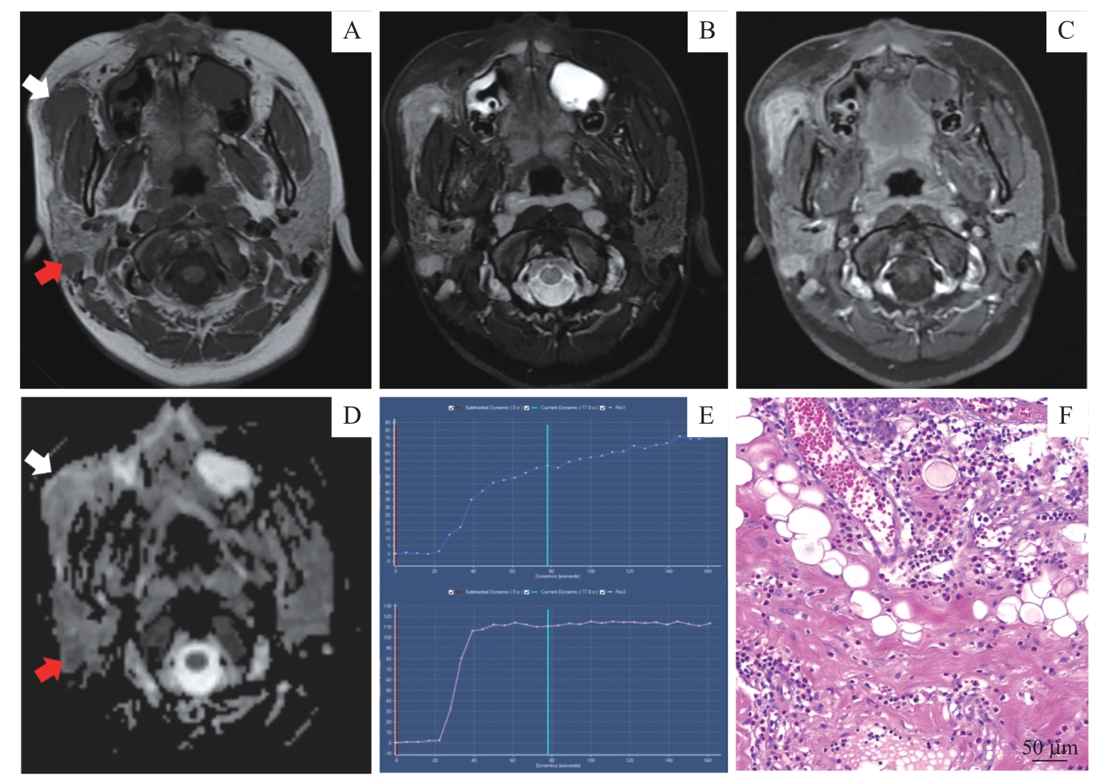
Fig 2 Imaging and histopathological manifestations of a nodular lesion in the right buccal subcutaneous tissues of a patient
| Item | Lymph node lesion | P value | |
|---|---|---|---|
| ADC/(×10 -3 mm 2·s -1) | 1.04 (0.92, 1.32) | 0.67 (0.60, 0.75) | 0.000 |
| TIC/ n(%) | 0.000 | ||
| Type Ⅰ | 23 (57.5) | 0 (0) | |
| Type Ⅱ | 17 (42.5) | 84 (96.6) | |
| Type Ⅲ | 0 (0) | 3 (3.4) | |
| Wash-in rate/s -1 | 15.5 (4.7, 39.7) | 33.0 (16.1, 55.6) | 0.000 |
| TTP/s | 73.0 (52.5, 110.5) | 45.0 (34.0, 60.0) | 0.000 |
Tab 4 Comparison of functional MRI parameters between extra-nodal lesions and lymph node lesions
| Item | Lymph node lesion | P value | |
|---|---|---|---|
| ADC/(×10 -3 mm 2·s -1) | 1.04 (0.92, 1.32) | 0.67 (0.60, 0.75) | 0.000 |
| TIC/ n(%) | 0.000 | ||
| Type Ⅰ | 23 (57.5) | 0 (0) | |
| Type Ⅱ | 17 (42.5) | 84 (96.6) | |
| Type Ⅲ | 0 (0) | 3 (3.4) | |
| Wash-in rate/s -1 | 15.5 (4.7, 39.7) | 33.0 (16.1, 55.6) | 0.000 |
| TTP/s | 73.0 (52.5, 110.5) | 45.0 (34.0, 60.0) | 0.000 |
| 1 | CHAN J K, HUI P K, NG C S, et al. Epithelioid haemangioma (angiolymphoid hyperplasia with eosinophilia) and Kimura′s disease in Chinese[J]. Histopathology, 1989, 15(6): 557-574. |
| 2 | LI T J, CHEN X M, WANG S Z, et al. Kimura′s disease: a clinicopathologic study of 54 Chinese patients[J]. Oral Surg Oral Med Oral Pathol Oral Radiol Endod, 1996, 82(5): 549-555. |
| 3 | STUCZYŃSKI S K, MURAS-SZWEDZIAK K, NOWICKI M. Diagnostic challenges in Kimura′s disease[J]. Pol Merkur Lekarski, 2022, 50(296): 128-130. |
| 4 | KAKEHI E, KOTANI K, OTSUKA Y, et al. Kimura′s disease: effects of age on clinical presentation[J]. QJM, 2020, 113(5): 336-345. |
| 5 | PARK S W, KIM H J, SUNG K J, et al. Kimura disease: CT and MR imaging findings[J]. AJNR Am J Neuroradiol, 2012, 33(4): 784-788. |
| 6 | KIM W J, KIM H K. Current concepts of Kimura disease: pathophysiology and evolution of treatment[J]. Arch Craniofac Surg, 2022, 23(6): 249-255. |
| 7 | CHEN H, THOMPSON L D R, AGUILERA N S I, et al. Kimura disease: a clinicopathologic study of 21 cases[J]. Am J Surg Pathol, 2004, 28(4): 505-513. |
| 8 | KELLY H R, CURTIN H D. Chapter 2 squamous cell carcinoma of the head and neck-imaging evaluation of regional lymph nodes and implications for management[J]. Semin Ultrasound CT MR, 2017, 38(5): 466-478. |
| 9 | HORIKOSHI T, MOTOORI K, UEDA T, et al. Head and neck MRI of Kimura disease[J]. Br J Radiol, 2011, 84(1005): 800-804. |
| 10 | HASHIDA Y, HIGUCHI T, NAKAJIMA K, et al. Human polyomavirus 6 with the Asian-Japanese genotype in cases of Kimura disease and angiolymphoid hyperplasia with eosinophilia[J]. J Invest Dermatol, 2020, 140(8): 1650-1653.e4. |
| 11 | KING R L, TAN B, CRAIG F E, et al. Reactive eosinophil proliferations in tissue and the lymphocytic variant of hypereosinophilic syndrome[J]. Am J Clin Pathol, 2021, 155(2): 211-238. |
| 12 | SATO R, BANDOH N, GOTO T, et al. Kimura disease presenting with buccal mass: a case report and literature review[J]. Head Neck Pathol, 2021, 15(2): 657-662. |
| 13 | LEE C C, YU K H, CHAN T M. Kimura′s disease: a clinicopathological study of 23 cases[J]. Front Med, 2022, 9: 1069102. |
| 14 | ZHANG G L, LI X M, SUN G B, et al. Clinical analysis of Kimura′s disease in 24 cases from China[J]. BMC Surg, 2020, 20(1): 1. |
| 15 | SANGWAN A, GOYAL A, BHALLA A S, et al. Kimura disease: a case series and systematic review of clinico-radiological features[J]. Curr Probl Diagn Radiol, 2022, 51(1): 130-142. |
| 16 | ZHU W X, ZHANG Y Y, SUN Z P, et al. Differential diagnosis of immunoglobulin G4-related sialadenitis and Kimura′s disease of the salivary gland: a comparative case series[J]. Int J Oral Maxillofac Surg, 2021, 50(7): 895-905. |
| 17 | TAKEISHI M, MAKINO Y, NISHIOKA H, et al. Kimura disease: diagnostic imaging findings and surgical treatment[J]. J Craniofac Surg, 2007, 18(5): 1062-1067. |
| 18 | WANG J, TANG Z H, FENG X Y, et al. Preliminary study of diffusion-weighted imaging and magnetic resonance spectroscopy imaging in Kimura disease[J]. J Craniofac Surg, 2014, 25(6): 2147-2151. |
| 19 | KIM S Y, BORNER U, LEE J H, et al. Magnetic resonance imaging of parotid gland tumors: a pictorial essay[J]. BMC Med Imaging, 2022, 22(1): 191. |
| 20 | MURAYAMA Y, KAMITANI T, SAGIYAMA K, et al. Evaluation of MR imaging findings differentiating parotid basal cell adenomas from other parotid tumors[J]. Eur J Radiol, 2021, 144: 109980. |
| 21 | SUROV A, MEYER H J, WIENKE A. Apparent diffusion coefficient for distinguishing between malignant and benign lesions in the head and neck region: a systematic review and meta-analysis[J]. Front Oncol, 2020, 9: 1362. |
| 22 | BAIK J, BAEK H J, RYU K H, et al. MALT lymphoma of the tongue in a patient with Sjögren′s syndrome: a case report and literature review[J]. Diagnostics, 2021, 11(9): 1715. |
| [1] | SUN Lei, DAI Shifeng, CHEN Yuhua, XU Xinyi, JIANG Kele, LI Xiaowen, LI Chengjing, WU Tingting. Quantitative analysis of the distance between articular disc and condyle in patients with temporomandibular disorders [J]. Journal of Shanghai Jiao Tong University (Medical Science), 2025, 45(6): 684-692. |
| [2] | LI Zhuohang, YU Xindi, REN Jingya, SHEN Jia, DONG Suzhen, WANG Wei. Postoperative neurodevelopmental outcomes of end-to-side anastomosis for coarctation of the aorta [J]. Journal of Shanghai Jiao Tong University (Medical Science), 2025, 45(6): 753-759. |
| [3] | LU Xiaobing, YUE Jiang, HE Shengyun, DONG Ying, LU Qing, MA Jing. Effect of intramuscular adipose tissue in the skeletal muscle of thigh on glucose metabolism in male patients with obesity [J]. Journal of Shanghai Jiao Tong University (Medical Science), 2023, 43(9): 1169-1174. |
| [4] | CHEN Jiaye, ZHANG Huifeng, LU Wenxian, PENG Daihui. Research progress of brain magnetic resonance imaging related to suicide in bipolar disorder patients [J]. Journal of Shanghai Jiao Tong University (Medical Science), 2023, 43(7): 939-944. |
| [5] | GU Wenxi, JIA Huan, WU Hao. Clinical values and advances in computed tomography evaluation after cochlear implantation [J]. Journal of Shanghai Jiao Tong University (Medical Science), 2023, 43(12): 1463-1469. |
| [6] | LIU Siyu, WU Bing, LI Xiaomin, ZHAO Lulu, CHEN Jun, AI Songtao. Preliminary exploration of diffusion-weighted imaging in pre-surgical planning of dermatofibrosarcoma protuberans [J]. Journal of Shanghai Jiao Tong University (Medical Science), 2022, 42(8): 1095-1102. |
| [7] | CHEN Liqi, XUE Zhuowei, WU Qingkai. Review of MRI-based three-dimensional digital model reconstruction of female pelvic floor organs [J]. Journal of Shanghai Jiao Tong University (Medical Science), 2022, 42(3): 381-386. |
| [8] | Xuehong WANG, Xuzhuo CHEN, Yi MAO, Da SHEN, Shanyong ZHANG. Difference in recurrence rates after temporomandibular joint disc repositioning surgery with miniscrew anchor at different developmental stages in adolescents [J]. JOURNAL OF SHANGHAI JIAOTONG UNIVERSITY (MEDICAL SCIENCE), 2022, 42(2): 173-177. |
| [9] | Cui CHEN, Ye JIN, Lin WANG, Hongli LI, Caifeng WAN, Lixin JIANG. Comparative analysis of 30 cases of metaplastic carcinoma of the breast [J]. JOURNAL OF SHANGHAI JIAOTONG UNIVERSITY (MEDICAL SCIENCE), 2022, 42(1): 70-76. |
| [10] | Yihuan WANG, Ruokun LI, Huanhuan CHONG, Fuhua YAN. Research progress of Gd-EOB-DTPA-enhanced magnetic resonance imaging in the evaluation of biological behavior of hepatocellular carcinoma [J]. JOURNAL OF SHANGHAI JIAOTONG UNIVERSITY (MEDICAL SCIENCE), 2022, 42(1): 130-134. |
| [11] | JI Ying-ying, XUE Bin, HUANG Yue, ZHANG Jian-wei. Efficacy and safety of oral midazolam in combination with intranasal dexmedetomidine for paediatric magnetic resonance imaging sedation [J]. JOURNAL OF SHANGHAI JIAOTONG UNIVERSITY (MEDICAL SCIENCE), 2020, 40(8): 1098-1102. |
| [12] | YANG Tao, CHEN Jun, FANG Yi-ru. Advances in magnetic resonance imaging study of bipolar Ⅰdisorder [J]. JOURNAL OF SHANGHAI JIAOTONG UNIVERSITY (MEDICAL SCIENCE), 2020, 40(12): 1660-1664. |
| [13] | LI Xiao-min1, QU Yang1, WU Wen2, ZHAO Liang3, ZHANG Shao-ting3, HAO Yong-qiang2, DAI Ke-rong2, AI Song-tao1. Preliminary application of MR imaging-pathology co-localization by 3D printing box in pelvic tumor assessment [J]. JOURNAL OF SHANGHAI JIAOTONG UNIVERSITY (MEDICAL SCIENCE), 2020, 40(10): 1408-1413. |
| [14] | YUE Xiu-hui, KONG Wei-dan, REN Ji-liang, YUAN Ying#, TAO Xiao-feng#. Value of 3.0-T MR diffusion-weighted imaging combined with dynamic contrast-enhanced imaging in differentiating benign and malignant thyroid nodules [J]. JOURNAL OF SHANGHAI JIAOTONG UNIVERSITY (MEDICAL SCIENCE), 2020, 40(10): 1393-1397. |
| [15] | WANG Tao, ZHANG Chen-cheng, LI Dian-you, SUN Bo-min, FU Meng. Imaging law of postoperative electrode locations in deep brain stimulation for Parkinsons disease [J]. , 2020, 40(1): 64-. |
| Viewed | ||||||
|
Full text |
|
|||||
|
Abstract |
|
|||||