上海交通大学学报(医学版) ›› 2024, Vol. 44 ›› Issue (8): 944-950.doi: 10.3969/j.issn.1674-8115.2024.08.002

• 牙颌面畸形专题 • 上一篇    下一篇

翼腭缝宏观解剖及其发育模式研究

章文益(), 郑美里(), 谢羽番, 江凌勇()   

  1. 上海交通大学医学院附属第九人民医院口腔颅颌面科·正颌正畸中心,上海交通大学口腔医学院,国家口腔医学中心,国家口腔疾病临床医学研究中心,上海市口腔医学重点实验室,上海市口腔医学研究所,中国医学科学院口腔颌面再生医学创新单元,上海 200011
  • 收稿日期:2024-03-01 接受日期:2024-05-11 出版日期:2024-08-28 发布日期:2024-08-27
  • 通讯作者: 江凌勇 E-mail:2020sjtuzwy@sjtu.edu.cn;miri_jung@hotmail.com;jianglingyong@sjtu.edu.cn
  • 作者简介:章文益(2002—),女,本科生;电子信箱:2020sjtuzwy@sjtu.edu.cn
    郑美里 (1993—),女,韩国,博士生;电子信箱:miri_jung@hotmail.com
  • 基金资助:
    上海市重中之重研究中心项目(2022ZZ01017);中央高校基本科研业务费专项资金资助(YG2023ZD14);中国医学科学院医学与健康科技创新工程基金(2019-12M-5-037);上海交通大学医学院附属第九人民医院生物样本库项目(YBKB202216);上海交通大学医学院“双百人-研究型医师”项目(20221809);国家重点领域大学生创新训练计划项目(1723Z501);上海市科技创新行动计划-国际科技合作项目(23410713600)

Study on macroscopic anatomy and developmental model of pterygopalatine suture

ZHANG Wenyi(), CHUNG Miri(), XIE Yufan, JIANG Lingyong()   

  1. Center of Craniofacial Orthodontics, Department of Oral and Craniomaxillofacial Surgery, Shanghai Ninth People's Hospital, Shanghai Jiao Tong University School of Medicine; College of Stomatology, Shanghai Jiao Tong University; National Center for Stomatology; National Clinical Research Center for Oral Diseases; Shanghai Key Laboratory of Stomatology; Shanghai Research Institute of Stomatology; Research Unit of Oral and Maxillofacial Regenerative Medicine, Chinese Academy of Medical Sciences, Shanghai 200011, China
  • Received:2024-03-01 Accepted:2024-05-11 Online:2024-08-28 Published:2024-08-27
  • Contact: JIANG Lingyong E-mail:2020sjtuzwy@sjtu.edu.cn;miri_jung@hotmail.com;jianglingyong@sjtu.edu.cn
  • Supported by:
    Project of Shanghai's Top Priority Research Center(2022ZZ01017);Fundamental Research Funds for the Central Universities(YG2023ZD14);Chinese Academy of Medical Sciences Innovation Fund for Medical Sciences(2019-I2M-5-037);Project of Biobank of Shanghai Ninth People's Hospital, Shanghai Jiao Tong University School of Medicine(YBKB202216);“Two-hundred Talents” Program of Shanghai Jiao Tong University School of Medicine(20221809);National Innovation Training Program for Undergraduates in Key Fields(1723Z501);Shanghai Science and Technology Innovation Action Plan-International Science and Technology Cooperation Program(23410713600)

摘要:

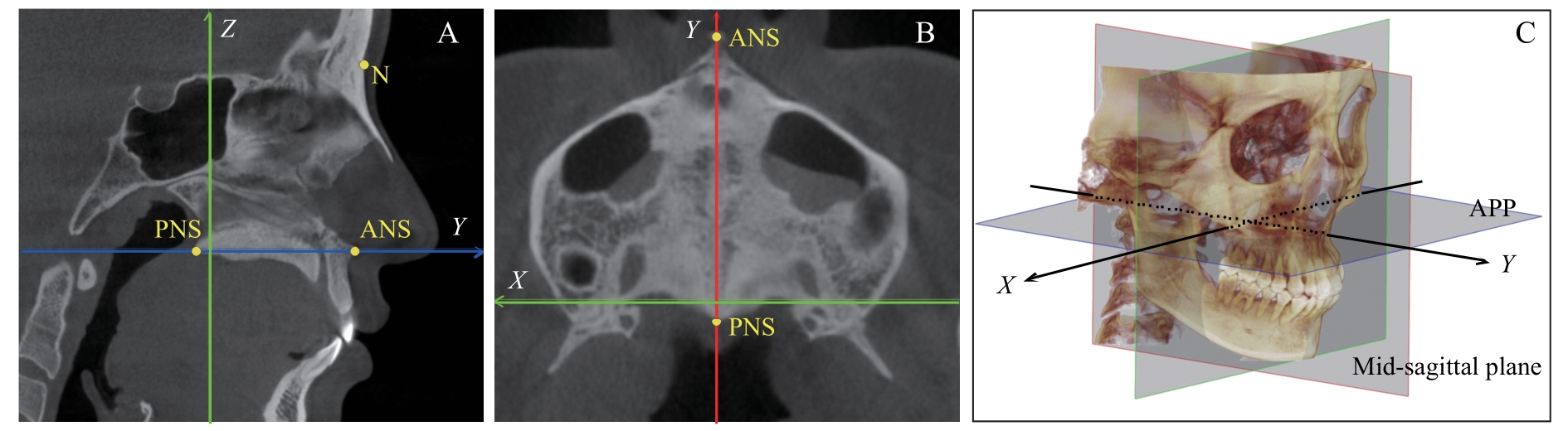
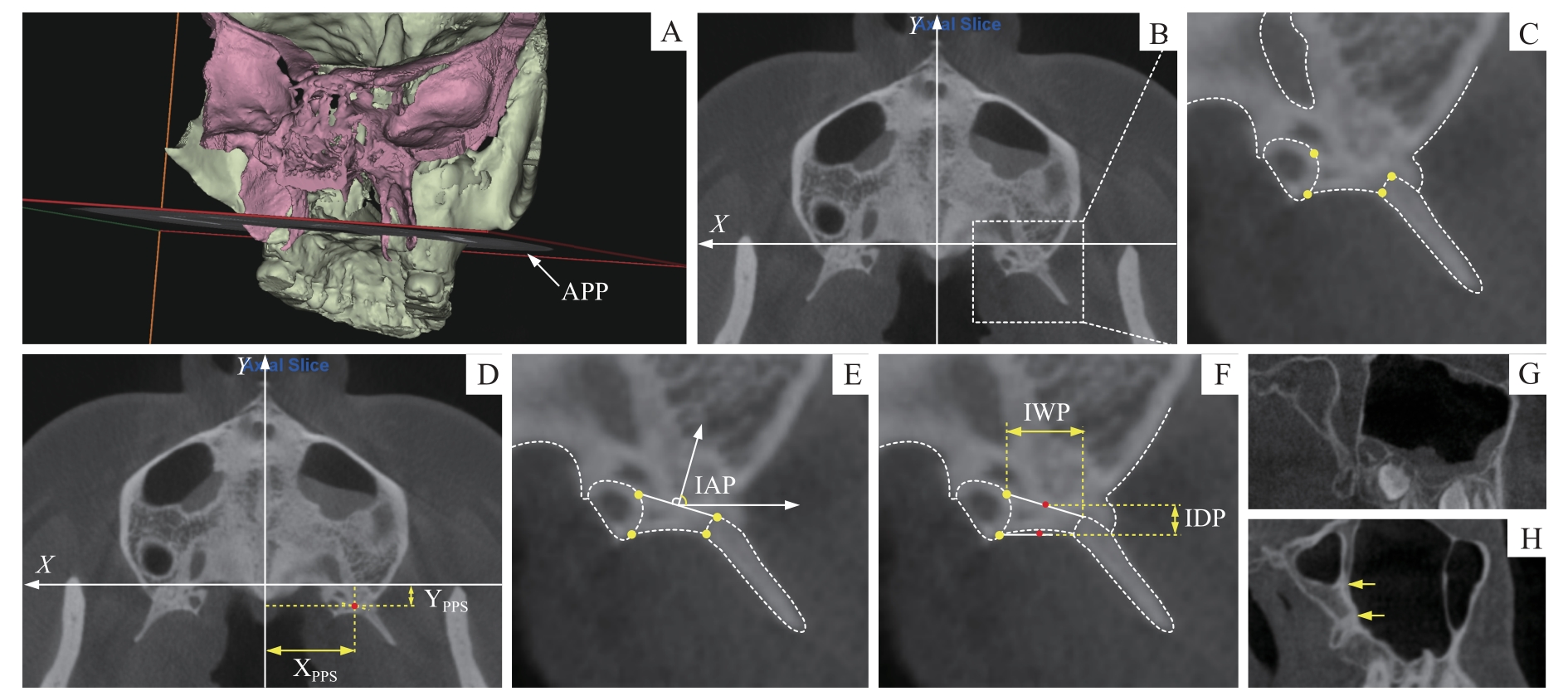
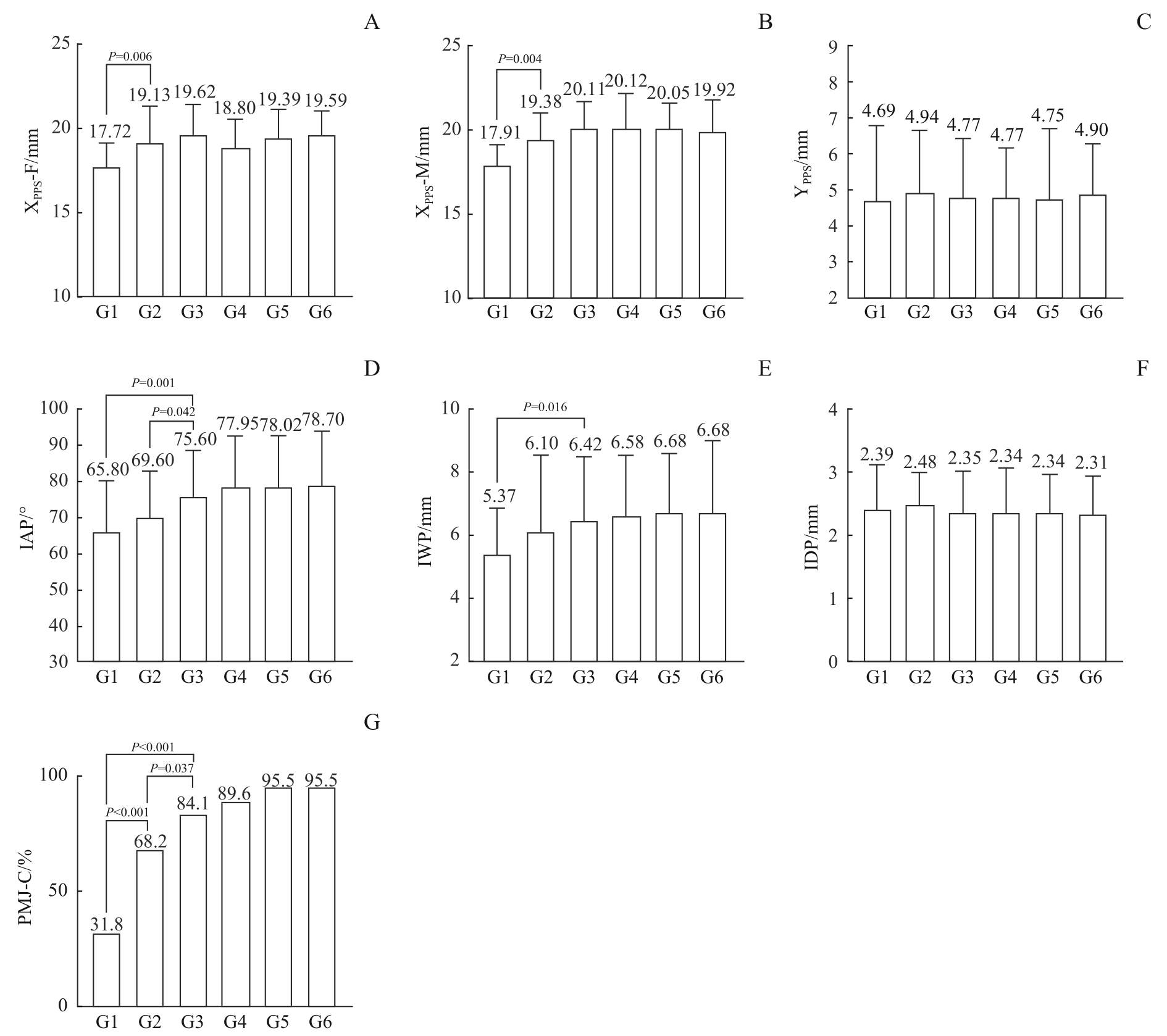
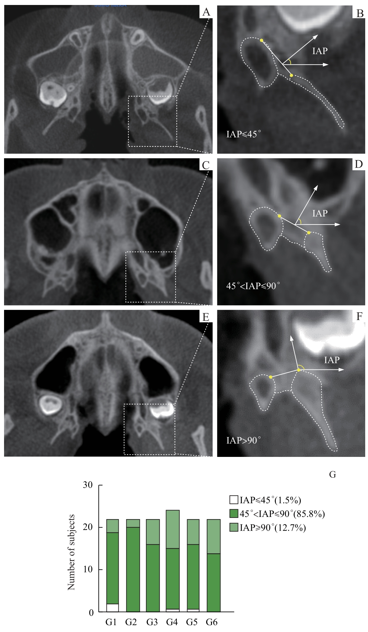
目的·利用锥形束计算机断层扫描(cone-beam computed tomography,CBCT)技术分析中国人群中翼腭缝(pterygopalatine suture,PPS)的解剖及宏观形态特征,并初步探讨其发育模式及其与矫正上颌发育不足之间的关联。方法·纳入2023年7月—8月在上海交通大学医学院附属第九人民医院拍摄的134例CBCT影像资料,根据年龄分为6组。以PPS标志点为依据,计算得出PPS整体的横向位置(transverse position of PPS,XPPS),PPS整体的矢状向位置(sagittal position of PPS,YPPS),以及PPS的锥突嵌入角(insertion angle of PPS,IAP)、锥突嵌入宽度(insertion width of pyramidal process,IWP)和锥突嵌入深度(insertion depth of pyramidal process,IDP),并通过多平面观察确定翼上颌联合情况。采用回归分析评估各参数与年龄、性别的相关性,并通过两两比较确定参数变化的稳定年龄段。采用配对t检验及配对χ2检验分析双侧参数的差异性。结果·YPPS、IDP与年龄无显著相关性,而XPPS、IAP、IWP、翼上颌连接的发生率均与年龄呈显著正相关(P<0.01)。性别差异仅在XPPS中显著,男性大于女性(P<0.01)。参数的年龄变化趋势分析表明:XPPS在组1(6岁≤年龄<9岁)和组2(9岁≤年龄<12岁)间差异显著(女性:P=0.006;男性:P=0.004);IAP在组2与组3(12岁≤年龄<15岁)间差异显著(P=0.042),98.5%样本的IAP大于45o;IWP在组1与组3间差异显著(P=0.016);翼上颌联合情况在组1、2、3间差异显著(组1、2间P<0.001,组2、3间P=0.037,组1、3间P<0.001),成人发生率高于90%。各项指标双侧比较均未见统计学差异。结论·6岁以后YPPS及IDP未表现出明显改变,上颌骨与翼突趋于融合。PPS整体横向位置在12岁左右趋于稳定,而IAP及IWP持续增长,至15岁左右达到稳定且翼腭缝锥突嵌入翼切迹的方向偏向矢状向。

关键词: 翼腭缝, 形态学, 生长发育, 上颌扩弓, 上颌前方牵引

Abstract:

Objective ·To analyze the anatomical and macroscopic characteristics of the pterygopalatine suture (PPS) in Chinese population using cone-beam computed tomography (CBCT) technology, and to preliminarily investigate its developmental pattern and its association with the correction of maxillary underdevelopment. Methods ·A total of 134 CBCT images, taken from July to August 2023 at Shanghai Ninth People's Hospital, Shanghai Jiao Tong University School of Medicine, were categorized into six age groups. The PPS landmarks were utilized to determine the overall transverse position (XPPS), sagittal position (YPPS), insertion angle (IAP), insertion width of the pyramidal process (IWP), and insertion depth of the pyramidal process (IDP). The pterygomaxillary junction was identified through multiplanar observations. Regression analysis was performed to assess the correlation of these parameters with age and gender, and pairwise comparisons were made to determine the stable age range for parameter changes. Paired t-tests and paired chi-squared tests were conducted to analyze the bilateral parameter differences. Results ·Results showed that YPPS and IDP did not exhibit significant correlation with age, whereas XPPS, IAP, IWP, and the incidence of pterygomaxillary fusion were significantly positively correlated with age (P<0.01). Gender differences were only significant for XPPS, with males demonstrating greater values than females (P<0.01). Analysis of age-related trends indicated significant differences in XPPS between group 1 (6 years≤age<9 years) and group 2 (9 years≤age<12 years) (female: P=0.006, male: P=0.004); significant differences in IAP were observed between group 2 and group 3 (12 years≤age<15 years) (P=0.042), with 98.5% of samples having an IAP greater than 45 degrees; IWP differences were significant between group 1 and group 3 (P=0.016), and the pterygomaxillary fusion incidence was significantly different among group 1, 2, and 3 (group 1 vs. 2: P<0.001, group 2 vs 3: P=0.037, group 1 vs 3: P<0.001), with an incidence rate exceeding 90% in adults. No significant bilateral differences were found for all parameters. Conclusion ·No significant changes were observed in YPPS and IDP after the age of 6, indicating a trend towards fusion of the maxilla with the pterygoid process. The overall transverse position of the PPS tends to stabilize around 12 years of age, while the IAP and IWP continue to increase and reach stability around 15 years of age,with a sagittal insertion orientation of the pyramidal process into the pterygoid notch.

Key words: pterygopalatine suture, morphology, growth and development, maxillary expansion, maxillary protraction

中图分类号: