| 1 |
KLEIN A P. Pancreatic cancer epidemiology: understanding the role of lifestyle and inherited risk factors[J]. Nat Rev Gastroenterol Hepatol, 2021, 18(7): 493-502.
|
| 2 |
PEREIRA S P, OLDFIELD L, NEY A, et al. Early detection of pancreatic cancer[J]. Lancet Gastroenterol Hepatol, 2020, 5(7): 698-710.
|
| 3 |
SIEGEL R L, MILLER K D, WAGLE N S, et al. Cancer statistics, 2023[J]. CA Cancer J Clin, 2023, 73(1): 17-48.
|
| 4 |
FERRONE C R, BRENNAN M F, GONEN M, et al. Pancreatic adenocarcinoma: the actual 5-year survivors[J]. J Gastrointest Surg, 2008, 12(4): 701-706.
|
| 5 |
FERRONE C R, PIERETTI-VANMARCKE R, BLOOM J P, et al. Pancreatic ductal adenocarcinoma: long-term survival does not equal cure[J]. Surgery, 2012, 152(3 Suppl 1): S43-S49.
|
| 6 |
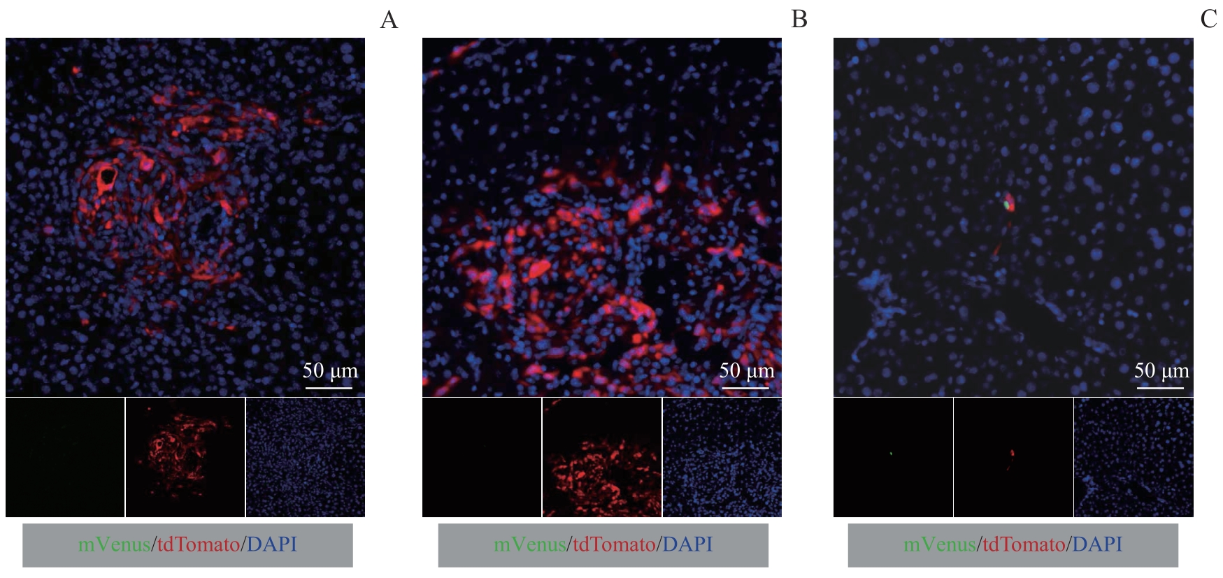
HE J, AHUJA N, MAKARY M A, et al. 2 564 resected periampullary adenocarcinomas at a single institution: trends over three decades[J]. HPB, 2014, 16(1): 83-90.
|
| 7 |
KATZ M H, WANG H M, FLEMING J B, et al. Long-term survival after multidisciplinary management of resected pancreatic adenocarcinoma[J]. Ann Surg Oncol, 2009, 16(4): 836-847.
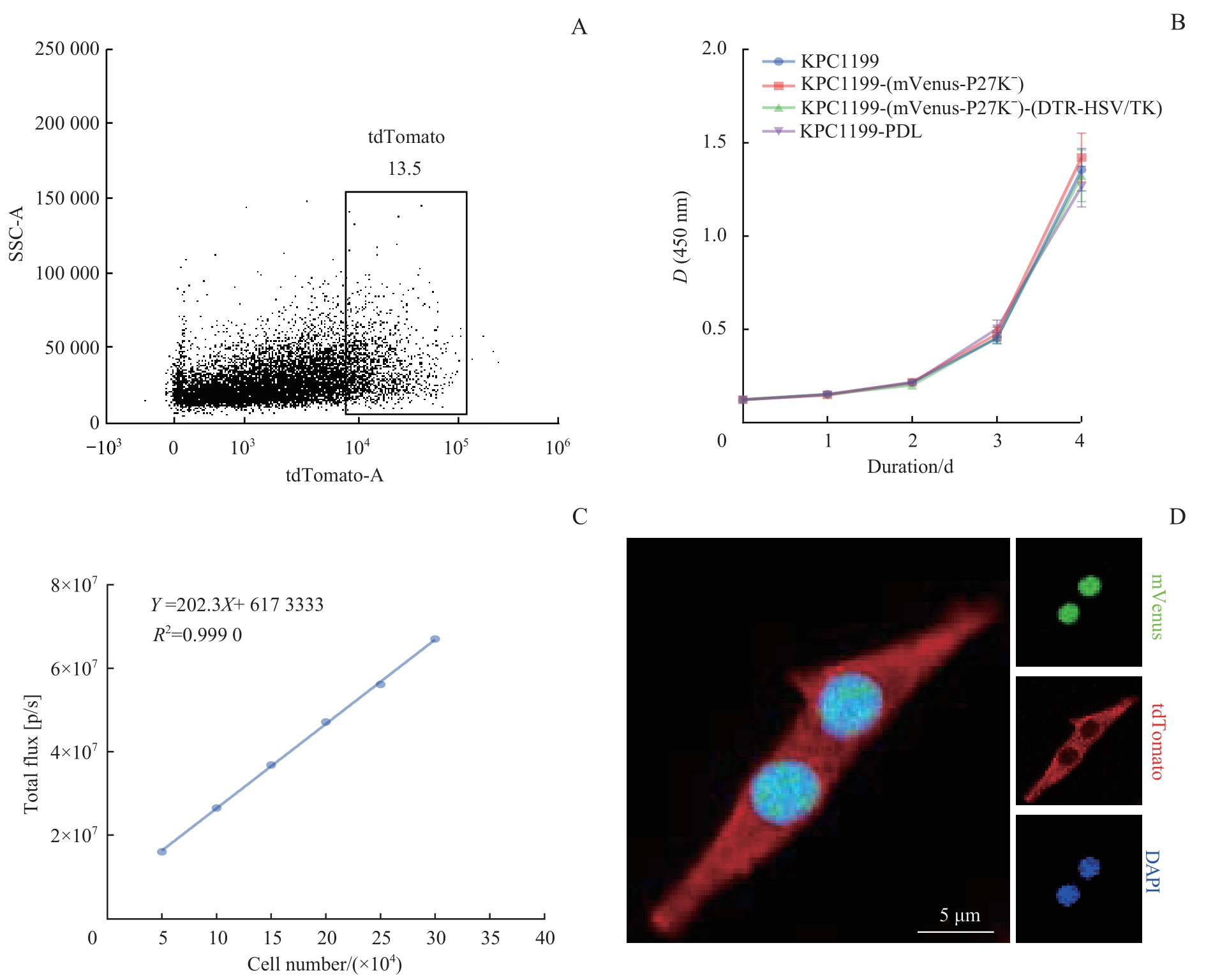
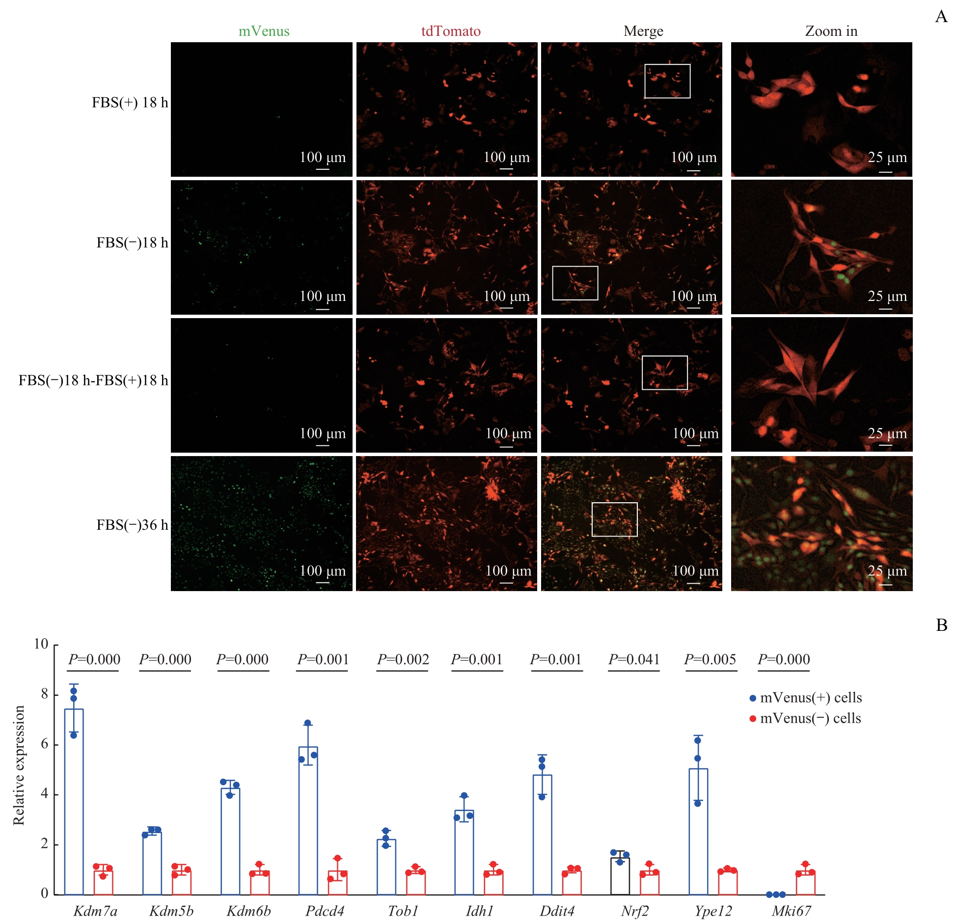
|
| 8 |
RHIM A D, MIREK E T, AIELLO N M, et al. EMT and dissemination precede pancreatic tumor formation[J]. Cell, 2012, 148(1/2): 349-361.
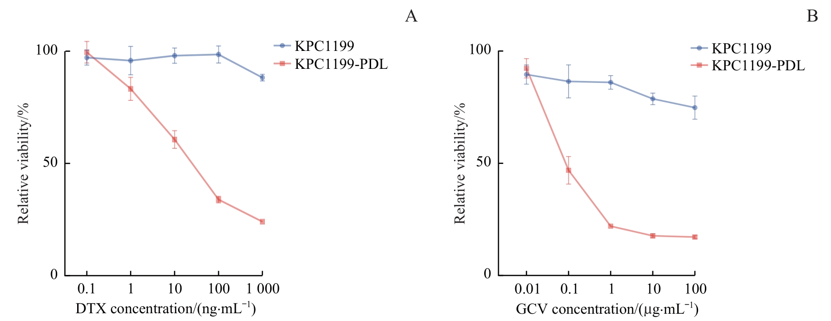
|
| 9 |
GHAJAR C M. Metastasis prevention by targeting the dormant niche[J]. Nat Rev Cancer, 2015, 15(4): 238-247.
|
| 10 |
MASSAGUÉ J, GANESH K. Metastasis-initiating cells and ecosystems[J]. Cancer Discov, 2021, 11(4): 971-994.
|
| 11 |
ALBRENGUES J, SHIELDS M A, NG D, et al. Neutrophil extracellular traps produced during inflammation awaken dormant cancer cells in mice[J]. Science, 2018, 361(6409): eaao4227.
|
| 12 |
AGUIRRE-GHISO J A. Models, mechanisms and clinical evidence for cancer dormancy[J]. Nat Rev Cancer, 2007, 7(11): 834-846.
|
| 13 |
POMMIER A, ANAPARTHY N, MEMOS N, et al. Unresolved endoplasmic reticulum stress engenders immune-resistant, latent pancreatic cancer metastases[J]. Science, 2018, 360(6394): eaao4908.
|
| 14 |
MALLADI S, MACALINAO D G, JIN X, et al. Metastatic latency and immune evasion through autocrine inhibition of WNT[J]. Cell, 2016, 165(1): 45-60.
|
| 15 |
PANTEL K, SCHLIMOK G, KUTTER D, et al. Frequent down-regulation of major histocompatibility class Ⅰ antigen expression on individual micrometastatic carcinoma cells[J]. Cancer Res, 1991, 51(17): 4712-4715.
|
| 16 |
BALDOMINOS P, BARBERA-MOURELLE A, BARREIRO O, et al. Quiescent cancer cells resist T cell attack by forming an immunosuppressive niche[J]. Cell, 2022, 185(10): 1694-1708.e19.
|
| 17 |
HU J, SÁNCHEZ-RIVERA F J, WANG Z H, et al. STING inhibits the reactivation of dormant metastasis in lung adenocarcinoma[J]. Nature, 2023, 616(7958): 806-813.
|
| 18 |
CONNOR A A, DENROCHE R E, JANG G H, et al. Association of distinct mutational signatures with correlates of increased immune activity in pancreatic ductal adenocarcinoma[J]. JAMA Oncol, 2017, 3(6): 774-783.
|
| 19 |
FEIG C, JONES J O, KRAMAN M, et al. Targeting CXCL12 from FAP-expressing carcinoma-associated fibroblasts synergizes with anti-PD-L1 immunotherapy in pancreatic cancer[J]. Proc Natl Acad Sci U S A, 2013, 110(50): 20212-20217.
|
| 20 |
OKI T, NISHIMURA K, KITAURA J, et al. A novel cell-cycle-indicator, mVenus-p27K-, identifies quiescent cells and visualizes G0-G1 transition[J]. Sci Rep, 2014, 4: 4012.
|
| 21 |
POSCHKE I, FARYNA M, BERGMANN F, et al. Identification of a tumor-reactive T-cell repertoire in the immune infiltrate of patients with resectable pancreatic ductal adenocarcinoma[J]. Oncoimmunology, 2016, 5(12): e1240859.
|
| 22 |
SAUVAGEAU M, SAUVAGEAU G. Polycomb group proteins: multi-faceted regulators of somatic stem cells and cancer[J]. Cell Stem Cell, 2010, 7(3): 299-313.
|
| 23 |
RUBSAM L Z, BOUCHER P D, MURPHY P J, et al. Cytotoxicity and accumulation of ganciclovir triphosphate in bystander cells cocultured with herpes simplex virus type 1 thymidine kinase-expressing human glioblastoma cells[J]. Cancer Res, 1999, 59(3): 669-675.
|
| 24 |
PROPPER D J, BALKWILL F R. Harnessing cytokines and chemokines for cancer therapy[J]. Nat Rev Clin Oncol, 2022, 19(4): 237-253.
|
| 25 |
VESELY M D, ZHANG T X, CHEN L P. Resistance mechanisms to anti-PD cancer immunotherapy[J]. Annu Rev Immunol, 2022, 40: 45-74.
|
| 26 |
SAXENA M, VAN DER BURG S H, MELIEF C J M, et al. Therapeutic cancer vaccines[J]. Nat Rev Cancer, 2021, 21(6): 360-378.
|
| 27 |
SHALHOUT S Z, MILLER D M, EMERICK K S, et al. Therapy with oncolytic viruses: progress and challenges[J]. Nat Rev Clin Oncol, 2023, 20(3): 160-177.
|
| 28 |
YEH A C, RAMASWAMY S. Mechanisms of cancer cell dormancy: another hallmark of cancer?[J]. Cancer Res, 2015, 75(23): 5014-5022.
|
| 29 |
GERSTBERGER S, JIANG Q W, GANESH K. Metastasis[J]. Cell, 2023, 186(8): 1564-1579.
|
| 30 |
PHAN T G, CROUCHER P I. The dormant cancer cell life cycle[J]. Nat Rev Cancer, 2020, 20(7): 398-411.
|
| 31 |
MIN H Y, LEE H Y. Cellular dormancy in cancer: mechanisms and potential targeting strategies[J]. Cancer Res Treat, 2023, 55(3): 720-736.
|
 ), 王俊杰1, 钱云臻1, 陈溯源1, 邵达2, 张志刚1(