上海交通大学学报(医学版) ›› 2022, Vol. 42 ›› Issue (6): 768-777.doi: 10.3969/j.issn.1674-8115.2022.06.011
吴靖1( ), 赵正宜2, 邹多宏1(
 ), 杨驰1, 张志愿1
收稿日期:2022-04-12
接受日期:2022-06-08
出版日期:2022-06-28
发布日期:2022-08-19
通讯作者:
邹多宏,电子信箱:zouduohongyy@163.com。作者简介:吴靖(1994—),男,住院医师,硕士;电子信箱:wujingsjtu2021@163.com。
基金资助:
WU Jing1( ), ZHAO Zhengyi2, ZOU Duohong1(
 ), YANG Chi1, ZHANG Zhiyuan1
Received:2022-04-12
Accepted:2022-06-08
Online:2022-06-28
Published:2022-08-19
Contact:
ZOU Duohong, E-mail: zouduohongyy@163.com.Supported by:摘要:
目的·探究帐篷钉技术在牙槽骨重度缺损修复重建中的作用。方法·选取2018年1月—2021年1月在上海交通大学医学院附属第九人民医院口腔外科接受帐篷钉技术完成牙槽骨重度缺损修复的30例患者作为研究对象,通过分析与重建患者术前以及术后8个月的锥形束计算机断层扫描(cone-beam computed tomography,CBCT)影像数据,统计患者牙槽骨水平、垂直以及混合型骨缺损的修复重建效果。术后8个月时,在患者接受二期种植手术过程中利用取骨环钻在待种植区域取一圆柱状骨组织样本,通过显微计算机断层扫描(micro-computed tomography,Micro-CT)数据分析样本骨小梁数量(trabecular bone number,Tb. N)、骨小梁厚度(trabecular bone thickness,Tb. Th)、骨体积分数(bone volume/total volume,BV/TV)以及骨骼矿物质密度(bone mineral density,BMD)以明确骨再生效果。通过硬组织切片观察术后组织样本内的新骨形成率及材料残留率以分析新骨形成情况。结果·术后8个月患者牙槽骨垂直骨增量为4.81(1.58,7.66)mm,水平骨增量为3.96(2.38,5.67)mm,骨增量区域的骨增量体积为2 157.22(776.59,2 831.63)mm3。术后8个月骨组织样本Micro-CT分析显示帐篷钉植骨区域Tb. N为(3.09±0.68)/mm,Tb. Th为(0.08±0.01)μm,BV/TV为(25.24±5.60)%,BMD为(0.24±0.05)g/cm3;硬组织切片显示新骨形成率为(16.30±3.57)%,材料残留率为34%(31.75%,38.25%)。上述研究数据表明骨修复与再生良好。患者术中、术后均未出现并发症以及严重不良事件。结论·基于以“稳定为核心”的牙槽骨骨增量理念,使用帐篷钉、膜钉及包袋结构,在规范化操作下,帐篷钉技术可以有效完成牙槽骨重度骨缺损的修复重建,并且术后并发症发生率低,骨增量效果可预测性高,有利于后期口腔种植修复治疗的顺利开展。
中图分类号:
吴靖, 赵正宜, 邹多宏, 杨驰, 张志愿. 帐篷钉技术在牙槽骨重度缺损修复重建中的应用:30例临床病例回顾性分析与总结[J]. 上海交通大学学报(医学版), 2022, 42(6): 768-777.
WU Jing, ZHAO Zhengyi, ZOU Duohong, YANG Chi, ZHANG Zhiyuan. Application of a tent-pole screw technology in reconstruction of severe alveolar bone defect: a retrospective study of 30 patients[J]. Journal of Shanghai Jiao Tong University (Medical Science), 2022, 42(6): 768-777.

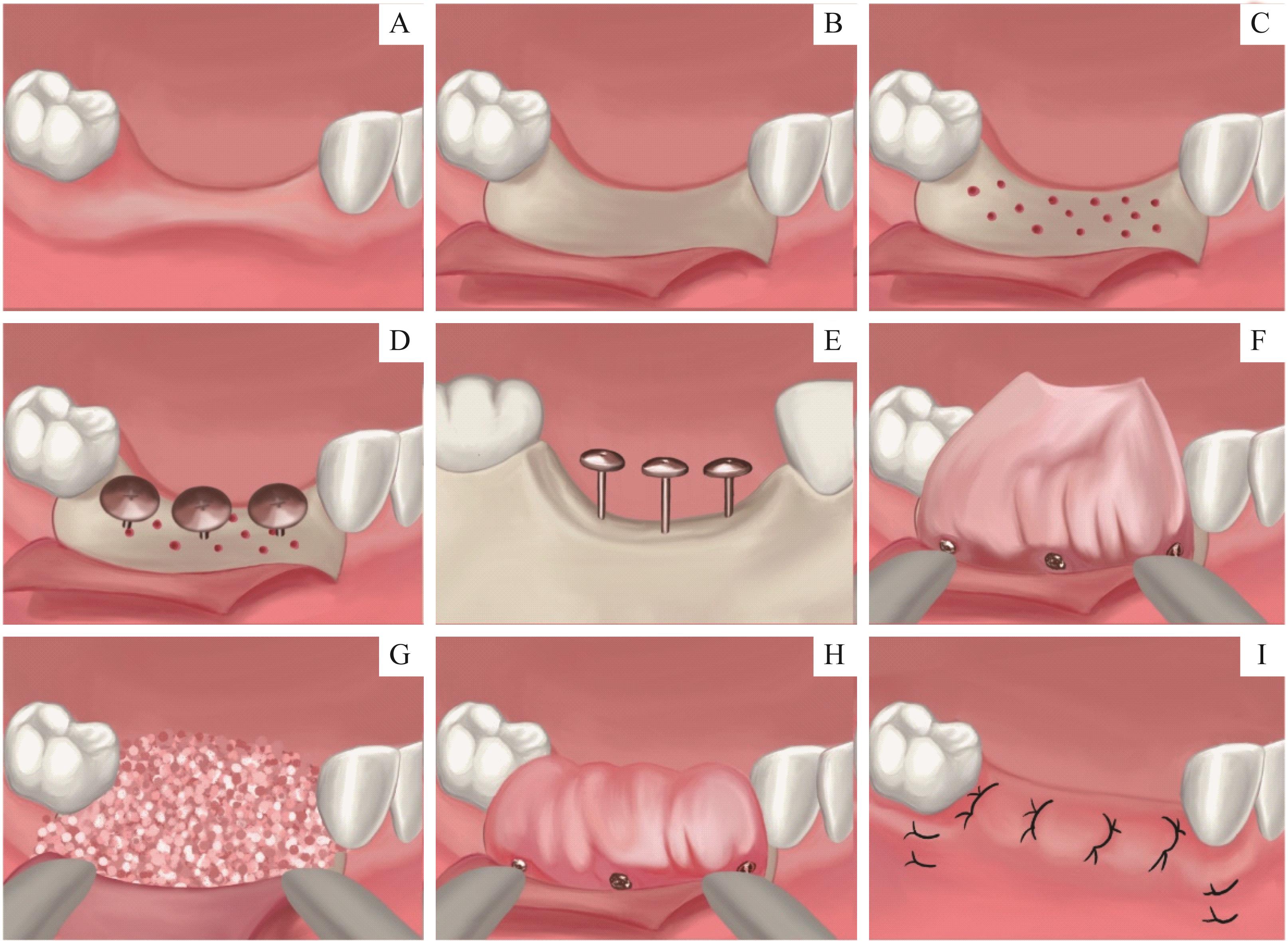
图1 手术流程示意图Note:A. Preoperative evaluation of severe bone defects. B/C. A mesial-distal crestal incision was made (B), and vertical incisions were made 5~10 mm from the bone defects (C). Full thickness flap was reflected. After eliminating the remaining soft tissue on bone surface, cortical perforation was performed to enhance revascularization. D/E. Tent-peg and pins were selected according to the size of bone defect (D) and the extent of expected bone augment (E). Tent pegs were placed on the occlusal side of the alveolar ridge. F. The GBR membranes were anchored to the buccal surface of alveolar bone to form a packaging structure with membrane pins. G. Filling the bone defect with fully mixed anorganic bovine bone-derived mineral. H. GBR membrane was tightly covered on anorganic bovine bone-derived mineral. I. After releasing soft-tissue tension of mucoperiosteal flap, tension-free wound closure with mattress suture and interrupted sutures after periosteal release were performed.
Fig 1 Flow chart of the surgical operation

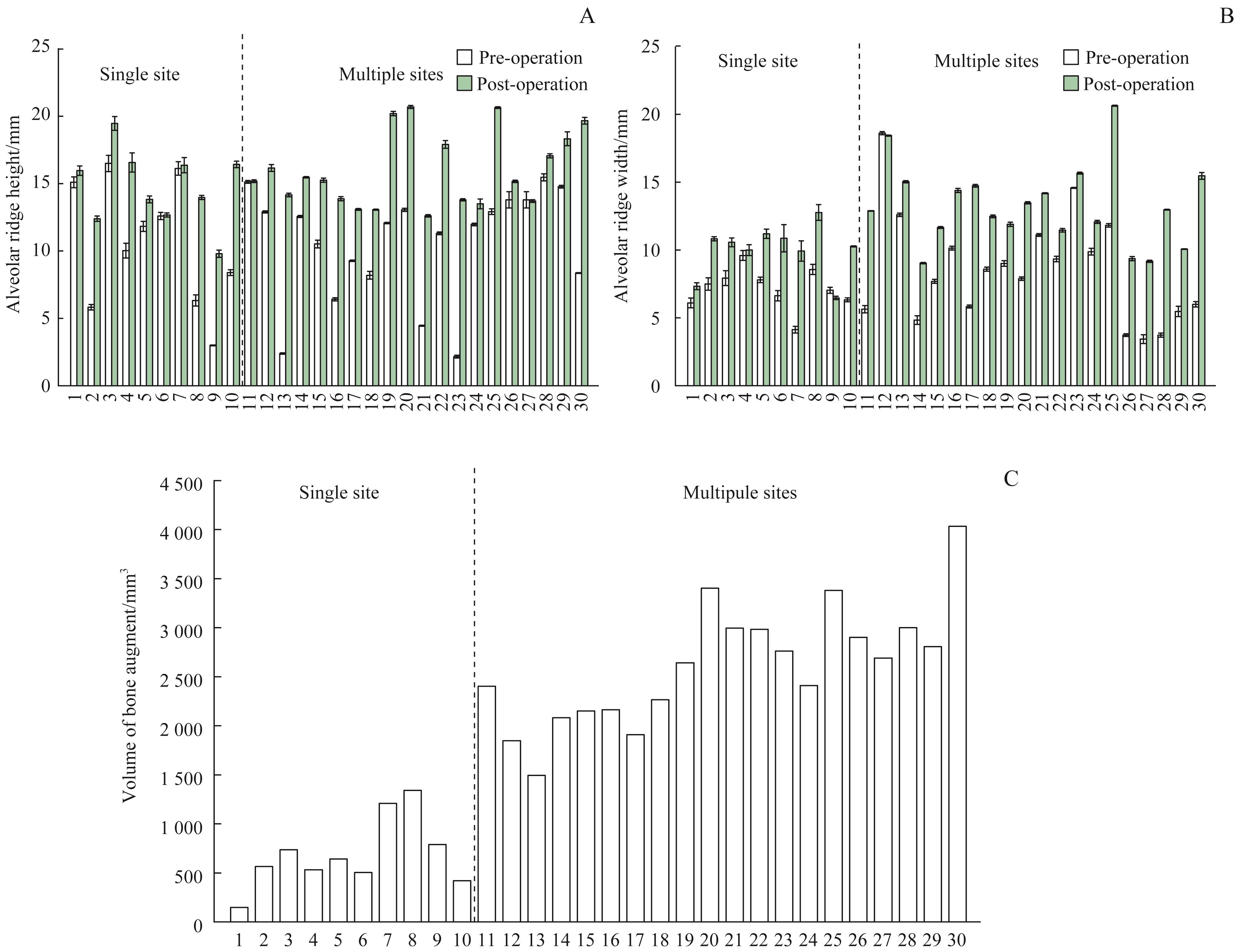
图3 30例患者术后8个月的骨增量数据Note: Alveolar ridge height (A) and width (B), and the bone volume gain (C) after tent peg surgery for the 30 augmentation sites.
Fig 3 Bone gain of 30 patients after tent peg surgery for 8 months

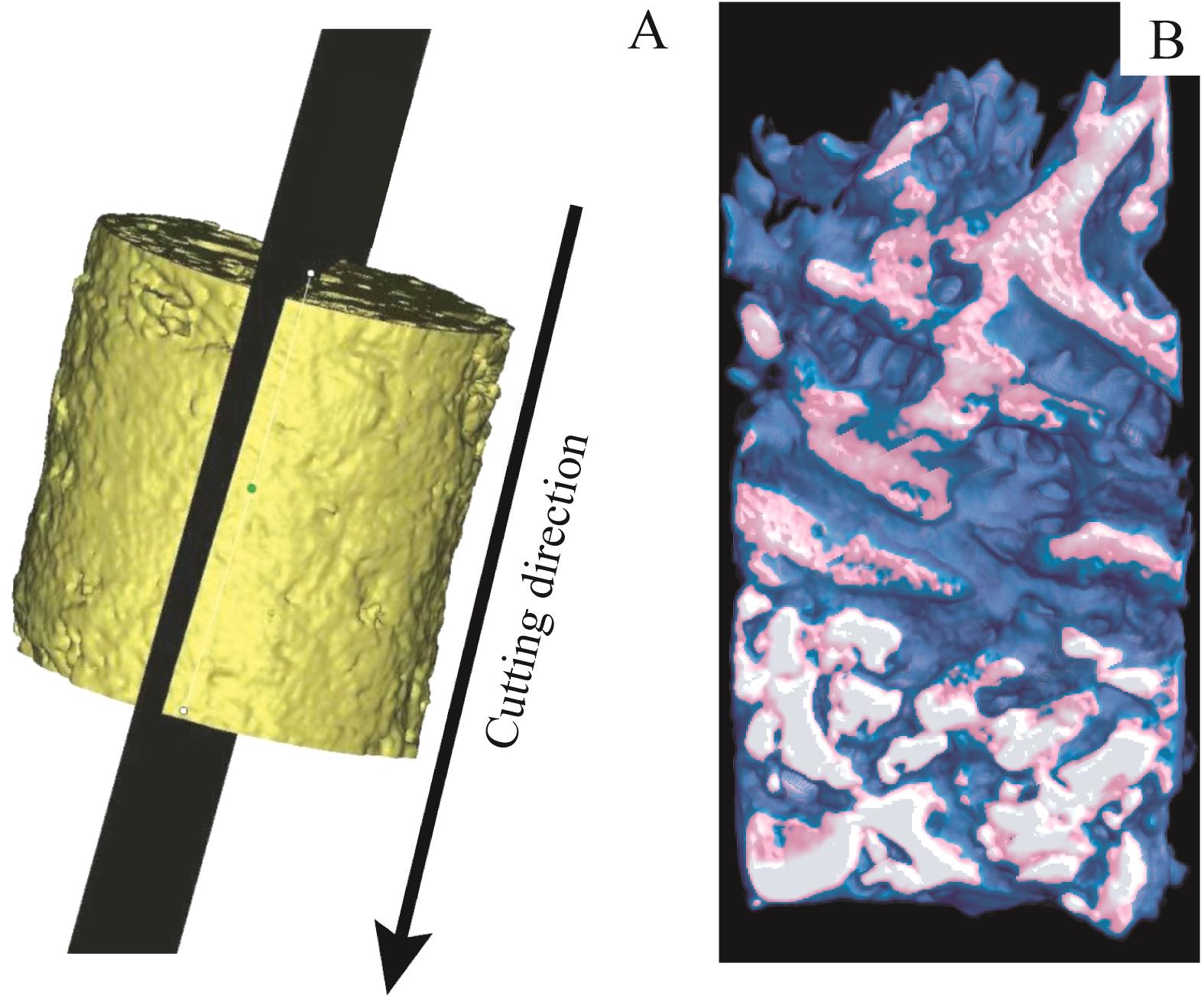
图4 术后8个月牙槽骨的Micro-CT重建方式Note: A. Cutting orientation of Micro-CT. B. Three-dimensional reconstruction of alveolar bone.
Fig 4 Micro-CT reconstruction of alveolar bone for 8 months after operation

图5 术后8个月牙槽骨Micro-CT重建结果Note: A. Trabecular bone number (Tb. N). B. Trabecular bone thickness (Tb. Th). C. Bone volume/total volume (BV/TV). D. Bone mineral density (BMD).
Fig 5 Alveolar bone data analysis of Micro-CT reconstruction for 8 months after operation

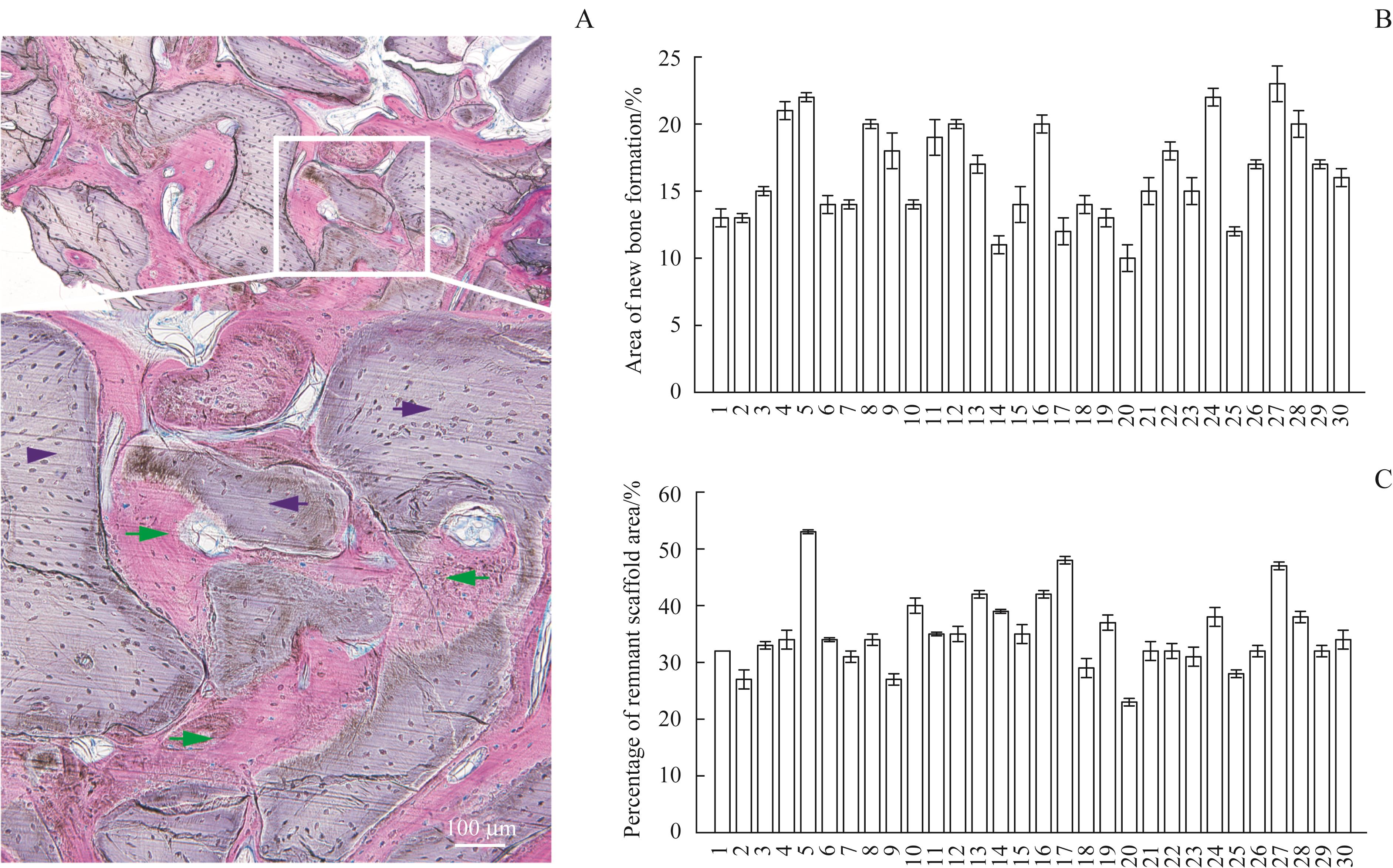
图6 术后8个月牙槽骨样本组织切片结果Note:A. H-E staining (×40) of the regenerated alveolar bone. Green arrow represents new regenerated bone, and blue arrow represents host bone. B. Area of new bone formation (%). C. Percentage of remnant scaffold area (%).
Fig 6 Histological analysis of the regenerated alveolar bone at 8 months post-operatively

图7 帐篷钉手术患者整体修复流程Note:A. Preoperative sagittal computed tomograms showed severe ridge defects. Red line indicates inferior alveolar nerve canal. Yellow row indicates bone defects. B/C. A mesial-distal crestal incision was made (B) and a full thickness flap was reflected (C). D. Image of tent peg used in the operation. E. Tent pegs were placed on the palatal side of the region of the right upper. F. Blood was collected from the patients. G. Fully mixed anorganic bovine bone-derived mineral and collected blood were put into the site. H. Tension-free wound closure with non-absorbable sutures after periosteal release was performed. I/J. Eight months after the tent peg placement, CBCT showed excellent alveolar ridge expansion (I and J showed different sections of the regenerated alveolar ridge). K. Augmented region after the tent peg placement. A buccal bony wall was formed after the eight months of placement of tent pegs. L/M. Preparation of drill holes (L) and placement of 2 implants (M). N. Inducing gingival formation with healing abutment. O. Results of induced gingival formation. P. Results of alveolar ridge reconstruction. Q. Prosthetic rehabilitation.
Fig 7 Whole reconstruction flow chart of the patient who underwent a tent-pole screw
| 1 | SIMION M, SCARANO A, GIONSO L, et al. Guided bone regeneration using resorbable and nonresorbable membranes: a comparative histologic study in humans[J]. Int J Oral Maxillofac Implants, 1996, 11(6): 735-742. |
| 2 | BRUNEL G, BROCARD D, DUFFORT J F, et al. Bioabsorbable materials for guided bone regeneration prior to implant placement and 7-year follow-up: report of 14 cases[J]. J Periodontol, 2001, 72(2): 257-264. |
| 3 | APRILE P, LETOURNEUR D, SIMON-YARZA T. Membranes for guided bone regeneration: a road from bench to bedside[J]. Adv Healthc Mater, 2020, 9(19): e2000707. |
| 4 | DESHPANDE S, DESHMUKH J, DESHPANDE S, et al. Vertical and horizontal ridge augmentation in anterior maxilla using autograft, xenograft and titanium mesh with simultaneous placement of endosseous implants[J]. J Indian Soc Periodontol, 2014, 18(5): 661-665. |
| 5 | LE B, ROHRER M D, PRASAD H S. Screw “tent-pole” grafting technique for reconstruction of large vertical alveolar ridge defects using human mineralized allograft for implant site preparation[J]. J Oral Maxillofac Surg, 2010, 68(2): 428-435. |
| 6 | XIAO T, ZHAO Y, LUO E, et al. “Tent-pole” for reconstruction of large alveolar defects: a case report[J]. J Oral Maxillofac Surg, 2016, 74(1): 55-67. |
| 7 | CHIAPASCO M, CASENTINI P, ZANIBONI M. Bone augmentation procedures in implant dentistry[J]. Int J Oral Maxillofac Implants, 2009, 24(Suppl): 237-259. |
| 8 | PEÑARROCHA-DIAGO M, GÓMEZ-ADRIÁN M D, GARCÍA-GARCÍA A, et al. Vertical mandibular alveolar bone distraction and dental implant placement: a case report[J]. J Oral Implantol, 2006, 32(3): 137-141. |
| 9 | SOEHARDI A, MEIJER G J, BERGE S J, et al. Lower border bone onlays to augment the severely atrophic (class Ⅵ) mandible in preparation for implants: a preliminary report[J]. Int J Oral Maxillofac Surg, 2014, 43(12): 1493-1499. |
| 10 | FELICE P, BARAUSSE C, PISTILLI R, et al. Guided “sandwich” technique: a novel surgical approach for safe osteotomies in the treatment of vertical bone defects in the posterior atrophic mandible: a case report[J]. Implant Dent, 2014, 23(6): 738-744. |
| 11 | KIYOKAWA K, RIKIMARU H, KIYOKAWA M, et al. Treatment outcomes of implants performed after regenerative treatment of absorbed alveolar bone due to the severe periodontal disease and endoscopic surgery for maxillary sinus lift without bone grafts[J]. J Craniofacial Surg, 2013, 24(5): 1599-1602. |
| 12 | POLI P P, BERETTA M, CICCIU M, et al. Alveolar ridge augmentation with titanium mesh. A retrospective clinical study[J]. Open Dent J, 2014, 8: 148-158. |
| 13 | BUSER D, DULA K, BELSER U, et al. Localized ridge augmentation using guided bone regeneration. 1. Surgical procedure in the maxilla[J]. Int J Periodontics Restor Dent, 1993, 13(1): 29-45. |
| 14 | URBAN I A, JOVANOVIC S A, LOZADA J L. Vertical ridge augmentation using guided bone regeneration (GBR) in three clinical scenarios prior to implant placement: a retrospective study of 35 patients 12 to 72 months after loading[J]. Int J Oral Maxillofac Implants, 2009, 24(3): 502-510. |
| 15 | URBAN I A, MONJE A, LOZADA J L, et al. Long-term evaluation of peri-implant bone level after reconstruction of severely atrophic edentulous maxilla via vertical and horizontal guided bone regeneration in combination with sinus augmentation: a case series with 1 to 15 years of loading[J]. Clin Implant Dent Relat Res, 2017, 19(1): 46-55. |
| 16 | SAGHEB K, SCHIEGNITZ E, MOERGEL M, et al. Clinical outcome of alveolar ridge augmentation with individualized CAD-CAM-produced titanium mesh[J]. Int J Implant Dent, 2017, 3(1): 36. |
| 17 | CIOCCA L, LIZIO G, BALDISSARA P, et al. Prosthetically CAD-CAM-guided bone augmentation of atrophic jaws using customized titanium mesh: preliminary results of an open prospective study[J]. J Oral Implantol, 2018, 44(2): 131-137. |
| 18 | ROCA-MILLAN E, JANÉ-SALAS E, ESTRUGO-DEVESA A, et al. Evaluation of bone gain and complication rates after guided bone regeneration with titanium foils: a systematic review[J]. Materials (Basel), 2020, 13(23): 5346. |
| 19 | CORINALDESI G, PIERI F, SAPIGNI L, et al. Evaluation of survival and success rates of dental implants placed at the time of or after alveolar ridge augmentation with an autogenous mandibular bone graft and titanium mesh: a 3- to 8-year retrospective study[J]. Int J Oral Maxillofac Implants, 2009, 24(6): 1119-1128. |
| 20 | MCALLISTER B S, HAGHIGHAT K. Bone augmentation techniques[J]. J Periodontol, 2007, 78(3): 377-396. |
| 21 | MACHTEI E E. The effect of membrane exposure on the outcome of regenerative procedures in humans: a meta-analysis[J]. J Periodontol, 2001, 72(4): 512-516. |
| 22 | MARX R E, SHELLENBERGER T, WIMSATT J, et al. Severely resorbed mandible: predictable reconstruction with soft tissue matrix expansion (tent pole) grafts[J]. J Oral Maxillofac Surg, 2002, 60(8): 878-888. |
| 23 | CHASIOTI E, CHIANG T F, DREW H J. Maintaining space in localized ridge augmentation using guided bone regeneration with tenting screw technology[J]. Quintessence Int, 2013, 44(10): 763-771. |
| 24 | KHOJASTEH A, BEHNIA H, SHAYESTEH Y S, et al. Localized bone augmentation with cortical bone blocks tented over different particulate bone substitutes: a retrospective study[J]. Int J Oral Maxillofac Implants, 2012, 27(6): 1481-1493. |
| 25 | 赵永强, 蒋练. 帐篷式骨增量技术在口腔种植中的应用研究进展[J]. 口腔疾病防治, 2020, 28(12): 811-816. |
| ZHAO Y Q, JIANG L. Application of the tent bone augmentation technique in dental implants[J]. J Prev & Treat Stomatol Dis, 2020, 28(12): 811-816. | |
| 26 | POURDANESH F, ESMAEELINEJAD M, AGHDASHI F. Clinical outcomes of dental implants after use of tenting for bony augmentation: a systematic review[J]. Br J Oral Maxillofac Surg, 2017, 55(10): 999-1007. |
| 27 | FARIAS D, CACERES F, SANZ A, et al. Horizontal bone augmentation in the posterior atrophic mandible and dental implant stability using the tenting screw technique[J]. Int J Periodontics Restorative Dent, 2021, 41(4): e147-e155. |
| 28 | CÉSAR NETO J B, CAVALCANTI M C, SAPATA V M, et al. The positive effect of tenting screws for primary horizontal guided bone regeneration: a retrospective study based on cone-beam computed tomography data[J]. Clin Oral Implants Res, 2020, 31(9): 846-855. |
| 29 | 王潇轶, 王国庆, 李芳, 等. “帐篷螺丝”技术在后牙种植区水平骨增量中的临床疗效观察[J]. 现代口腔医学杂志, 2022, 36(01): 27-29. |
| WANG X Y, WANG G Q, LI F, et al. Clinical observation of horizontal bone augmentation during posterior tooth implantation with tent-screw technique[J]. J Modern Stomatol, 2022, 36(01): 27-29. | |
| 30 | 邹多宏, 刘昌奎, 薛洋, 等. 帐篷钉技术在牙槽骨修复与再生中的临床应用及操作规范[J]. 中国口腔颌面外科杂志, 2021, 19(1): 1-5. |
| ZOU D H, LIU C K, XUE Y, et al. Clinical application of Tent-Peg technique in the reparation and regeneration of alveolar bone-standard operational practice[J]. China J Oral Maxillofac Surg, 2021, 19(1): 1-5. | |
| 31 | HURLEY L A, STINCHFIELD F E, BASSETT A L, et al. The role of soft tissues in osteogenesis. An experimental study of canine spine fusions[J]. J Bone Joint Surg Am, 1959, 41-A: 1243-1254. |
| 32 | LIU J, KERNS D G. Mechanisms of guided bone regeneration: a review[J]. Open Dent J, 2014, 8: 56-65. |
| 33 | RASIA-DAL POLO M, POLI P P, RANCITELLI D, et al. Alveolar ridge reconstruction with titanium meshes: a systematic review of the literature[J]. Med Oral Patol Oral Cir Bucal, 2014, 19(6): e639-e646. |
| 34 | MISCH C M, JENSEN O T, PIKOS M A, et al. Vertical bone augmentation using recombinant bone morphogenetic protein, mineralized bone allograft, and titanium mesh: a retrospective cone beam computed tomography study[J]. Int J Oral Maxillofac Implants, 2015, 30(1): 202-207. |
| 35 | RIBEIRO FILHO S A, FRANCISCHONE C E, DE OLIVEIRA J C, et al. Bone augmentation of the atrophic anterior maxilla for dental implants using rhBMP-2 and titanium mesh: histological and tomographic analysis[J]. Int J Oral Maxillofac Surg, 2015, 44(12): 1492-1498. |
| 36 | DAGA D, MEHROTRA D, MOHAMMAD S, et al. Tentpole technique for bone regeneration in vertically deficient alveolar ridges: a prospective study[J]. Oral Biol Craniofac Res, 2018, 8(1): 20-24. |
| 37 | DEEB G R, TRAN D, CARRICO C K, et al. How effective is the tent screw pole technique compared to other forms of horizontal ridge augmentation?[J]. J Oral Maxillofac Surg, 2017, 75(10): 2093-2098. |
| 38 | WANG H L, BOYAPATI L. "PASS" principles for predictable bone regeneration[J]. Implant Dent, 2006, 15(1): 8-17. |
| 39 | SCHWARZ F, ROTHAMEL D, HERTEN M, et al. Immunohis-tochemical characterization of guided bone regeneration at a dehiscence-type defect using different barrier membranes: an experimental study in dogs[J]. Clin Oral Implants Res, 2008, 19(4): 402-415. |
| 40 | OH T J, MERAW S J, LEE E J, et al. Comparative analysis of collagen membranes for the treatment of implant dehiscence defects[J]. Clin Oral Implants Res, 2003, 14(1): 80-90. |
| 41 | LI X, WANG X, ZHAO T, et al. Guided bone regeneration using chitosan-collagen membranes in dog dehiscence-type defect model[J]. J Oral Maxillofac Surg, 2014, 72(2): 304. e1-e14. |
| 42 | CLAVERO J, LUNDGREN S. Ramus or chin grafts for maxillary sinus inlay and local onlay augmentation: comparison of donor site morbidity and complications[J]. Clin Implant Dent Relat Res, 2003, 5(3): 154-160. |
| 43 | YAMAUCHI K, TAKAHASHI T, FUNAKI K, et al. Histological and histomorphometrical comparative study of β-tricalcium phosphate block grafts and periosteal expansion osteogenesis for alveolar bone augmentation[J]. Int J Oral Maxillofac Surg, 2010, 39(10): 1000-1006. |
| 44 | KLINGE B, ALBERIUS P, ISAKSSON S, et al. Osseous response to implanted natural bone mineral and synthetic hydroxylapatite ceramic in the repair of experimental skull bone defects[J]. J Oral Maxillofac Surg, 1992, 50(3): 241-249. |
| 45 | QUIÑONES C R, HÜRZELER M B, SCHÜPBACH P, et al. Maxillary sinus augmentation using different grafting materials and dental implants in monkeys. Part Ⅳ. Evaluation of hydroxyapatite-coated implants[J]. Clin Oral Implants Res, 1997, 8(6): 497-505. |
| 46 | PIATTELLI M, FAVERO G A, SCARANO A, et al. Bone reactions to anorganic bovine bone (Bio-Oss) used in sinus augmentation procedures: a histologic long-term report of 20 cases in humans[J]. Int J Oral Maxillofac Implants, 1999, 14(6): 835-840. |
| 47 | HALLMAN M, SENNERBY L, LUNDGREN S. A clinical and histologic evaluation of implant integration in the posterior maxilla after sinus floor augmentation with autogenous bone, bovine hydroxyapatite, or a 20:80 mixture[J]. Int J Oral Maxillofac Implants, 2002, 17(5): 635-643. |
| 48 | BLOCK M S, DEGEN M. Horizontal ridge augmentation using human mineralized particulate bone: preliminary results[J]. J Oral Maxillofac Surg, 2004, 62(9 Suppl 2): 67-72. |
| [1] | 魏祥, 魏凌飞, 徐纯峰, 高玉洁, 聂萍, 于德栋. 负载牛磺熊去氧胆酸的光交联明胶水凝胶支架在兔膝关节软骨缺损修复中的效能[J]. 上海交通大学学报(医学版), 2025, 45(7): 829-837. |
| [2] | 赵萌, 江莉婷, 高益鸣. 基于锥形线束CT的上颌后牙区牙槽骨增龄性变化研究[J]. 上海交通大学学报(医学版), 2024, 44(10): 1273-1278. |
| [3] | 李晨琳, 李岩, 徐光宙. 预防性拔除下颌第三磨牙牙胚的临床研究[J]. 上海交通大学学报(医学版), 2022, 42(7): 893-897. |
| [4] | 陆叶平, 陈敏洁. 改良位点保留法拔除复杂下颌阻生第三磨牙对临床成骨愈合的作用初探[J]. 上海交通大学学报(医学版), 2022, 42(6): 729-734. |
| [5] | 董玉山, 张文杰. 细胞因子治疗骨关节炎的研究进展[J]. 上海交通大学学报(医学版), 2022, 42(11): 1627-1632. |
| [6] | 袁剑鸣, 魏斌, 许卫星, 张磊. 锥形束CT研究不同基台角度对上颌前牙区牙槽骨改变的影响[J]. 上海交通大学学报(医学版), 2021, 41(3): 339-343. |
| [7] | 李 元,史俊宇,张 枭,赖红昌. 上颌前牙区牙槽骨缺损形态学特征与引导骨再生手术效果的相关性研究[J]. 上海交通大学学报(医学版), 2020, 40(10): 1414-1419. |
| [8] | 林歆妍1,周文洁1,王 凤2,黄 伟2,王 震3,曲行舟3#,吴轶群1#. 颧种植技术重建上颌骨缺损的疗效评估[J]. 上海交通大学学报(医学版), 2020, 40(10): 1382-1387. |
| [9] | 沈舜尧 1, 2,柳稚旭 1, 2,姜腾飞 1,余婧爽 1, 2,袁灏 1, 2,张雷 1, 2,史俊 1, 2,王旭东 1, 2. 逆向工程技术在腓骨瓣修复复杂下颌骨缺损中的应用[J]. 上海交通大学学报(医学版), 2019, 39(9): 1060-. |
| [10] | 喻金凤,胡赟,黄明娜,陈军,明叶,郑雷蕾. 骨性Ⅲ类错 伴下颌偏斜患者第一磨牙及其周围牙槽骨对称性的三维研究[J]. 上海交通大学学报(医学版), 2018, 38(3): 288-. |
| [11] | 周迪,吴艳,王云霁,范小平. 上颌前牙不同内收方式下相关牙槽骨改建变化的锥形束CT研究[J]. 上海交通大学学报(医学版), 2018, 38(11): 1375-. |
| [12] | 贾朗,黄荣忠,王愉乐,贾功伟. 不同强度体外冲击波联合骨髓间充质干细胞移植对大鼠骨缺损的修复效果[J]. 上海交通大学学报(医学版), 2016, 36(12): 1706-. |
| [13] | 易诚青, 马春辉, 张国桥, 等. CXCL13和CXCR5在骨缺损成骨微环境中的表达特征[J]. , 2012, 32(12): 1536-. |
| 阅读次数 | ||||||
|
全文 |
|
|||||
|
摘要 |
|
|||||