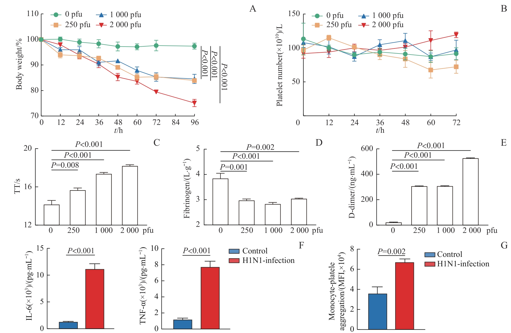
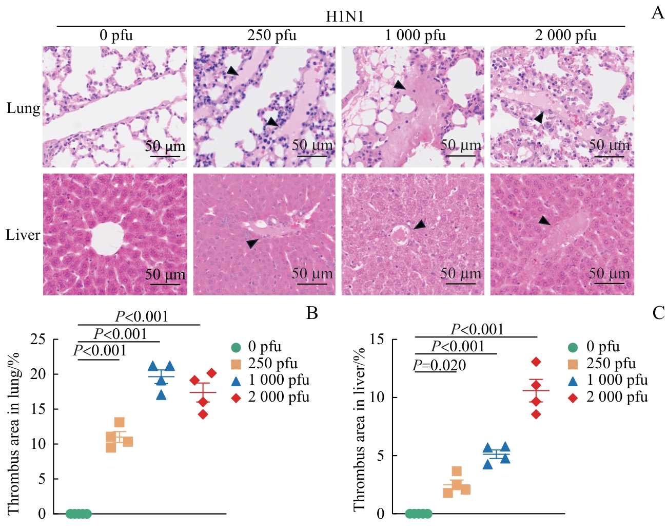
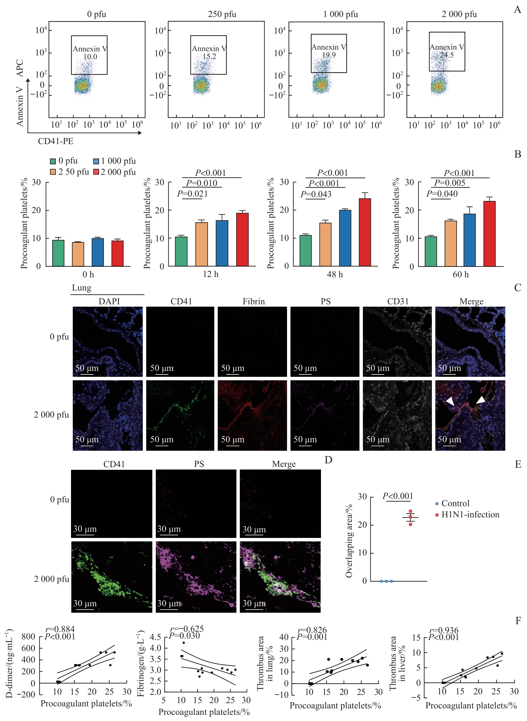
| [1] |
Ksiazek T G, Erdman D, Goldsmith C S, et al. A novel coronavirus associated with severe acute respiratory syndrome[J]. N Engl J Med, 2003, 348(20): 1953-1966.
|
| [2] |
Sullivan S J, Jacobson R M, Dowdle W R, et al. 2009 H1N1 influenza[J]. Mayo Clin Proc, 2010, 85(1): 64-76.
|
| [3] |
Van Wissen M, Keller T T, Ronkes B, et al. Influenza infection and risk of acute pulmonary embolism[J]. Thromb J, 2007, 5: 16.
|
| [4] |
Al-Samkari H, Karp Leaf R S, Dzik W H, et al. COVID-19 and coagulation: bleeding and thrombotic manifestations of SARS-CoV-2 infection[J]. Blood, 2020, 136(4): 489-500.
|
| [5] |
Evans P C, Rainger G E, Mason J C, et al. Endothelial dysfunction in COVID-19: a position paper of the ESC working group for atherosclerosis and vascular biology, and the ESC council of basic cardiovascular science[J]. Cardiovasc Res, 2020, 116(14): 2177-2184.
|
| [6] |
Mackman N, Antoniak S, Wolberg A S, et al. Coagulation abnormalities and thrombosis in patients infected with SARS-CoV-2 and other pandemic viruses[J]. Arterioscler Thromb Vasc Biol, 2020, 40(9): 2033-2044.
|
| [7] |
GóMez-Mesa J E, Galindo-Coral S, Montes M C, et al. Thrombosis and coagulopathy in COVID-19[J]. Curr Probl Cardiol, 2021, 46(3): 100742.
|
| [8] |
Zerangian N, Erabi G, Poudineh M, et al. Venous thromboembolism in viral diseases: a comprehensive literature review[J]. Health Sci Rep, 2023, 6(2): e1085.
|
| [9] |
Avnon L S, Munteanu D, Smoliakov A, et al. Thromboembolic events in patients with severe pandemic influenza A/H1N1[J]. Eur J Intern Med, 2015, 26(8): 596-598.
|
| [10] |
Repsold L, Joubert A M. Platelet function, role in thrombosis, inflammation, and consequences in chronic myeloproliferative disorders[J]. Cells, 2021, 10(11): 3034.
|
| [11] |
Gremmel T, Frelinger A L, Michelson A D. Platelet physiology[J]. Semin Thromb Hemost, 2024, 50(8): 1173-1186.
|
| [12] |
Koupenova M, Kehrel B E, Corkrey H A, et al. Thrombosis and platelets: an update[J]. Eur Heart J, 2017, 38(11): 785-791.
|
| [13] |
Holinstat M. Normal platelet function[J]. Cancer Metastasis Rev, 2017, 36(2): 195-198.
|
| [14] |
Ali M A M, Spinler S A. COVID-19 and thrombosis: from bench to bedside[J]. Trends Cardiovasc Med, 2021, 31(3): 143-160.
|
| [15] |
Ding J, Hostallero D E, El Khili M R, et al. A network-informed analysis of SARS-CoV-2 and hemophagocytic lymphohistiocytosis genes′ interactions points to neutrophil extracellular traps as mediators of thrombosis in COVID-19[J]. PLoS Comput Biol, 2021, 17(3): e1008810.
|
| [16] |
Ackermann M, Verleden S E, Kuehnel M, et al. Pulmonary vascular endothelialitis, thrombosis, and angiogenesis in covid-19[J]. N Engl J Med, 2020, 383(2): 120-128.
|
| [17] |
Heemskerk J M, Mattheij N A, Cosemans J M. Platelet-based coagulation: different populations, different functions[J]. J Thromb Haemost, 2013, 11(1): 2-16.
|
| [18] |
Reddy E C, Rand M L. Procoagulant phosphatidylserine-exposing platelets in vitro and in vivo[J]. Front Cardiovasc Med, 2020, 7: 15.
|
| [19] |
Bevers E M, Williamson P L. Getting to the outer leaflet: physiology of phosphatidylserine exposure at the plasma membrane[J]. Physiol Rev, 2016, 96(2): 605-645.
|
| [20] |
Millington-Burgess S L, Harper M T. Cytosolic and mitochondrial Ca2+ signaling in procoagulant platelets[J]. Platelets, 2021, 32(7): 855-862.
|
| [21] |
Lentz B R. Exposure of platelet membrane phosphatidylserine regulates blood coagulation[J]. Prog Lipid Res, 2003, 42(5): 423-438.
|
| [22] |
Colicchia M, Schrottmaier W C, Perrella G, et al. S100A8/A9 drives the formation of procoagulant platelets through GPIbα[J]. Blood, 2022, 140(24): 2626-2643.
|
| [23] |
Meyer N J, Gattinoni L, Calfee C S. Acute respiratory distress syndrome[J]. Lancet, 2021, 398(10300): 622-637.
|
| [24] |
Kwong J C, Schwartz K L, Campitelli M A, et al. Acute myocardial infarction after laboratory-confirmed influenza infection[J]. N Engl J Med, 2018, 378(4): 345-353.
|
| [25] |
Goldberg R, Ye W, Johns K, et al. Comparison of thrombotic and clinical outcomes in SARS-CoV-2-pneumonia versus other viral pneumonia in an urban academic medical center[J]. Heart Lung, 2023, 61: 153-157.
|
| [26] |
Snell J. SARS-CoV-2 infection and its association with thrombosis and ischemic stroke: a review[J]. Am J Emerg Med, 2021, 40: 188-192.
|
| [27] |
Mandel J, Casari M, Stepanyan M, et al. Beyond hemostasis: platelet innate immune interactions and thromboinflammation[J]. Int J Mol Sci, 2022, 23(7): 3868.
|
| [28] |
Thomas M R, Storey R F. The role of platelets in inflammation[J]. Thromb Haemost, 2015, 114(3): 449-458.
|
| [29] |
Franco A T, Corken A, Ware J. Platelets at the interface of thrombosis, inflammation, and cancer[J]. Blood, 2015, 126(5): 582-588.
|
| [30] |
Jenne C N, Kubes P. Platelets in inflammation and infection[J]. Platelets, 2015, 26(4): 286-292.
|
| [31] |
Li S P, Lu Z F, Wu S Y, et al. The dynamic role of platelets in cancer progression and their therapeutic implications[J]. Nat Rev Cancer, 2024, 24(1): 72-87.
|
| [32] |
Koupenova M, Corkrey H A, Vitseva O, et al. The role of platelets in mediating a response to human influenza infection[J]. Nat Commun, 2019, 10(1): 1780.
|
| [33] |
Rolling C C, Barrett T J, Berger J S. Platelet-monocyte aggregates: molecular mediators of thromboinflammation[J]. Front Cardiovasc Med, 2023, 10: 960398.
|
| [34] |
Liang H, Duan Z J, Li D, et al. Higher levels of circulating monocyte-platelet aggregates are correlated with viremia and increased sCD163 levels in HIV-1 infection[J]. Cell Mol Immunol, 2015, 12(4): 435-443.
|
| [35] |
Shantsila E, Lip G Y H. The role of monocytes in thrombotic disorders. Insights from tissue factor, monocyte-platelet aggregates and novel mechanisms[J]. Thromb Haemost, 2009, 102(5): 916-924.
|
| [36] |
Kappelmayer J, Kunapuli S P, Wyshock E G, et al. Characterization of monocyte-associated factor V[J]. Thromb Haemost, 1993, 70(2): 273-280.
|
| [37] |
Le Guyader A, Davis-Gorman G, Copeland J G, et al. A flow cytometric method for determining the binding of coagulation factor X to monocytes in whole human blood[J]. J Immunol Methods, 2004, 292(1/2): 207-215.
|
| [38] |
Toti F, Satta N, Fressinaud E, et al. Scott syndrome, characterized by impaired transmembrane migration of procoagulant phosphatidylserine and hemorrhagic complications, is an inherited disorder[J]. Blood, 1996, 87(4): 1409-1415.
|
| [39] |
Yuan Y P, Alwis I, Wu M C L, et al. Neutrophil macroaggregates promote widespread pulmonary thrombosis after gut ischemia[J]. Sci Transl Med, 2017, 9(409): eaam5861.
|
 ), 徐艳艳3,4(