| 1 |
GETGOOD A, BROOKS R, FORTIER L, et al. Articular cartilage tissue engineering: today′s research, tomorrow′s practice?[J]. J Bone Joint Surg Br, 2009, 91(5): 565-576.
|
| 2 |
STEINWACHS M R, GUGGI T, KREUZ P C. Marrow stimulation techniques[J]. Injury, 2008, 39 (Suppl 1): S26-S31.
|
| 3 |
HANGODY L, VÁSÁRHELYI G, HANGODY L R, et al. Autologous osteochondral grafting: technique and long-term results[J]. Injury, 2008, 39 (Suppl 1): S32-S39.
|
| 4 |
REVELL C M, ATHANASIOU K A. Success rates and immunologic responses of autogenic, allogenic, and xenogenic treatments to repair articular cartilage defects[J]. Tissue Eng Part B Rev, 2009, 15(1): 1-15.
|
| 5 |
XIE X, WANG Y, ZHAO C, et al. Comparative evaluation of MSCs from bone marrow and adipose tissue seeded in PRP-derived scaffold for cartilage regeneration[J]. Biomaterials, 2012, 33(29): 7008-7018.
|
| 6 |
KESIKBURUN S. Intra-articular platelet-rich plasma injections were not superior to viscosupplementation for early knee degeneration[J]. Ann Transl Med, 2015, 3(16): 228.
|
| 7 |
STELLER D, HERBST N, PRIES R, et al. Impact of incubation method on the release of growth factors in non-Ca2+-activated PRP, Ca2+-activated PRP, PRF and A-PRF[J]. J Cranio Maxillo Facial Surg, 2019, 47(2): 365-372.
|
| 8 |
DOHAN EHRENFEST D M, RASMUSSON L, ALBREKTSSON T. Classification of platelet concentrates: from pure platelet-rich plasma (P-PRP) to leucocyte- and platelet-rich fibrin (L-PRF)[J]. Trends Biotechnol, 2009, 27(3): 158-167.
|
| 9 |
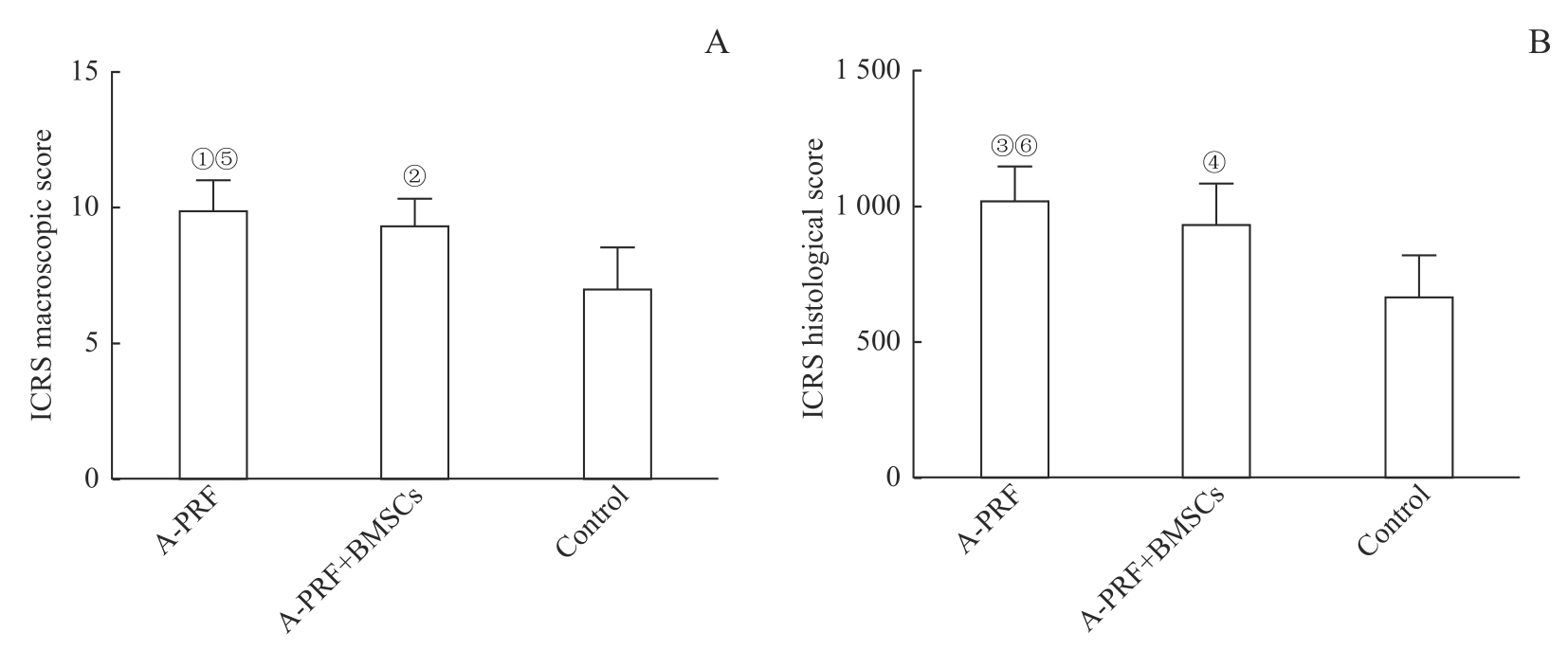
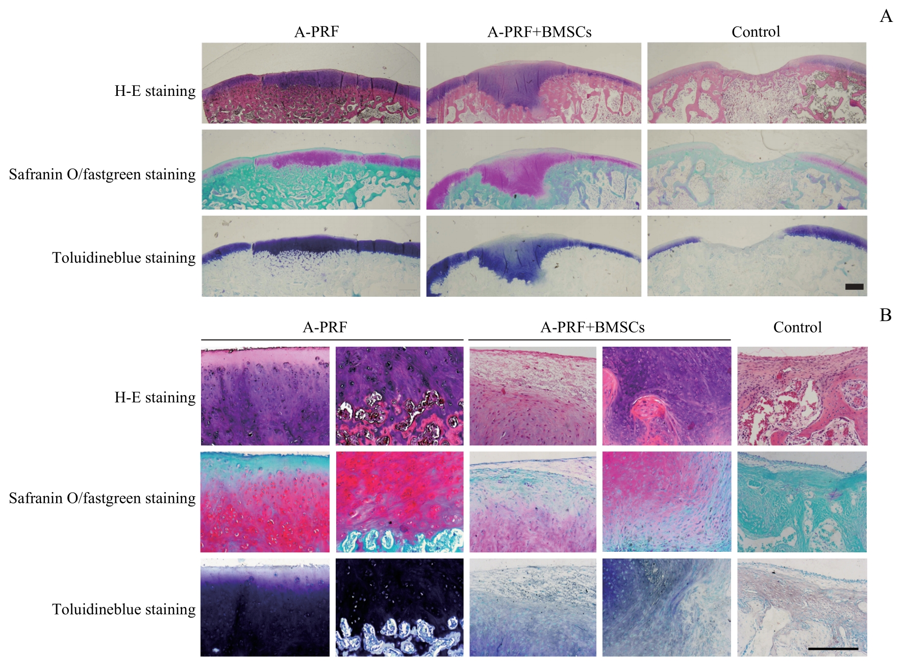
CLARK D, RAJENDRAN Y, PAYDAR S, et al. Advanced platelet-rich fibrin and freeze-dried bone allograft for ridge preservation: a randomized controlled clinical trial[J]. J Periodontol, 2018, 89(4): 379-387.
|
| 10 |
GHANAATI S, BOOMS P, ORLOWSKA A, et al. Advanced platelet-rich fibrin: a new concept for cell-based tissue engineering by means of inflammatory cells[J]. J Oral Implantol, 2014, 40(6): 679-689.
|
| 11 |
EGLE K, SALMA I, DUBNIKA A. From blood to regenerative tissue: how autologous platelet-rich fibrin can be combined with other materials to ensure controlled drug and growth factor release[J]. Int J Mol Sci, 2021, 22(21): 11553.
|
| 12 |
MASUKI H, OKUDERA T, WATANEBE T, et al. Growth factor and pro-inflammatory cytokine contents in platelet-rich plasma (PRP), plasma rich in growth factors (PRGF), advanced platelet-rich fibrin (A-PRF), and concentrated growth factors (CGF)[J]. Int J Implant Dent, 2016, 2(1): 19.
|
| 13 |
YU M, WANG X, LIU Y, et al. Correction to: cytokine release kinetics of concentrated growth factors in different scaffolds[J]. Clin Oral Investig, 2019, 23(4): 1999.
|
| 14 |
WONG C C, OU K L, LIN Y H, et al. Platelet-rich fibrin facilitates one-stage cartilage repair by promoting chondrocytes viability, migration, and matrix synthesis[J]. Int J Mol Sci, 2020, 21(2): E577.
|
| 15 |
NARAYANASWAMY R, PATRO B P, JEYARAMAN N, et al. Evolution and clinical advances of platelet-rich fibrin in musculoskeletal regeneration[J]. Bioengineering (Basel), 2023, 10(1): 58.
|
| 16 |
WONG C C, KUO T F, YANG T L, et al. Platelet-rich fibrin facilitates rabbit meniscal repair by promoting meniscocytes proliferation, migration, and extracellular matrix synthesis[J]. Int J Mol Sci, 2017, 18(8): E1722.
|
| 17 |
WONG C C, CHEN C H, CHAN W P, et al. Single-stage cartilage repair using platelet-rich fibrin scaffolds with autologous cartilaginous grafts[J]. Am J Sports Med, 2017, 45(13): 3128-3142.
|
| 18 |
DA SILVA L M P, SÁVIO D S F, DE ÁVILA F C, et al. Comparison of the effects of platelet concentrates produced by high and low-speed centrifugation protocols on the healing of critical-size defects in rat calvaria: a microtomographic and histomorphometric study[J]. Platelets, 2022, 33(8): 1175-1184.
|
| 19 |
ENGLER-PINTO A, SIESSERE S, CALEFI A, et al. Effects of leukocyte‐and platelet‐rich fibrin associated or not with bovine bone graft on the healing of bone defects in rats with osteoporosis induced by ovariectomy[J]. Clin Oral Implants Res, 2019, 30(10): 962-976.
|
| 20 |
FERREIRA SÁVIO D S, SILVA L M P D, REIS G G D, et al. Effects of platelet-rich fibrin produced by three centrifugation protocols on bone neoformation in defects created in rat calvaria[J]. Platelets, 2023, 34(1): 2228417.
|
| 21 |
TSUKIOKA T, HIRATSUKA T, NAKAMURA M, et al. An on-site preparable, novel bone-grafting complex consisting of human platelet-rich fibrin and porous particles made of a recombinant collagen-like protein[J]. J Biomed Mater Res Part B Appl Biomater, 2019, 107(5): 1420-1430.
|
| 22 |
SHEU S Y, WANG C H, PAO Y H, et al. The effect of platelet-rich fibrin on autologous osteochondral transplantation: an in vivo porcine model[J]. Knee, 2017, 24(6): 1392-1401.
|
| 23 |
SUMARTA N P M, KAMADJAJA D B, HENDRIJANTINI N, et al. Human umbilical cord mesenchymal stem cells over platelet rich fibrin scaffold for mandibular cartilage defects regenerative medicine[J]. Pesq Bras Odontoped Clin Integr, 2021, 21: e0034.
|
| 24 |
ABD EL RAOUF M, WANG X, MIUSI S, et al. Injectable-platelet rich fibrin using the low speed centrifugation concept improves cartilage regeneration when compared to platelet-rich plasma[J]. Platelets, 2019, 30(2): 213-221.
|
| 25 |
VAN DEN BORNE M P J, RAIJMAKERS N J H, VANLAUWE J, et al. International Cartilage Repair Society (ICRS) and Oswestry macroscopic cartilage evaluation scores validated for use in Autologous Chondrocyte Implantation (ACI) and microfracture[J]. Osteoarthr Cartil, 2007, 15(12): 1397-1402.
|
| 26 |
MAINIL-VARLET P, VAN DAMME B, NESIC D, et al. A new histology scoring system for the assessment of the quality of human cartilage repair: icrs Ⅱ[J]. Am J Sports Med, 2010, 38(5): 880-890.
|
| 27 |
ZHANG Z J, MCCAFFERY J M, SPENCER R G S, et al. Growth and integration of neocartilage with native cartilage in vitro[J]. J Orthop Res, 2005, 23(2): 433-439.
|
| 28 |
MIRHAJ M, TAVAKOLI M, VARSHOSAZ J, et al. Preparation of a biomimetic bi-layer chitosan wound dressing composed of A-PRF/sponge layer and L-arginine/nanofiber[J]. Carbohydr Polym, 2022, 292: 119648.
|
| 29 |
KOBAYASHI E, FLÜCKIGER L, FUJIOKA-KOBAYASHI M, et al. Comparative release of growth factors from PRP, PRF, and advanced-PRF[J]. Clin Oral Investig, 2016, 20(9): 2353-2360.
|
| 30 |
PARMAR P A, CHOW L W, ST-PIERRE J P, et al. Collagen-mimetic peptide-modifiable hydrogels for articular cartilage regeneration[J]. Biomaterials, 2015, 54: 213-225.
|
| 31 |
DOMÍNGUEZ PÉREZ J M, FERNÁNDEZ-SARMIENTO J A, AGUILAR GARCÍA D, et al. Cartilage regeneration using a novel autologous growth factors-based matrix for full-thickness defects in sheep[J]. Knee Surg Sports Traumatol Arthrosc, 2019, 27(3): 950-961.
|
| 32 |
LEE C R, GRODZINSKY A J, SPECTOR M. The effects of cross-linking of collagen-glycosaminoglycan scaffolds on compressive stiffness, chondrocyte-mediated contraction, proliferation and biosynthesis[J]. Biomaterials, 2001, 22(23): 3145-3154.
|
| 33 |
PARK H, TEMENOFF J S, HOLLAND T A, et al. Delivery of TGF-beta1 and chondrocytes via injectable, biodegradable hydrogels for cartilage tissue engineering applications[J]. Biomaterials, 2005, 26(34): 7095-7103.
|
| 34 |
BAE S E, CHOI D H, HAN D K, et al. Effect of temporally controlled release of dexamethasone on in vivo chondrogenic differentiation of mesenchymal stromal cells[J]. J Control Release, 2010, 143(1): 23-30.
|
| 35 |
CUI Y, HUANG R, WANG Y, et al. Down-regulation of LGR6 promotes bone fracture recovery using bone marrow stromal cells[J]. Biomed Pharmacother, 2018, 99: 629-637.
|
| 36 |
CHENG B, TU T, SHI X, et al. A novel construct with biomechanical flexibility for articular cartilage regeneration[J]. Stem Cell Res Ther, 2019, 10(1): 298.
|
 ), 吕成奇, 刘旭凌, 陈昱璐, 邹德荣, 陆家瑜(