上海交通大学学报(医学版) ›› 2023, Vol. 43 ›› Issue (6): 699-708.doi: 10.3969/j.issn.1674-8115.2023.06.006

• 论著 · 基础研究 • 上一篇    下一篇

miR-185-5p通过抑制巨噬细胞自噬促进胞内分枝杆菌生长

吴淇琦1(), 汪豪2, 林砺1, 晏博3(), 张舒林1,3()   

  1. 1.上海交通大学医学院免疫学与微生物学系,上海 200025
    2.武汉轻工大学生命科学与技术学院,武汉 430023
    3.复旦大学附属公共卫生临床中心,上海 201508
  • 收稿日期:2023-02-03 接受日期:2023-04-24 出版日期:2023-06-28 发布日期:2023-06-28
  • 通讯作者: 晏博,张舒林 E-mail:wuqiqi0223@163.com;bo.yan@shphc.org.cn;shulinzhang@sjtu.edu.cn
  • 作者简介:吴淇琦(1997—),女,硕士生;电子信箱:wuqiqi0223@163.com
  • 基金资助:
    国家自然科学基金(81871613)

miR-185-5p facilitates intracellular Mycobacterium growth via inhibiting macrophage autophagy

WU Qiqi1(), WANG Hao2, LIN Li1, YAN Bo3(), ZHANG Shulin1,3()   

  1. 1.Department of Immunology and Microbiology, Shanghai Jiao Tong University School of Medicine, Shanghai 200025, China
    2.School of Life Science and Technology, Wuhan Polytechnic University, Wuhan 430023, China
    3.Shanghai Public Health Clinical Center, Fudan University, Shanghai 201508, China
  • Received:2023-02-03 Accepted:2023-04-24 Online:2023-06-28 Published:2023-06-28
  • Contact: YAN Bo,ZHANG Shulin E-mail:wuqiqi0223@163.com;bo.yan@shphc.org.cn;shulinzhang@sjtu.edu.cn
  • Supported by:
    National Natural Science Foundation of China(81871613)

摘要:

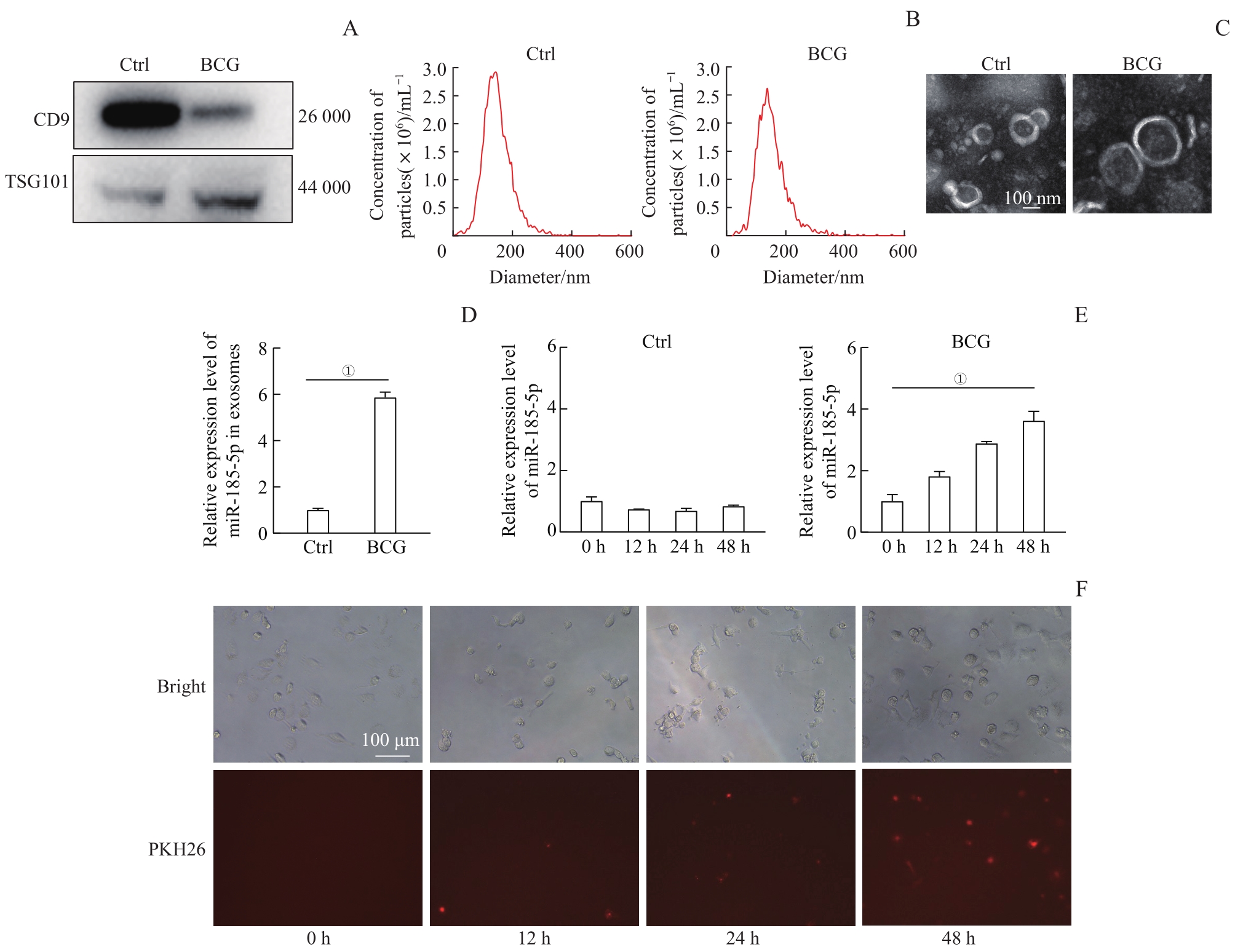
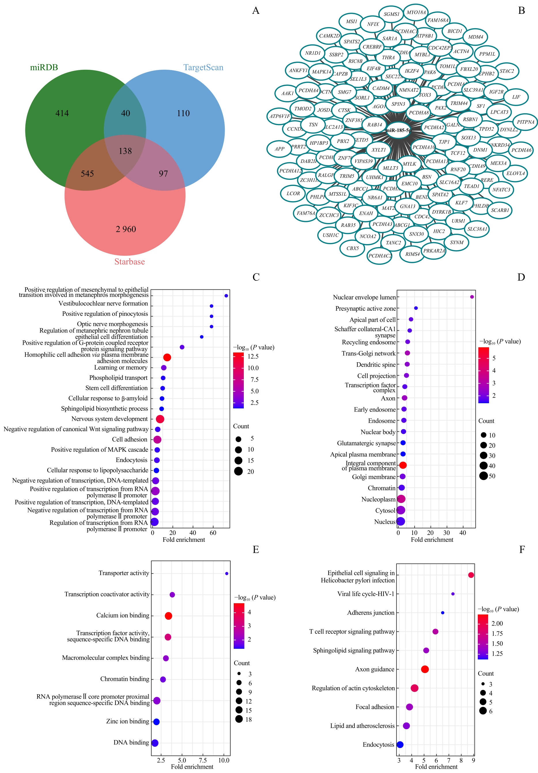
目的·探究miR-185-5p在分枝杆菌感染的巨噬细胞中对自噬的调控作用和对胞内分枝杆菌生存的影响。方法·使用卡介苗(Bacillus Calmette‐Guérin,BCG)感染人单核细胞白血病细胞(THP-1)诱导分化的巨噬细胞,通过超速离心法分离细胞培养上清液中的外泌体;通过Western blotting、纳米颗粒跟踪分析(nanoparticle tracking analysis,NTA)、透射电子显微镜(transmission electron microscopy,TEM)分别对外泌体的表面标志蛋白、大小和形态进行鉴定,通过实时荧光定量PCR(quantitative polymerase chain reaction,qPCR)检测BCG感染的巨噬细胞内及外泌体中miR-185-5p的表达水平。外泌体使用PKH26染料进行荧光染色并与巨噬细胞共培养,观察巨噬细胞对外泌体的吞噬情况。通过模拟物(mimic)和抑制剂(inhibitor)实现miR-185-5p在巨噬细胞内的过表达和抑制作用,通过菌落形成单位(colony-forming unit,CFU)实验验证miR-185-5p对巨噬细胞内BCG生长存活的影响。通过Western blotting检测自噬标志物微管相关蛋白1轻链3(microtubule-associated protein 1 light chain 3,LC3)蛋白的表达,研究过表达、抑制miR-185-5p以及使用自噬抑制剂氯喹(chloroquine,CQ)对巨噬细胞自噬的影响;进一步使用SensGFP-StubRFP-LC3自噬双荧光慢病毒检测miR-185-5p对自噬流的影响。使用TargetScan、miRDB和Starbase数据库对miR-185-5p的靶基因进行预测,并使用注释、可视化和综合发现数据库(Database for Annotation,Visualization and Integrated Discovery,DAVID)工具进行基因本体(Gene Ontology,GO)分析和京都基因与基因组百科全书(Kyoto Encyclopedia of Genes and Genomes,KEGG)功能富集分析。结果·超速离心法成功分离出巨噬细胞外泌体,Western blotting成功检测到外泌体标志蛋白白细胞分化抗原9(cluster differentiation 9,CD9)、肿瘤易感基因101(tumor susceptibility gene 101,TSG101);通过TEM、NTA鉴定,外泌体为双层膜结构,直径约150 nm。BCG感染后,miR-185-5p在巨噬细胞内和外泌体中的表达均显著上调(P=0.000),且其在BCG感染的巨噬细胞内的表达水平呈时间依赖性升高。外泌体与巨噬细胞共培养,可被巨噬细胞吞噬。与mimic和inhibitor的各自对照相比,mimic显著上调miR-185-5p的表达(P=0.000),而inhibitor抑制miR-185-5p的表达(P=0.002)。使用mimic引起巨噬细胞内BCG的CFU数量显著增加(P=0.000),而inhibitor则使CFU数量减少(P=0.041)。BCG感染使巨噬细胞中LC3Ⅱ蛋白的表达上调,而mimic可降低LC3Ⅱ的表达,inhibitor可升高LC3Ⅱ的表达。在CQ作用下,使用inhibitor抑制miR-185-5p仍能显著增强巨噬细胞中LC3Ⅱ的表达。自噬流检测结果显示,相比于各自的对照组,mimic组自噬体的生成明显减少(P=0.034),而inhibitor组自噬体的生成明显增加(P=0.042)。通过靶基因预测以及GO和KEGG功能富集分析发现,miR-185-5p可能通过靶向核受体亚家族1组D成员1(nuclear receptor subfamily 1 group D member 1,NR1D1Ras相关蛋白Rab-35(Ras-related protein Rab-35,RAB35细胞分裂控制蛋白42同源物(cell division control protein 42 homolog,CDC42)等基因发挥自噬调节作用。结论·分枝杆菌感染诱导巨噬细胞内及外泌体中miR-185-5p表达上调,miR-185-5p通过抑制自噬体的生成抑制巨噬细胞的自噬过程,从而促进细胞内分枝杆菌的生长及存活。

关键词: 分枝杆菌感染, 巨噬细胞, miR-185-5p, 自噬, 外泌体

Abstract:

Objective ·To explore the regulatory effect of miR-185-5p on autophagy in Mycobacterium-infected macrophages and its influence on the survival of intracellular Mycobacterium. Methods ·Macrophages derived from human monocytic-leukemia (THP-1) cells were infected with Bacillus Calmette-Guérin (BCG) strain. Exosomes from cell culture supernatant were isolated by ultracentrifugation, and exosome surface marker proteins, size and morphology were detected by Western blotting, nanoparticle tracking analysis (NTA) and transmission electron microscopy (TEM), respectively. The expression level of miR-185-5p in both macrophages and exosomes was detected by real time quantitative polymerase chain reaction (qPCR). The exosomes were labeled with the fluorescent dye PKH26 and co-cultured with macrophages to observe the phagocytosis of exosomes by macrophages. Subsequently, the overexpression and inhibition of miR-185-5p in macrophages were achieved by transfecting cells with miR-185-5p mimic and inhibitor, and the influence of miR-185-5p on intracellular BCG survival was detected by colony-forming units (CFU) assay. Autophagy marker microtubule-associated protein 1 light chain 3 (LC3) protein was detected by Western blotting.The effects of overexpression and inhibition of miR-185-5p and the use of autophagy inhibitor chloroquine (CQ) on macrophage autophagy were studied. The SensGFP-StubRFP-LC3 lentvirus was further employed to detect the effect of miR-185-5p on autophagic flux. Finally, TargetScan, miRDB and Starbase databases were used to predict the target genes of miR-185-5p, and the Database for Annotation, Visualization and Integrated Discovery (DAVID) was used for Gene Ontology (GO) analysis and Kyoto Encyclopedia of Genes and Genomes (KEGG) functional enrichment analysis of these predicted target genes. Results ·The macrophage exosomes were isolated by ultracentrifugation. Exosome marker proteins cluster differentiation 9 (CD9) and tumor susceptibility gene 101 (TSG101) were successfully detected by Western blotting. The exosomes had a bilayer membrane with a diameter of approximately 150 nm through TEM and NTA identification. After BCG infection, the expression of miR-185-5p was up-regulated in both macrophages and exosomes (P=0.000), and showed a time-dependent increase in macrophages. Exosomes, which were co-cultured with macrophages, can be phagocytized by macrophages. Compared with respective control of mimic and inhibitor, mimic significantly up-regulated miR-185-5p expression (P=0.000), while inhibitor inhibited miR-185-5p expression (P=0.002). miR-185-5p mimic significantly increased the number of CFU of intracellular BCG (P=0.000), while miR-185-5p inhibitor decreased the number of CFU (P=0.041). BCG infection up-regulated the expression of autophagy protein LC3Ⅱ in macrophages, while LC3Ⅱ expression was decreased by miR-185-5p mimic and increased by miR-185-5p inhibitor. CQ treatment combined with miR-185-5p inhibitor transfection still significantly increased LC3Ⅱ expression in macrophages. The SensGFP-StubRFP-LC3 lentivirus was further used to detect autophagy flux, and it was suggested that, compared with that in the respective control group, mimic significantly reduced the formation of autophagosomes (P=0.034), while inhibitor increased the formation of autophagosomes (P=0.042). Through target genes prediction and GO and KEGG functional enrichment analysis, it was found that miR-185-5p might play a role in autophagy regulation by targeting nuclear receptor subfamily 1 group D member 1 (NR1D1), Ras-related protein Rab-35 (RAB35), cell division control protein 42 homolog (CDC42), etc. Conclusion ·Mycobacterium infection can induce the up-regulation of miR-185-5p in both macrophages and exosomes, and miR-185-5p can inhibit the autophagy process of macrophages by inhibiting the formation of autophagosomes, which can promote the growth and survival of intracellular Mycobacterium.

Key words: Mycobacterium infection, macrophage, miR-185-5p, autophagy, exosome

中图分类号: