上海交通大学学报(医学版) ›› 2023, Vol. 43 ›› Issue (4): 406-416.doi: 10.3969/j.issn.1674-8115.2023.04.002
李旭冉1,2,3( ), 陶诗聪1,2,3(), 郭尚春1,2,3()
收稿日期:2022-12-23
接受日期:2023-03-27
出版日期:2023-04-28
发布日期:2023-04-28
通讯作者:
陶诗聪,电子信箱:sctao@shsmu.edu.cn。作者简介:李旭冉(1999—),男,硕士生;电子信箱:15737905921@163.com。
基金资助:
LI Xuran1,2,3(), TAO Shicong1,2,3(), GUO Shangchun1,2,3()
Received:2022-12-23
Accepted:2023-03-27
Online:2023-04-28
Published:2023-04-28
Contact:
TAO Shicong, E-mail: sctao@shsmu.edu.cn.Supported by:摘要:
目的·探究人骨髓间充质干细胞(bone marrow mesenchymal stem cell,BMSC)来源的小细胞外囊泡(small extracellular vesicle,sEV)对小鼠破骨细胞分化和巨噬细胞极化的调控作用,以及对骨质疏松症小鼠的影响。方法·培养BMSC并通过差速离心法提取sEV,通过透射电子显微镜(transmission electron microscope,TEM)及纳米颗粒跟踪分析技术(nanoparticle tracking analysis,NTA)鉴定得到的sEV。通过巨噬细胞集落刺激因子(macrophage colony-stimulating factor,M-CSF)及核因子κB受体激活蛋白配体(receptor activator of nuclear factor-κB ligand,RANKL)刺激RAW264.7细胞以诱导形成破骨细胞,通过抗酒石酸酸性磷酸酶(tartrate-resistant acid phosphatase,TRAP)染色及鬼笔环肽染色检测sEV对破骨细胞分化的调控作用。通过荧光定量PCR检测sEV对破骨细胞标志基因环磷腺苷效应元件结合蛋白(cAMP-response element binding protein,CREB)、组织蛋白酶K(cathepsin K,CTSK)及c-Jun(Jun proto-oncogene)mRNA表达量的影响。使用脂多糖刺激RAW264.7细胞极化为M1型巨噬细胞;使用白细胞介素-4(interleukin-4,IL-4)及IL-13刺激RAW264.7细胞极化为M2型巨噬细胞。利用流式细胞术检测sEV对M1及M2型巨噬细胞极化的影响。通过微计算机断层扫描成像(micro-computed tomography,micro-CT)及TRAP染色观察sEV对骨质疏松症小鼠模型腰椎骨组织的影响。结果·TEM及NTA结果显示分离得到的sEV具有典型的球状结构,直径为30~150 nm。TRAP染色及鬼笔环肽染色结果显示,BMSC来源的sEV能够有效抑制RAW264.7细胞融合形成破骨细胞。PCR结果表明sEV能够降低CREB、CTSK和c-Jun mRNA的表达量(均P<0.05)。流式细胞术分析表明,BMSC来源的sEV能够抑制RAW264.7细胞极化为M1型巨噬细胞,促进其极化为M2型巨噬细胞。Micro-CT检测结果显示,sEV干预后模型小鼠腰椎骨小梁数量和骨体积分数显著高于未干预小鼠(均P<0.05);TRAP染色结果显示,sEV干预后腰椎组织中的破骨细胞数量减少。结论·人BMSC来源的sEV可以延缓骨质疏松小鼠的骨质流失,这可能与其抑制小鼠破骨细胞分化及促进M2型巨噬细胞极化的作用有关。
中图分类号:
李旭冉, 陶诗聪, 郭尚春. 骨髓间充质干细胞来源小细胞外囊泡对骨质疏松症的改善作用[J]. 上海交通大学学报(医学版), 2023, 43(4): 406-416.
LI Xuran, TAO Shicong, GUO Shangchun. Ameliorative effects on osteoporosis of small extracellular vesicles derived from bone marrow mesenchymal stem cells[J]. Journal of Shanghai Jiao Tong University (Medical Science), 2023, 43(4): 406-416.
| Primer | Sequence |
|---|---|
| β-actin forward | 5'-CCTCTATGCCAACACAGT-3' |
| β-actin reverse | 5'-AGCCACCAATCCACACAG-3' |
| CREB forward | 5'-CCTTGCTTTCCGAATCCTC-3' |
| CREB reverse | 5'-CACTTTGGCTGGACATCTTG-3' |
| c-Jun forward | 5'-AGCAACTTTCCTGACCCAGAG-3' |
| c-Jun reverse | 5'-TCTTTACAGTCTCGGTGGCAG-3' |
| CTSK forward | 5'-CCAGAATCTTGTGGACTGTGT-3' |
| CTSK reverse | 5'-CATCTTCAGAGTCAATGCCTC-3' |
表1 荧光定量PCR引物序列
Tab 1 Primer sequences for qPCR
| Primer | Sequence |
|---|---|
| β-actin forward | 5'-CCTCTATGCCAACACAGT-3' |
| β-actin reverse | 5'-AGCCACCAATCCACACAG-3' |
| CREB forward | 5'-CCTTGCTTTCCGAATCCTC-3' |
| CREB reverse | 5'-CACTTTGGCTGGACATCTTG-3' |
| c-Jun forward | 5'-AGCAACTTTCCTGACCCAGAG-3' |
| c-Jun reverse | 5'-TCTTTACAGTCTCGGTGGCAG-3' |
| CTSK forward | 5'-CCAGAATCTTGTGGACTGTGT-3' |
| CTSK reverse | 5'-CATCTTCAGAGTCAATGCCTC-3' |
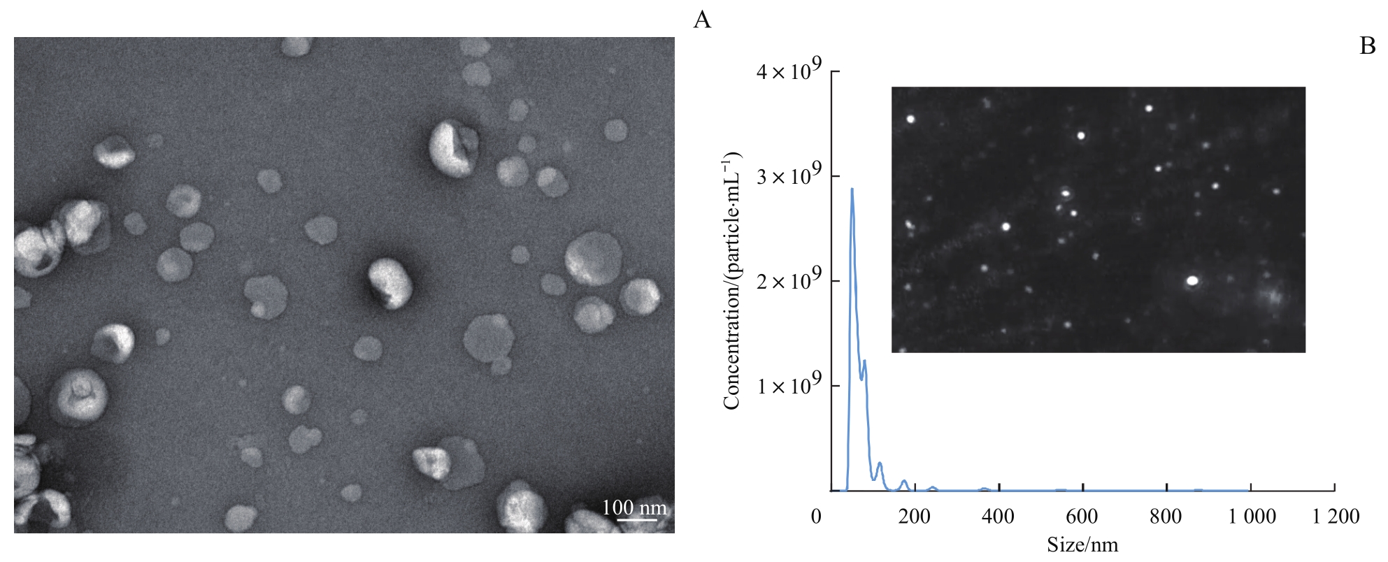
图1 通过TEM及NTA对BMSC来源sEV的鉴定Note: A. The typical morphology of sEVs in a TEM image (×20 000). B. The result and a typical image of NTA.
Fig 1 Identification of sEVs from BMSCs by TEM and NTA
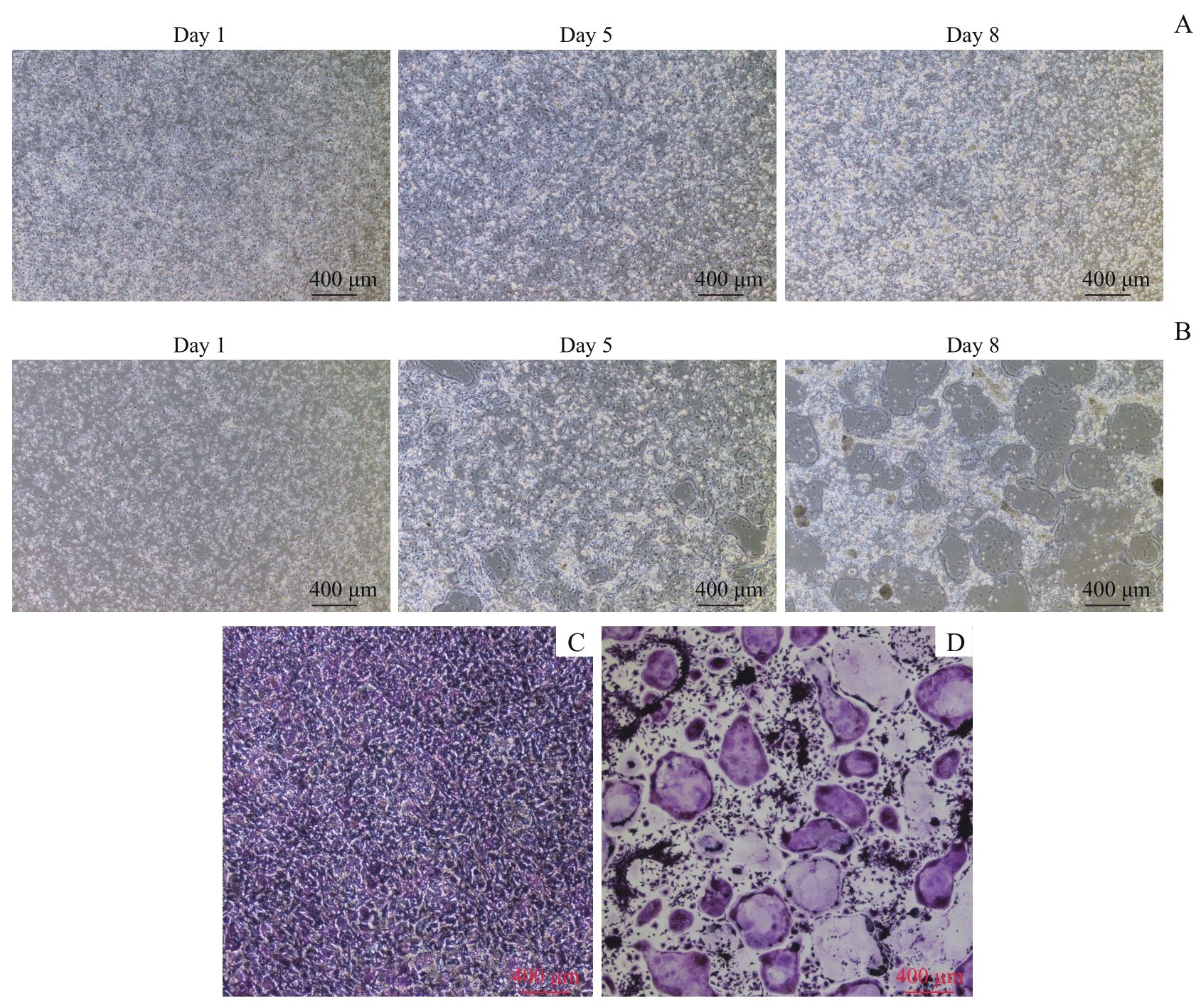
图2 诱导破骨细胞分化及TRAP染色结果Note: A/B. The RAW264.7 cells were seeded at a high density (1.5×10⁶ per well, A) and a low density (7.5×10⁵ per well, B) and the formation of osteoclasts was observed at different time-points (×40). C/D. The results of TRAP staining of the cells seeded at a high density (C) and a low density (D) (×40).
Fig 2 Formation of osteoclasts and the results of TRAP staining
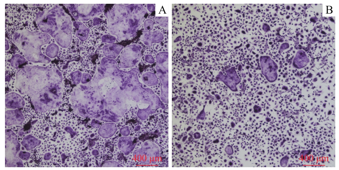
图3 2组细胞向破骨细胞分化后的TRAP染色结果 (×40)Note: A. The control group. B. The sEV group.
Fig 3 TRAP staining results of two groups of cells differentiated into osteoclasts (×40)
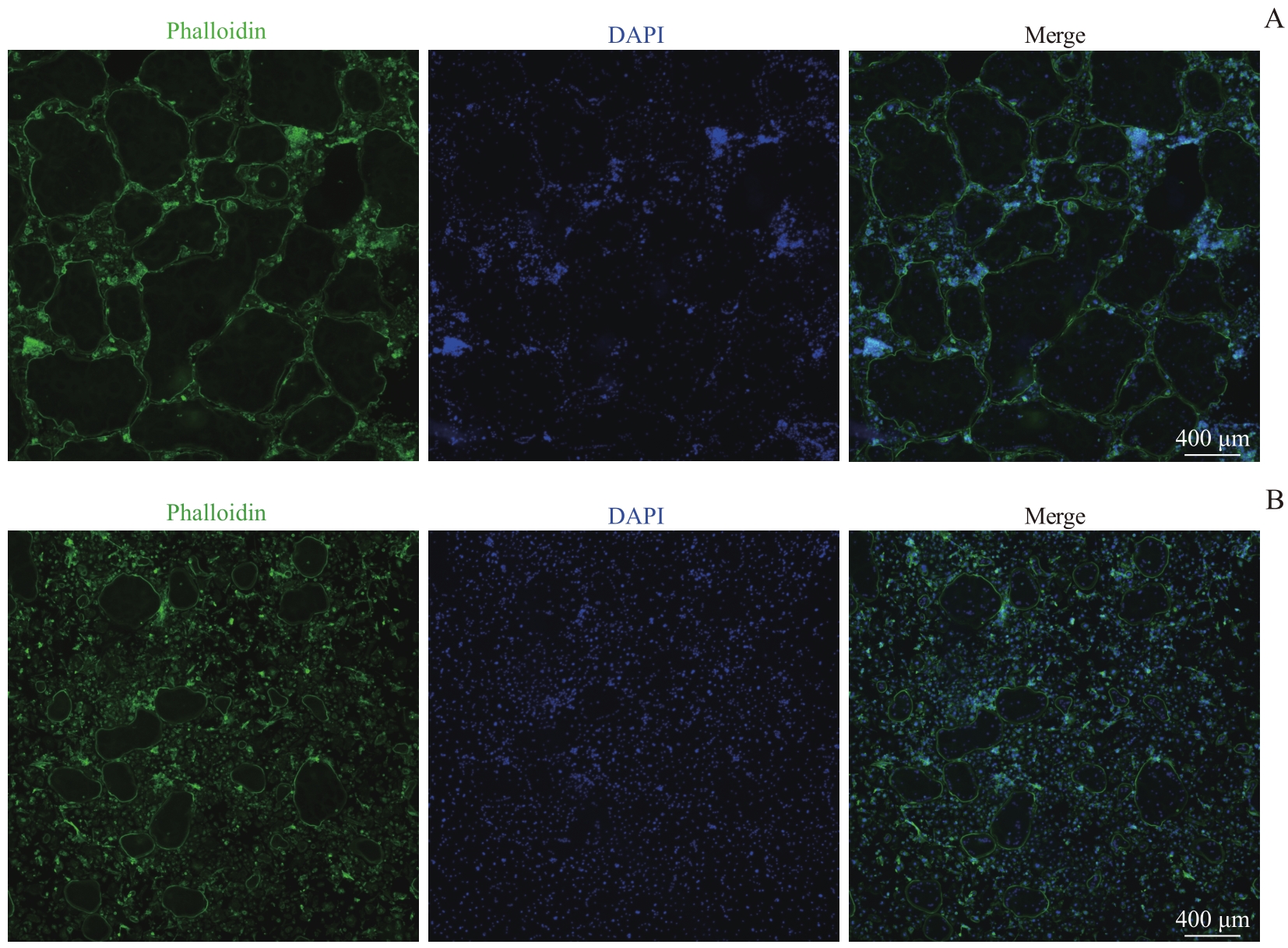
图4 2组细胞向破骨细胞分化后的鬼笔环肽染色结果 (×40)Note: A. The control group. B. The sEV group.
Fig 4 Phalloidin staining results of two groups of cells differentiated into osteoclasts (×40)
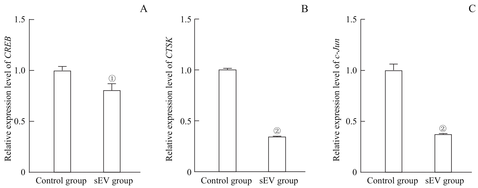
图5 荧光定量PCR检测2组细胞向破骨细胞分化后的 CREB (A)、 CTSK (B)与 c-Jun (C)的mRNA水平Note: ①P=0.011, ②P=0.000, compared with the control group.
Fig 5 mRNA levels of CREB (A), CTSK (B) and c-Jun (C) detected by qPCR after the cells differentiated into osteoclasts in the two groups
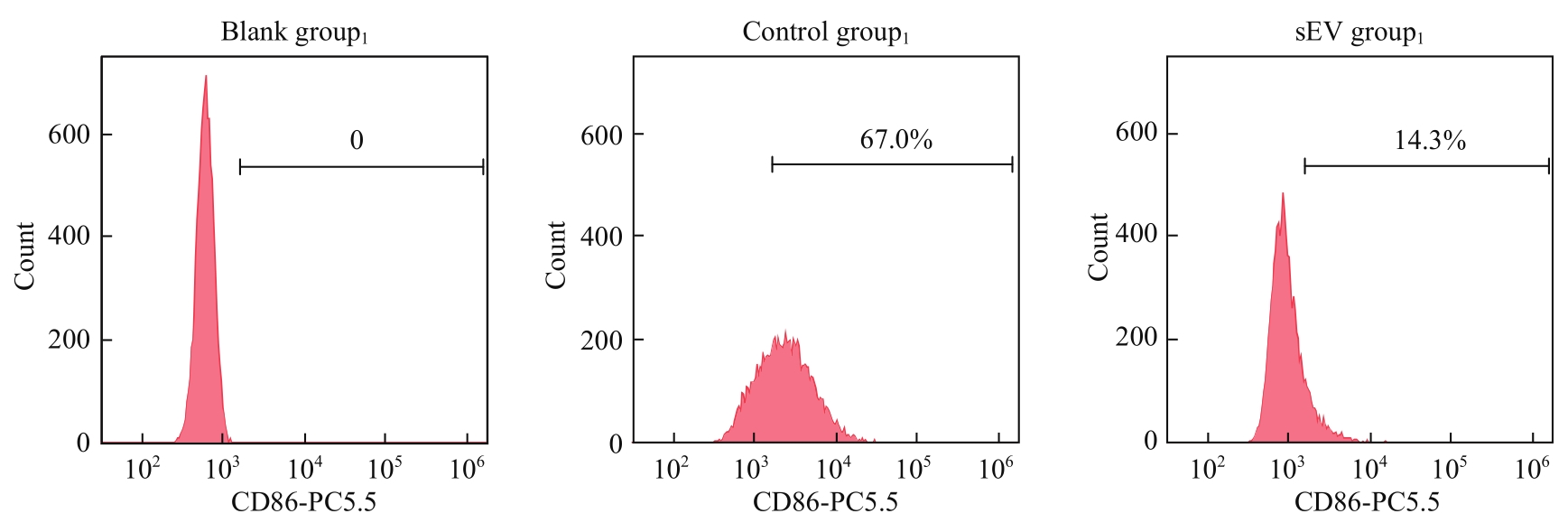
图6 流式细胞术检测M1型巨噬细胞标志物(CD86)表达水平
Fig 6 Expression level of M1 macrophage marker (CD86) detected by flow cytometry
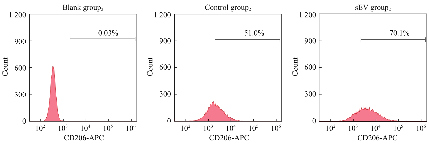
图7 流式细胞术检测M2型巨噬细胞标志物(CD206)表达水平
Fig 7 Expression level of M2 macrophage marker (CD206) detected by flow cytometry
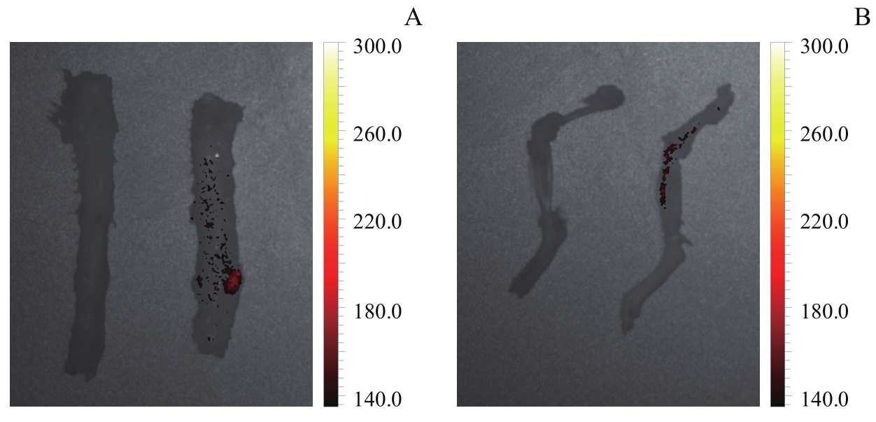
图8 活体成像观察小鼠骨骼中sEV分布情况Note: A. The live imaging of the spines (left: the control group; right: the sEV group). B. The live imaging of the femurs and the tibias (left: the control group; right: the sEV group).
Fig 8 Observation of sEVs distribution in mouse bones by living imaging
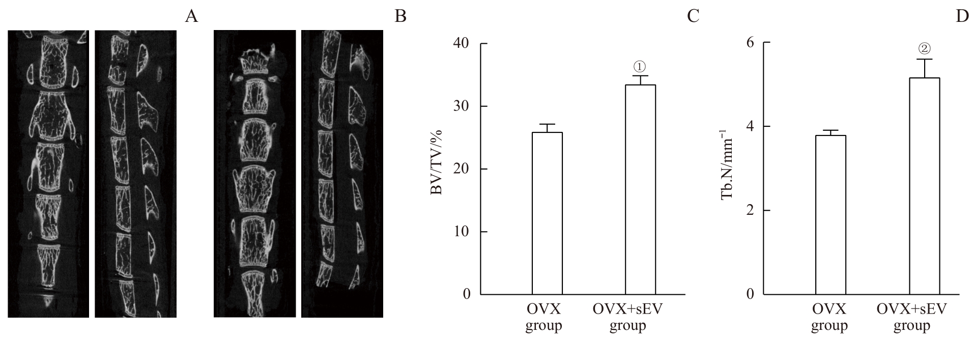
图9 骨质疏松症小鼠及sEV干预小鼠的micro-CT影像学分析及骨参数Note: A. The lumbar spine of the OVX group. B. The lumbar spine of the OVX+sEV group. C. The BV/TV values of 2 groups. D. The Tb.N values of 2 groups. ①P=0.002, ②P=0.005, compared with the OVX group.
Fig 9 Micro-CT imaging analysis and bone parameters of the osteoporosis mice and the sEV-intervened mice
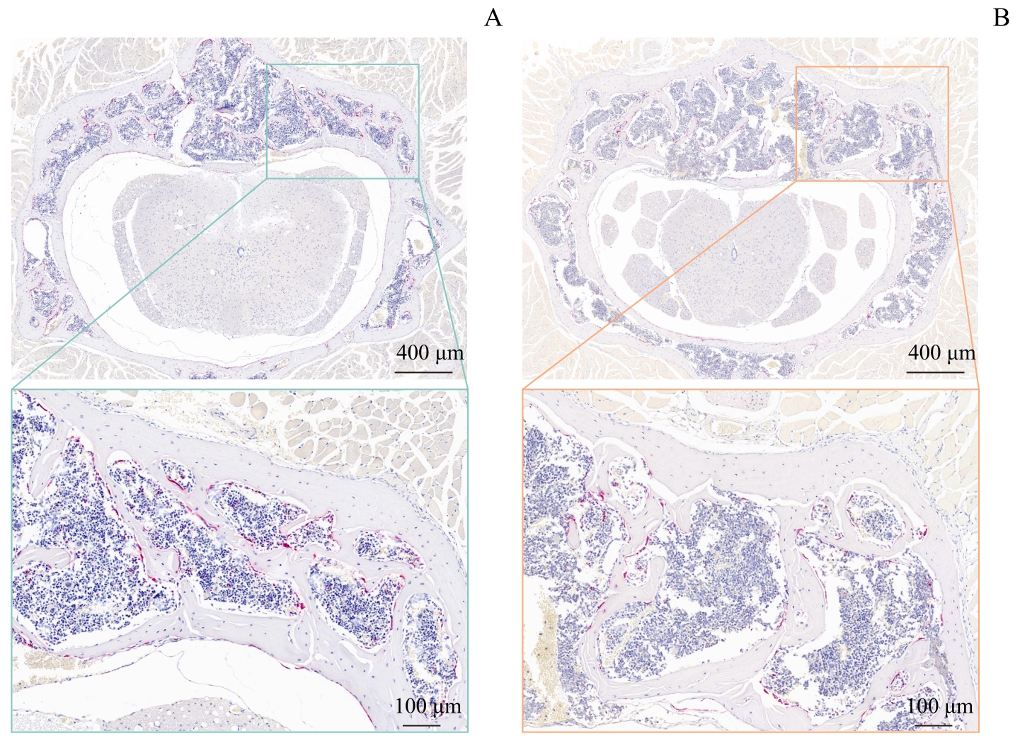
图10 骨质疏松症小鼠及sEV干预小鼠腰椎的TRAP染色结果Note: A. The OVX group. B. The OVX+sEV group. Above (×5); below (×30).
Fig 10 TRAP staining images of the osteoporosis mice and the sEV-intervened mice
| 1 | CHEATHAM S W, HANNEY W, KOLBER M, et al. Osteoporosis: exercise programming insight for the sports medicine professional[J]. Strength Cond J, 2017, 39: 2-13. |
| 2 | SI L, WINZENBERG T M, JIANG Q, et al. Projection of osteoporosis-related fractures and costs in China: 2010‒2050[J]. Osteoporos Int, 2015, 26(7): 1929-1937. |
| 3 | YU F, XIA W B. The epidemiology of osteoporosis, associated fragility fractures, and management gap in China[J]. Arch Osteoporos, 2019, 14(1): 32. |
| 4 | LEE C W, LIN H C, WANG B Y, et al. Ginkgolide B monotherapy reverses osteoporosis by regulating oxidative stress-mediated bone homeostasis[J]. Free Radic Biol Med, 2021, 168: 234-246. |
| 5 | LIU P, LEE S, KNOLL J, et al. Loss of menin in osteoblast lineage affects osteocyte-osteoclast crosstalk causing osteoporosis[J]. Cell Death Differ, 2017, 24(4): 672-682. |
| 6 | LEE K M, LEE C Y, ZHANG G, et al. Methylglyoxal activates osteoclasts through JNK pathway leading to osteoporosis[J]. Chem Biol Interact, 2019, 308: 147-154. |
| 7 | BARSONY J, XU Q, VERBALIS J G. Hyponatremia elicits gene expression changes driving osteoclast differentiation and functions[J]. Mol Cell Endocrinol, 2022, 554: 111724. |
| 8 | JACOME-GALARZA C E, PERCIN G I, MULLER J T, et al. Developmental origin, functional maintenance and genetic rescue of osteoclasts[J]. Nature, 2019, 568(7753): 541-545. |
| 9 | PESCE VIGLIETTI A I, GIAMBARTOLOMEI G H, DELPINO M V. Endocrine modulation of Brucella abortus-infected osteocytes function and osteoclastogenesis via modulation of RANKL/OPG[J]. Microbes Infect, 2019, 21(7): 287-295. |
| 10 | KIM J M, LIN C J, STAVRE Z, et al. Osteoblast-osteoclast communication and bone homeostasis[J]. Cells, 2020, 9(9): 2073. |
| 11 | QIAN J, HE Y, ZHAO J, et al. IL4/IL4R signaling promotes the osteolysis in metastatic bone of CRC through regulating the proliferation of osteoclast precursors[J]. Mol Med, 2021, 27(1): 152. |
| 12 | YAO Z, GETTING S J, LOCKE I C. Regulation of TNF-induced osteoclast differentiation[J]. Cells, 2021, 11(1): 132. |
| 13 | KANG M Y, HUANG C C, LU Y, et al. Bone regeneration is mediated by macrophage extracellular vesicles[J]. Bone, 2020, 141: 115627. |
| 14 | CAI F Y, LIU S L, LEI Y X, et al. Epigallocatechin-3 gallate regulates macrophage subtypes and immunometabolism to ameliorate experimental autoimmune encephalomyelitis[J]. Cell Immunol, 2021, 368: 104421. |
| 15 | ZHANG Z G, ZHANG C Y, ZHANG S R. Irisin activates M1 macrophage and suppresses Th2-type immune response in rats with pelvic inflammatory disease[J]. Evid Based Complement Alternat Med, 2022, 2022: 5215915. |
| 16 | EOM J, YOO J, KIM J J, et al. Viperin deficiency promotes polarization of macrophages and secretion of M1 and M2 cytokines[J]. Immune Netw, 2018, 18(4): e32. |
| 17 | ZHANG W J, GUAN N, ZHANG X M, et al. Study on the imbalance of M1/M2 macrophage polarization in severe chronic periodontitis[J]. Technol Health Care, 2023, 31(1): 117-124. |
| 18 | WANG W H, LIU H, LIU T, et al. Insights into the role of macrophage polarization in the pathogenesis of osteoporosis[J]. Oxid Med Cell Longev, 2022, 2022: 2485959. |
| 19 | YU L, HU M, CUI X, et al. M1 macrophage-derived exosomes aggravate bone loss in postmenopausal osteoporosis via a microRNA-98/DUSP1/JNK axis[J]. Cell Biol Int, 2021, 45(12): 2452-2463. |
| 20 | LU Y P, LIU S S, YANG P P, et al. Exendin-4 and eldecalcitol synergistically promote osteogenic differentiation of bone marrow mesenchymal stem cells through M2 macrophages polarization via PI3K/AKT pathway[J]. Stem Cell Res Ther, 2022, 13(1): 113. |
| 21 | CHEN M, LIN W M, YE R, et al. PPARβ/δ agonist alleviates diabetic osteoporosis via regulating M1/M2 macrophage polarization[J]. Front Cell Dev Biol, 2021, 9: 753194. |
| 22 | WEI H, CHEN Q, LIN L, et al. Regulation of exosome production and cargo sorting[J]. Int J Biol Sci, 2021, 17(1): 163-177. |
| 23 | LI M D, JIA J, LI S S, et al. Exosomes derived from tendon stem cells promote cell proliferation and migration through the TGF β signal pathway[J]. Biochem Biophys Res Commun, 2021, 536: 88-94. |
| 24 | WANG S W, JU T Y, WANG J J, et al. Migration of BEAS-2B cells enhanced by H1299 cell derived-exosomes[J]. Micron, 2021, 143: 103001. |
| 25 | SHARIATI NAJAFABADI S, AMIRPOUR N, AMINI S, et al. Human adipose derived stem cell exosomes enhance the neural differentiation of PC12 cells[J]. Mol Biol Rep, 2021, 48(6): 5033-5043. |
| 26 | YANG S D, GUO S, TONG S, et al. Promoting osteogenic differentiation of human adipose-derived stem cells by altering the expression of exosomal miRNA[J]. Stem Cells Int, 2019, 2019: 1351860. |
| 27 | ZHANG B B, ZHAXI D W, LI C, et al. M2 macrophagy-derived exosomal miRNA-26a-5p induces osteogenic differentiation of bone mesenchymal stem cells[J]. J Orthop Surg Res, 2022, 17(1): 137. |
| 28 | WEN X, HU G, XIAO X, et al. FGF2 positively regulates osteoclastogenesis via activating the ERK-CREB pathway[J]. Arch Biochem Biophys, 2022, 727: 109348. |
| 29 | ZHU G C, CHEN W, TANG C Y, et al. Knockout and double knockout of cathepsin K and Mmp9 reveals a novel function of cathepsin K as a regulator of osteoclast gene expression and bone homeostasis[J]. Int J Biol Sci, 2022, 18(14): 5522-5538. |
| 30 | HE F T, LUO S H, LIU S J, et al. Zanthoxylum bungeanum seed oil inhibits RANKL-induced osteoclastogenesis by suppressing ERK/c-JUN/NFATc1 pathway and regulating cell cycle arrest in RAW264.7 cells[J]. J Ethnopharmacol, 2022, 289: 115094. |
| 31 | KUMAR A, HUGHES T M, CRAFT S, et al. A novel approach to isolate brain-cell-derived exosomes from plasma to better understand pathogenesis of Alzheimer's disease[J]. Alzheimer's Dement, 2020, 16(Suppl 4): e044894. |
| 32 | LI K, WONG D K, HONG K Y, et al. Cushioned-density gradient ultracentrifugation (C-DGUC): a refined and high performance method for the isolation, characterization, and use of exosomes[J]. Methods Mol Biol, 2018, 1740: 69-83. |
| 33 | HELWA I, CAI J W, DREWRY M D, et al. A comparative study of serum exosome isolation using differential ultracentrifugation and three commercial reagents[J]. PLoS One, 2017, 12(1): e0170628. |
| 34 | DING M, WANG C, LU X L, et al. Comparison of commercial exosome isolation kits for circulating exosomal microRNA profiling[J]. Anal Bioanal Chem, 2018, 410(16): 3805-3814. |
| 35 | LIANG B, BURLEY G, LIN S, et al. Osteoporosis pathogenesis and treatment: existing and emerging avenues[J]. Cell Mol Biol Lett, 2022, 27(1): 72. |
| 36 | LI K, XIU C M, ZHOU Q, et al. A dual role of cholesterol in osteogenic differentiation of bone marrow stromal cells[J]. J Cell Physiol, 2019, 234(3): 2058-2066. |
| 37 | CHE Y T, YANG J Z, TANG F, et al. New function of cholesterol oxidation products involved in osteoporosis pathogenesis[J]. Int J Mol Sci, 2022, 23(4): 2020. |
| 38 | LI K Q, CHEN S H, CAI P Y, et al. MiRNA-483-5p is involved in the pathogenesis of osteoporosis by promoting osteoclast differentiation[J]. Mol Cell Probes, 2020, 49: 101479. |
| 39 | PARK E, LEE C G, LIM E, et al. Osteoprotective effects of loganic acid on osteoblastic and osteoclastic cells and osteoporosis-induced mice[J]. Int J Mol Sci, 2020, 22(1): 233. |
| 40 | LAI G H, ZHAO R L, ZHUANG W D, et al. BMSC-derived exosomal miR-27a-3p and miR-196b-5p regulate bone remodeling in ovariectomized rats[J]. PeerJ, 2022, 10: e13744. |
| 41 | SONG H Y, LI X Q, ZHAO Z C, et al. Reversal of osteoporotic activity by endothelial cell-secreted bone targeting and biocompatible exosomes[J]. Nano Lett, 2019, 19(5): 3040-3048. |
| 42 | CHEN X T, WAN Z, YANG L, et al. Exosomes derived from reparative M2-like macrophages prevent bone loss in murine periodontitis models via IL-10 mRNA[J]. J Nanobiotechnology, 2022, 20(1): 110. |
| 43 | ZHU L F, LI L, WANG X Q, et al. M1 macrophages regulate TLR4/AP1 via paracrine to promote alveolar bone destruction in periodontitis[J]. Oral Dis, 2019, 25(8): 1972-1982. |
| 44 | LIANG B L, WANG H C, WU D, et al. Macrophage M1/M2 polarization dynamically adapts to changes in microenvironment and modulates alveolar bone remodeling after dental implantation[J]. J Leukoc Biol, 2021, 110(3): 433-447. |
| 45 | SHI M S, WANG C, WANG Y L, et al. Deproteinized bovine bone matrix induces osteoblast differentiation via macrophage polarization[J]. J Biomed Mater Res A, 2018, 106(5): 1236-1246. |
| 46 | SHI C, YUAN F, LI Z L, et al. MSN@IL-4 sustainingly mediates macrophagocyte M2 polarization and relieves osteoblast damage via NF-κB pathway-associated apoptosis[J]. Biomed Res Int, 2022, 2022: 2898729. |
| 47 | Horibe K, Hara M, Nakamura H. M2-like macrophage infiltration and transforming growth factor-β secretion during socket healing process in mice[J]. Arch Oral Biol, 2021, 123: 105042. |
| 48 | WANG X Y, JI Q B, HU W H, et al. Isobavachalcone prevents osteoporosis by suppressing activation of ERK and NF-κB pathways and M1 polarization of macrophages[J]. Int Immunopharmacol, 2021, 94: 107370. |
| 49 | LI Z K, ZHU X D, XU R J, et al. Deacylcynaropicrin inhibits RANKL-induced osteoclastogenesis by inhibiting NF-κB and MAPK and promoting M2 polarization of macrophages[J]. Front Pharmacol, 2019, 10: 599. |
| 50 | YAO M Y, CUI B, ZHANG W H, et al. Exosomal miR-21 secreted by IL-1β-primed-mesenchymal stem cells induces macrophage M2 polarization and ameliorates sepsis[J]. Life Sci, 2021, 264: 118658. |
| 51 | MA J, CHEN L, ZHU X, et al. Mesenchymal stem cell-derived exosomal miR-21a-5p promotes M2 macrophage polarization and reduces macrophage infiltration to attenuate atherosclerosis[J]. Acta Biochim Biophys Sin (Shanghai), 2021, 53(9): 1227-1236. |
| 52 | LI R, LI D Z, WANG H N, et al. Exosomes from adipose-derived stem cells regulate M1/M2 macrophage phenotypic polarization to promote bone healing via miR-451a/MIF[J]. Stem Cell Res Ther, 2022, 13(1): 149. |
| 53 | LI R, ZHAO K C, RUAN Q, et al. Bone marrow mesenchymal stem cell-derived exosomal microRNA-124-3p attenuates neurological damage in spinal cord ischemia-reperfusion injury by downregulating Ern1 and promoting M2 macrophage polarization[J]. Arthritis Res Ther, 2020, 22(1): 75. |
| [1] | 王琳, 徐萍, 张乔婷, 田军, 娄晓丽, 王静. 胱天蛋白酶募集域蛋白9在重症急性胰腺炎巨噬细胞M1极化中的作用[J]. 上海交通大学学报(医学版), 2025, 45(8): 981-989. |
| [2] | 赵心雨, 张文超, 陈旭卓, 宋佳琪, 黄慧, 张善勇. 亚精胺对脂多糖诱导的小鼠颅骨炎症性骨溶解的作用研究[J]. 上海交通大学学报(医学版), 2025, 45(6): 673-683. |
| [3] | 刘媛琪, 孙思远, 代庆刚, 江凌勇, 沈国芳. 全反式视黄酸调控颌骨骨髓间充质干细胞成骨分化双向效应的体外研究[J]. 上海交通大学学报(医学版), 2024, 44(9): 1083-1093. |
| [4] | 牛媛媛, 汪龙德, 胥文娟, 李正菊, 张瑞婷, 吴毓谦. 巨噬细胞M1/M2型极化在不同肝病中的作用研究进展[J]. 上海交通大学学报(医学版), 2024, 44(4): 509-517. |
| [5] | 金芳全, 樊成虎, 唐晓栋, 陈彦同, 齐兵献. 线粒体功能障碍与骨质疏松症相关性研究进展[J]. 上海交通大学学报(医学版), 2023, 43(6): 761-767. |
| [6] | 刘辰骏, 尹博浩, 孙辉, 张伟. 非侵入性影像学技术在骨质疏松症中的应用[J]. 上海交通大学学报(医学版), 2023, 43(3): 385-390. |
| [7] | 刘宏强, 陆艳青, 高宇轩, 王一云, 王传东, 张晓玲. 构建高效载体OPEI沉默TRAF6促进骨关节炎软骨再生的研究[J]. 上海交通大学学报(医学版), 2022, 42(7): 846-857. |
| [8] | 王昕芃, 王君颖, 蔡佳翌, 付婉彬, 钟华. 低剂量地西他滨对免疫性血小板减少症患者来源的骨髓间充质干细胞生物学行为的影响[J]. 上海交通大学学报(医学版), 2022, 42(6): 758-767. |
| [9] | 郝磊, 金戈, 杨涌涛, 王军伟, 孙洋, 秦翠玲, 展群岭. 骨髓间充质干细胞外泌体介导miR-124-1对小胶质细胞M2型极化调控的影响[J]. 上海交通大学学报(医学版), 2022, 42(3): 323-330. |
| [10] | 童徐, 舒林径. miR-877-3p对骨质疏松症小鼠骨髓间充质干细胞增殖能力的影响[J]. 上海交通大学学报(医学版), 2021, 41(7): 884-890. |
| [11] | 蔡苗苗, 高艳虹. 肌少-骨质疏松症的研究进展[J]. 上海交通大学学报(医学版), 2021, 41(5): 678-683. |
| [12] | 罗 虹1, 2,武红彦1, 3,谭 玺1, 3,戴红卫1, 2, 3,黄 兰1, 2, 3. 肥胖和肥胖抵抗大鼠的正畸牙移动速率及压力侧骨改建差异研究[J]. 上海交通大学学报(医学版), 2020, 40(8): 1055-1062. |
| [13] | 王峰伟1,沈秋明1,拉巴仓拉1,施 悦1,张舒娴1,王沪雯1,常睿捷1,杨颖华2,万和平3,沈 恬1,蔡 泳1. 上海市社区中老年居民骨质疏松症预防相关路径分析[J]. 上海交通大学学报(医学版), 2020, 40(4): 525-. |
| [14] | 李子林1,顾文钦2,沈 恬1. 骨质疏松症与肠道菌群间联系的研究进展[J]. 上海交通大学学报(医学版), 2019, 39(10): 1214-. |
| [15] | 李晖 *,张术涛 *,岳冰. 小分子抗菌肽 KR -12-a5促进人骨髓间充质干细胞成骨分化的作用[J]. 上海交通大学学报(医学版), 2018, 38(8): 881-. |
| 阅读次数 | ||||||
|
全文 |
|
|||||
|
摘要 |
|
|||||