
上海交通大学学报(医学版) ›› 2026, Vol. 46 ›› Issue (8): 1037-1052.doi: 10.3969/j.issn.1674-8115.2026.08.005
• 论著 · 基础研究 • 上一篇
陈赟琪, 那迪娜·帕尔哈提, 张鹏善( ), 黄陈()
收稿日期:2026-03-02
接受日期:2026-05-18
出版日期:2026-08-13
发布日期:2026-08-13
通讯作者:
黄 陈,主任医师,博士;电子信箱:richard-hc@hotmail.com作者简介:第一联系人:陈赟琪负责实验设计、实验操作、数据整理、初稿撰写和作图,那迪娜·帕尔哈提负责实验操作、数据整理、作图和文章修改,张鹏善负责实验方法建立、稿件审阅与修改,黄陈负责对稿件进行最终的审阅和修改。所有作者均阅读并同意最终稿件的提交。
基金资助:
Chen Yunqi, Paerhati Nadina, Zhang Pengshan(), Huang Chen()
Received:2026-03-02
Accepted:2026-05-18
Online:2026-08-13
Published:2026-08-13
Contact:
Huang Chen, E-mail: richard-hc@hotmail.comAbout author:First author contact:Chen Yunqi was responsible for the experimental design, conducting the experiments, data organization, drafting the initial manuscript, and figure preparation. Nadina Paerhati was responsible for conducting the experiments, data organization, figure preparation, and manuscript revision. Zhang Pengshan was responsible for establishing the experimental methods, manuscript review, and revision. Huang Chen was responsible for the final review and revision of the manuscript. All authors have read the final version of the manuscript and consented to its submission.
Supported by:摘要:
目的·探讨去泛素化酶泛素特异性肽酶38(ubiquitin-specific peptidase 38,USP38)在胃癌中的表达特征、临床预后价值及其促癌分子机制,并鉴定下游关键靶点核糖核酸结合基序蛋白14(RNA-binding motif protein 14,RBM14)。方法·收集50例在上海交通大学医学院附属第一人民医院胃肠外科接受手术治疗的胃癌患者的肿瘤及癌旁组织标本,利用实时荧光定量PCR和蛋白质印迹法验证USP38在胃癌中的表达,并使用癌症基因组图谱(The Cancer Genome Atlas,TCGA)和基因表达综合数据库(Gene Expression Omnibus,GEO)分析胃癌患者临床特征与USP38表达水平的相关性;通过基因集富集分析(Gene Set Enrichment Analysis,GSEA)、基因本体论(Gene Ontology,GO)及京都基因与基因组百科全书(Kyoto Encyclopedia of Genes and Genomes,KEGG)预测USP38相关信号通路。结合文献报道与Pearson相关性分析结果筛选USP38的潜在底物。采用细胞计数试剂盒-8(cell counting kit-8,CCK-8)、5-乙炔基-2'-脱氧尿苷(5-ethynyl-2'- deoxyuridine,EdU)试剂盒、克隆形成及划痕实验评估USP38对AGS、HGC-27胃癌细胞增殖和迁移的影响;利用慢病毒载体包装靶向USP38的短发夹核糖核酸(short hairpin RNA,shRNA)构建稳定敲低细胞株,并通过功能回补(rescue)实验验证USP38-RBM14调控轴的生物学功能。结果·TCGA数据库分析显示,USP38在胃癌组织中显著高表达(P<0.001),其表达水平与患者较短的总生存期(Log-rank P<0.001,HR=1.36)及病理分期(P=0.017)呈正相关,GSEA分析发现USP38高表达组显著富集于G2/M期检查点、早期2因子(early 2 factor,E2F)靶点、核斑点及DNA损伤修复等通路(调整后P值均<0.001),且敲低USP38能够下调增殖及间质标志物的表达。功能实验证实敲低USP38可显著抑制胃癌细胞增殖与迁移(均P<0.05)。Pearson相关性分析显示,USP38与其潜在结合蛋白RBM14的表达呈显著正相关(R=0.22,P<0.001)。进一步研究发现,敲低USP38可降低RBM14蛋白水平,而蛋白酶体抑制剂及放线菌酮(cycloheximide,CHX)实验表明USP38在蛋白质翻译后水平稳定RBM14。功能回补实验显示,过表达RBM14可部分恢复USP38敲低对胃癌细胞的抑制效应,证实USP38通过稳定RBM14蛋白发挥促癌作用。结论·USP38在胃癌中高表达且与患者不良预后相关;敲低USP38能够抑制胃癌细胞增殖和迁移。USP38可通过稳定RBM14蛋白促进胃癌进展。
中图分类号:
陈赟琪, 那迪娜·帕尔哈提, 张鹏善, 黄陈. 去泛素化酶USP38通过稳定RBM14促进胃癌进展的机制研究[J]. 上海交通大学学报(医学版), 2026, 46(8): 1037-1052.
Chen Yunqi, Paerhati Nadina, Zhang Pengshan, Huang Chen. Deubiquitinase USP38 promotes gastric cancer progression by stabilizing RBM14[J]. Journal of Shanghai Jiao Tong University (Medical Science), 2026,(8): 1037-1052.
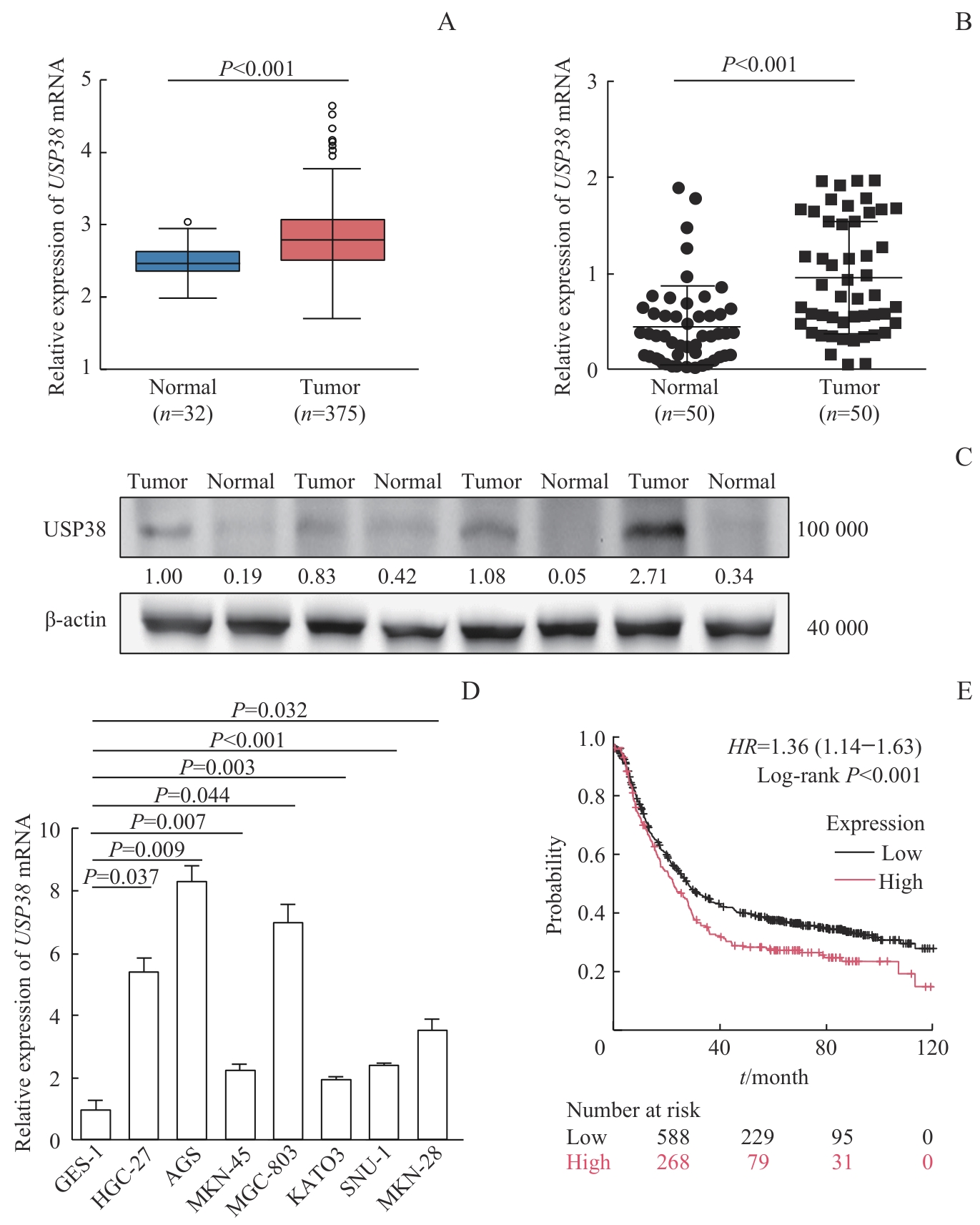
图1 USP38在胃癌组织及细胞中的表达模式及其对预后的影响Note: A. Expression of USP38 mRNAin gastric cancer (n=375) and normal (n=32) tissues from the TCGA-stomach adenocarcinoma (STAD) database. B. USP38 mRNA expression in 50 paired gastric cancer and adjacent normal tissues determined by qPCR. C. Detection of USP38 expression in gastric cancer (n=4) and adjacent normal (n=4) tissues detected by Western blotting. The numbers indicate relative protein expression levels. D. qPCR analysis of USP38 mRNA expression in different gastric cancer cell lines, compared with human normal gastric mucosal cells. E. Kaplan-Meier survival curves of gastric cancer patients (n=856) from GEO database according to USP38 expression.
Fig 1 Expression of USP38 in gastric cancer tissues and cells and its impact on prognosis
| Variable | Overall (n =458) | Low (n =229) | High (n =229) | P value |
|---|---|---|---|---|
| Age/year | 67.5 (58.0, 74.0) | 67.0 (58.0, 72.0) | 68.0 (60.0, 75.0) | 0.051 |
| Gender/n(%) | 0.005 | |||
| Female | 167 (36.5) | 69 (30.1) | 98 (42.8) | |
| Male | 291 (63.5) | 160 (69.9) | 131 (57.2) | |
| Histologic grade/n(%) | 0.600 | |||
| G1 | 13 (2.9) | 6 (2.7) | 7 (3.1) | |
| G2 | 168 (37.8) | 88 (40.0) | 80 (35.6) | |
| G3 | 264 (59.3) | 126 (57.3) | 138 (61.3) | |
| Pathologic stage/n(%) | 0.017 | |||
| Stage Ⅰ | 68 (16.2) | 34 (15.7) | 34 (16.7) | |
| Stage Ⅱ | 142 (33.8) | 88 (40.7) | 54 (26.5) | |
| Stage Ⅲ | 165 (39.3) | 73 (33.8) | 92 (45.1) | |
| Stage Ⅳ | 45 (10.7) | 21 (9.7) | 24 (11.8) | |
| T stage/n(%) | 0.066 | |||
| T1 | 25 (5.7) | 12 (5.2) | 13 (6.1) | |
| T2 | 104 (23.5) | 49 (21.4) | 55 (25.8) | |
| T3 | 201 (45.5) | 118 (51.5) | 83 (39.0) | |
| T4 | 112 (25.3) | 50 (21.8) | 62 (29.1) | |
| N stage/n(%) | 0.200 | |||
| N0 | 141 (32.9) | 81 (37.0) | 60 (28.6) | |
| N1 | 120 (28.0) | 61 (27.9) | 59 (28.1) | |
| N2 | 88 (20.5) | 38 (17.4) | 50 (23.8) | |
| N3 | 80 (18.6) | 39 (17.8) | 41 (19.5) | |
| M stage/n(%) | 0.800 | |||
| M0 | 402 (93.3) | 200 (93.0) | 202 (93.5) | |
| M1 | 29 (6.7) | 15 (7.0) | 14 (6.5) |
表1 USP38 表达水平与胃癌临床病理特征相关性分析
Tab 1 Association between USP38 expression and clinicopathological characteristics of gastric cancer
| Variable | Overall (n =458) | Low (n =229) | High (n =229) | P value |
|---|---|---|---|---|
| Age/year | 67.5 (58.0, 74.0) | 67.0 (58.0, 72.0) | 68.0 (60.0, 75.0) | 0.051 |
| Gender/n(%) | 0.005 | |||
| Female | 167 (36.5) | 69 (30.1) | 98 (42.8) | |
| Male | 291 (63.5) | 160 (69.9) | 131 (57.2) | |
| Histologic grade/n(%) | 0.600 | |||
| G1 | 13 (2.9) | 6 (2.7) | 7 (3.1) | |
| G2 | 168 (37.8) | 88 (40.0) | 80 (35.6) | |
| G3 | 264 (59.3) | 126 (57.3) | 138 (61.3) | |
| Pathologic stage/n(%) | 0.017 | |||
| Stage Ⅰ | 68 (16.2) | 34 (15.7) | 34 (16.7) | |
| Stage Ⅱ | 142 (33.8) | 88 (40.7) | 54 (26.5) | |
| Stage Ⅲ | 165 (39.3) | 73 (33.8) | 92 (45.1) | |
| Stage Ⅳ | 45 (10.7) | 21 (9.7) | 24 (11.8) | |
| T stage/n(%) | 0.066 | |||
| T1 | 25 (5.7) | 12 (5.2) | 13 (6.1) | |
| T2 | 104 (23.5) | 49 (21.4) | 55 (25.8) | |
| T3 | 201 (45.5) | 118 (51.5) | 83 (39.0) | |
| T4 | 112 (25.3) | 50 (21.8) | 62 (29.1) | |
| N stage/n(%) | 0.200 | |||
| N0 | 141 (32.9) | 81 (37.0) | 60 (28.6) | |
| N1 | 120 (28.0) | 61 (27.9) | 59 (28.1) | |
| N2 | 88 (20.5) | 38 (17.4) | 50 (23.8) | |
| N3 | 80 (18.6) | 39 (17.8) | 41 (19.5) | |
| M stage/n(%) | 0.800 | |||
| M0 | 402 (93.3) | 200 (93.0) | 202 (93.5) | |
| M1 | 29 (6.7) | 15 (7.0) | 14 (6.5) |
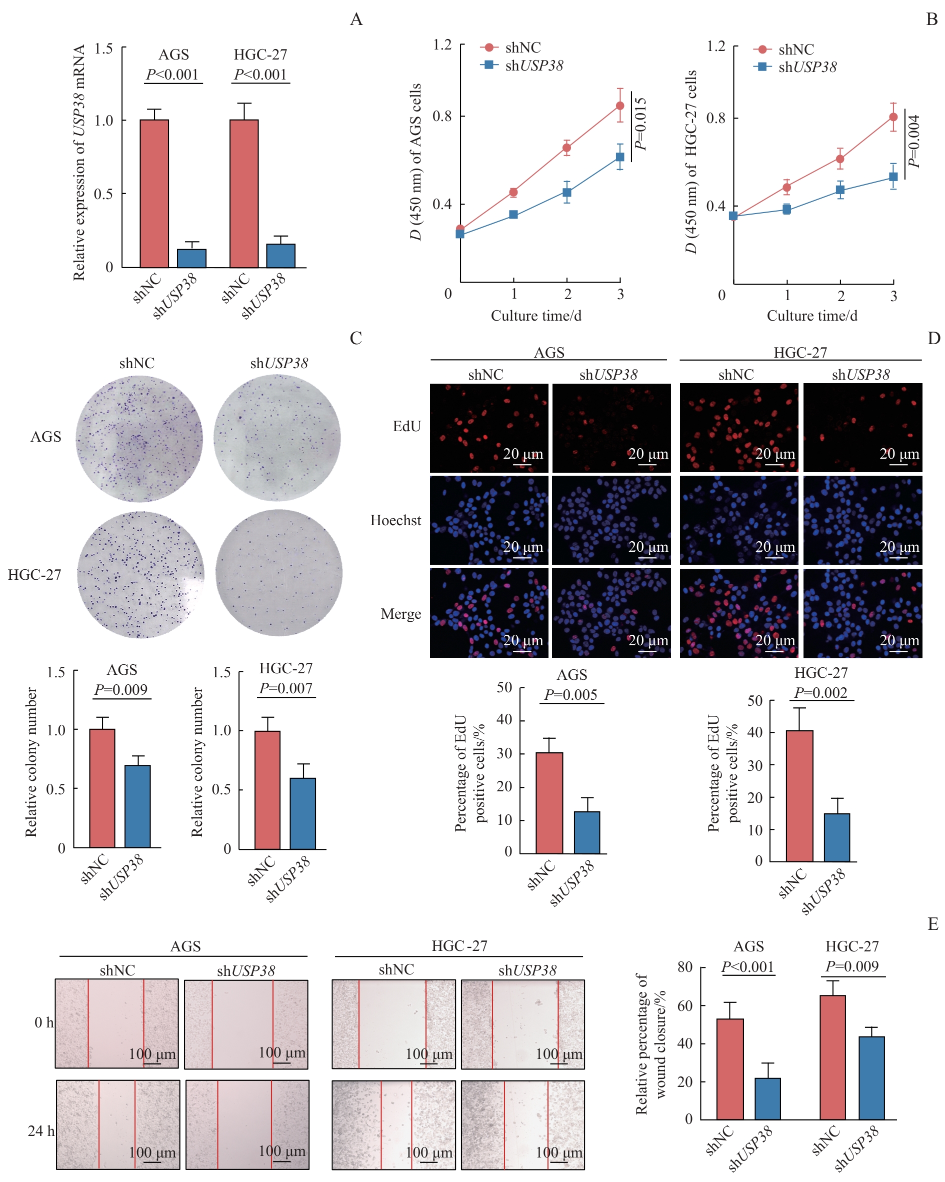
图2 USP38促进胃癌细胞增殖和迁移的作用Note: A. Validation of USP38 knockdown efficiency in AGS and HGC-27 gastric cancer cells by qPCR. B‒D. CCK-8 assay (B), colony formation assay (C), and EdU assay (D) assessing the effect of USP38 knockdown on the proliferation of AGS and HGC-27 gastric cancer cells. E. Wound healing assay examining the effect of USP38 knockdown on the migration of AGS and HGC-27 gastric cancer cells. Data are presented as mean±SD (unpaired t‑test, shUSP38vs shNC).
Fig 2 Promotive effects of USP38 on proliferation and migration of gastric cancer cells
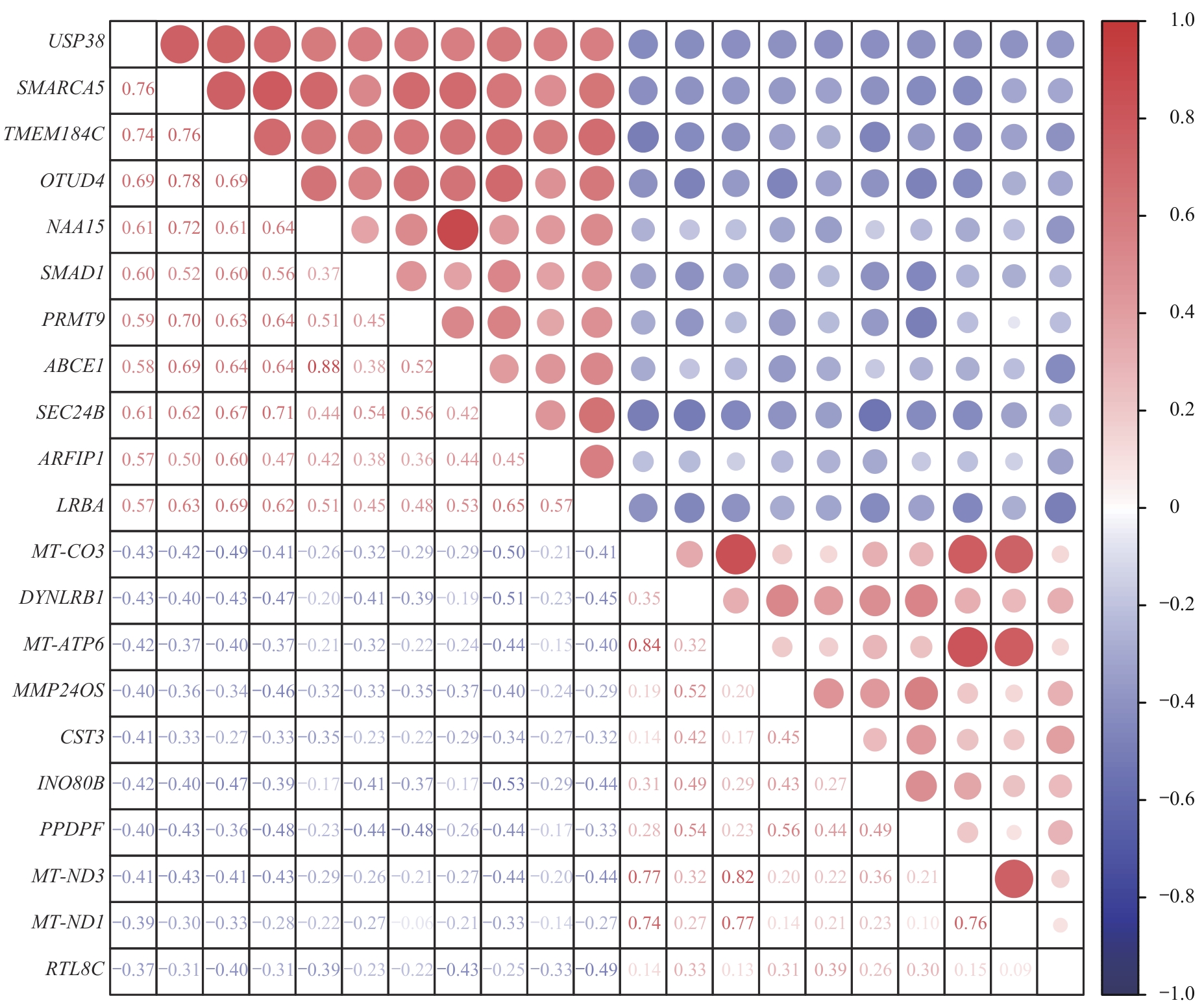
图3 与 USP38 表达水平相关的基因热图Note: Color represents the Pearson correlation coefficient, with red indicating positive correlation and blue indicating negative correlation. SMARCA5—SWI/SNF-related, matrix-associated, actin-dependent regulator of chromatin, subfamily A, member 5; TMEM184C—transmembrane protein 184C; OTUD4—OTU domain containing 4; NAA15—N-ɑ-acetyltransferase 15 NatA auxiliary subunit; SMAD1—SMAD family member 1; PRMT9—protein arginine methyltransferase 9; ABCE1—ATP-binding cassette subfamily E member 1; SEC24B—SEC24 homolog B COP Ⅱ coat complex component; DYNLRB1—dynein light chain roadblock-type 1; MMP24OS—MMP24 opposite strand; CST3—cystatin C; INO80B—INO80 complex subunit B; PPDPF—pancreatic progenitor cell differentiation and proliferation factor; RTL8C—retrotransposon Gag-like 8C; ARFIP1—ADP-ribosylation factor interacting protein 1; LRBA—lipopolysaccharide responsive beige-like anchor protein.
Fig 3 Heatmap of genes correlated with USP38 expression
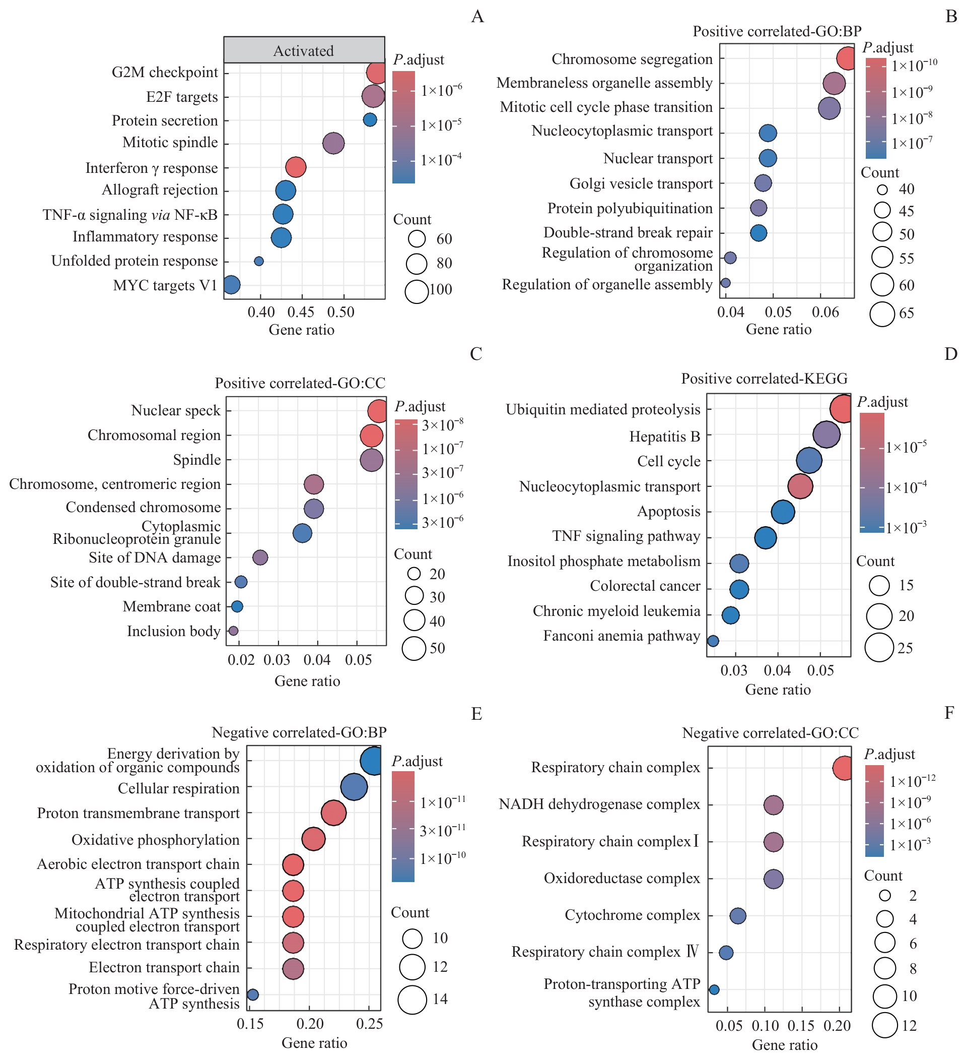
图4 USP38 相关基因在胃癌中的通路富集分析Note: A. GSEA bubble plot (TCGA-STAD) showing the top 10 hallmark pathways activated in the USP38-high expression group. B‒D. Enrichment analyses of genes positively correlated with USP38: GO biological process (B), GO cellular component (C), and KEGG pathway (D). E/F. Enrichment analyses of genes negatively correlated with USP38: GO biological process (E), and GO cellular component (F).
Fig 4 Pathway enrichment analysis of USP38-related genes in gastric cancer
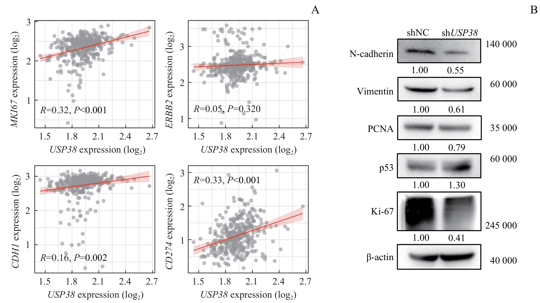
图5 USP38相关标志物在胃癌中的表达分析Note: A. Correlation of USP38 with gastric cancer biomarkers MKI67, ERBB2, CDH1, and CD274 (95% confidence intervals shaded). B. Western blotting analysis of USP38 knockdown effects on AGS cell proliferation and tumor-associated protein expression. The numbers indicate relative protein expression levels.
Fig 5 Expression analysis of USP38-associated biomarkers in gastric cancer
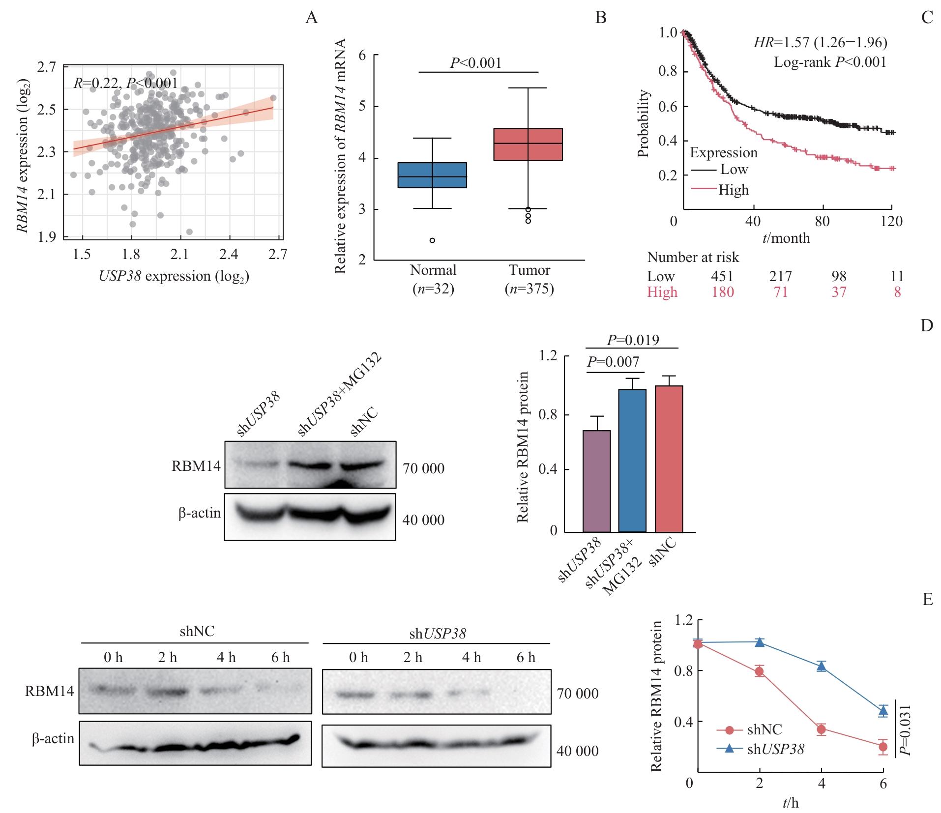
图6 USP38对RBM14蛋白的稳定作用Note: A. Correlation analysis showing a positive association between USP38 and RBM14 mRNA expression in the TCGA-STAD database. B. RBM14 expression in gastric cancer tissues (n=375) and normal tissues (n=32) in TCGA database. C. Kaplan-Meier survival curves associating high RBM14 expression with poor prognosis in gastric cancer patients (n=631) in GEO database. D. Western blotting analysis of RBM14 protein levels following USP38 knockdown with or without MG132 treatment. Comparison of RBM14 protein levels: shUSP38vs shNC and shUSP38vs shUSP38+MG132. E. CHX chase assay showing accelerated RBM14 protein degradation upon USP38 knockdown (shUSP38vs shNC). Data are presented as mean±SD.
Fig 6 Stabilizing effect of USP38 on RBM14 protein
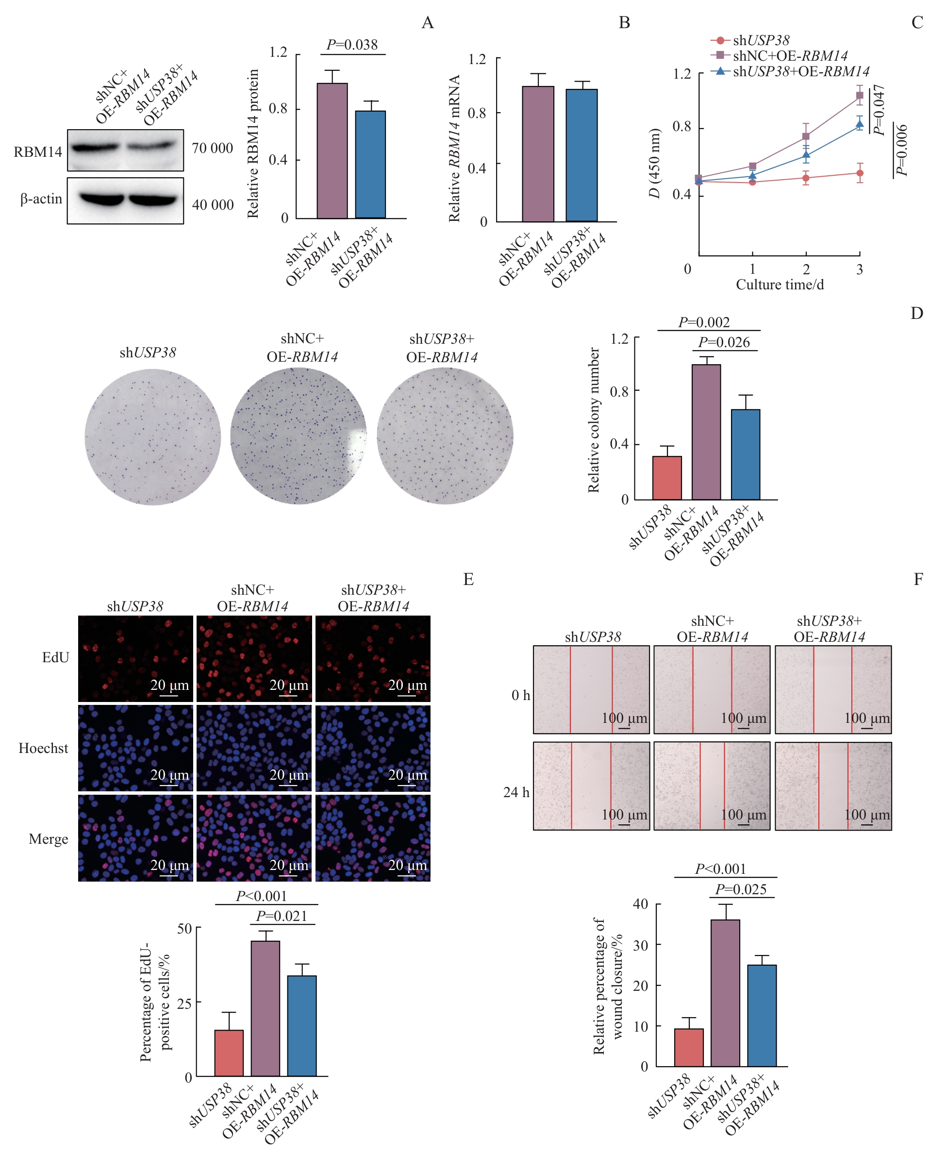
图7 USP38通过稳定RBM14蛋白对胃癌细胞增殖迁移的促进作用Note: A. Validation of RBM14 overexpression efficiency in shUSP38 cells by Western blotting. B. qPCR analysis of the effect of USP38 knockdown on RBM14 mRNA expression in gastric cancer cells. C. CCK-8 assay showing the rescue effect of RBM14 overexpression on the inhibition of cell proliferation induced by USP38 knockdown. D. Colony formation assay assessing the rescue effect of RBM14 overexpression on the clonogenic capacity of shUSP38 cells. E. EdU assay showing that RBM14 overexpression rescues the proliferation defect caused by USP38 knockdown. F. Wound healing assay assessing the rescue effect of RBM14 overexpression on cell migration. Data are presented as mean±SD. Comparisons were performed between the shUSP38+OE-RBM14 group and the shNC+OE-RBM14 group or the shUSP38 group.
Fig 7 Promotive effect of USP38 on the proliferation and migration of gastric cancer cells via stabilizing RBM14 protein
| [1] | Bray F, Laversanne M, Sung H, et al. Global cancer statistics 2022: GLOBOCAN estimates of incidence and mortality worldwide for 36 cancers in 185 countries[J]. CA Cancer J Clin, 2024, 74(3): 229-263. |
| [2] | 孙可欣, 李荔, 王少明, 等. 2024年中国分地区恶性肿瘤流行情况分析[J]. 中华肿瘤杂志, 2026, 48(3): 400-412. |
| Sun K X, Li L, Wang S M, et al. Cancer incidence and mortality across diverse geographical regions in China, 2024[J]. Chinese Journal of Oncology, 2026, 48(3): 400-412. | |
| [3] | Dewson G, Eichhorn P J A, Komander D. Deubiquitinases in cancer[J]. Nat Rev Cancer, 2023, 23(12): 842-862. |
| [4] | Han S Z, Wang R K, Zhang Y N, et al. The role of ubiquitination and deubiquitination in tumor invasion and metastasis[J]. Int J Biol Sci, 2022, 18(6): 2292-2303. |
| [5] | Li J Y, Zhong J H, Ye J M, et al. USP38: an important regulatory factor in tumor malignant progression[J]. Front Immunol, 2025, 16: 1612723. |
| [6] | Wang R, Cai X L, Li X, et al. USP38 promotes deubiquitination of K11-linked polyubiquitination of HIF1α at Lys769 to enhance hypoxia signaling[J]. J Biol Chem, 2024, 300(1): 105532. |
| [7] | Xiao Z, Dai C, Yu T T, et al. Ubiquitin specific protease 38 aggravates pathological cardiac remodeling by stabilizing phospho-TBK1[J]. Int J Biol Sci, 2024, 20(5): 1815-1832. |
| [8] | Yi X M, Li M, Chen Y D, et al. Reciprocal regulation of IL-33 receptor-mediated inflammatory response and pulmonary fibrosis by TRAF6 and USP38[J]. Proc Natl Acad Sci U S A, 2022, 119(10): e2116279119. |
| [9] | Wang Y C, Li Q, Hu D W, et al. USP38 inhibits zika virus infection by removing envelope protein ubiquitination[J]. Viruses, 2021, 13(10): 2029. |
| [10] | Zheng Z, Shang Y X, Xu R, et al. Ubiquitin specific peptidase 38 promotes the progression of gastric cancer through upregulation of fatty acid synthase[J]. Am J Cancer Res, 2022, 12(6): 2686-2696. |
| [11] | Yue Q Y, Li Z J, Du Y X, et al. Dehydrocostus lactone attenuates ulcerative colitis via USP38-mediated histone H2B deubiquitination to suppress NF-κB-driven inflammation[J]. Int Immunopharmacol, 2025, 164: 115346. |
| [12] | Hu Q Y, Chen Y H, Zhou Q R, et al. ADAR promotes USP38 auto-deubiquitylation and stabilization in an RNA editing-independent manner in esophageal squamous cell carcinoma[J]. J Biol Chem, 2024, 300(10): 107789. |
| [13] | Huang J, Zhou W M, Hao C, et al. The feedback loop of METTL14 and USP38 regulates cell migration, invasion and EMT as well as metastasis in bladder cancer[J]. PLoS Genet, 2022, 18(10): e1010366. |
| [14] | Yang Y F, Yang C Z, Li T T, et al. The deubiquitinase USP38 promotes NHEJ repair through regulation of HDAC1 activity and regulates cancer cell response to genotoxic insults[J]. Cancer Res, 2020, 80(4): 719-731. |
| [15] | Zhan W, Liao X, Liu J, et al. USP38 regulates the stemness and chemoresistance of human colorectal cancer via regulation of HDAC3[J]. Oncogenesis, 2020, 9(5): 48. |
| [16] | Zhang T, Su F, Wang B F, et al. Ubiquitin specific peptidase 38 epigenetically regulates KLF transcription factor 5 to augment malignant progression of lung adenocarcinoma[J]. Oncogene, 2024, 43(16): 1190-1202. |
| [17] | Zhao S Y, Liu X L, Luo R K, et al. USP38 functions as an oncoprotein by downregulating the p53 pathway through deubiquitination and stabilization of MDM2[J]. Cell Death Differ, 2025, 32(6): 1128-1141. |
| [18] | Li Z, Liao Y T, Tang C, et al. RBM14 promotes DNA end resection during homologous recombination repair[J]. Acta Biochim Biophys Sin, 2023, 55(12): 1864-1873. |
| [19] | Posta M, Győrffy B. Pathway-level mutational signatures predict breast cancer outcomes and reveal therapeutic targets[J]. Br J Pharmacol, 2025, 182(23): 5734-5747. |
| [20] | Jang Y, Elsayed Z, Eki R, et al. Intrinsically disordered protein RBM14 plays a role in generation of RNA: DNA hybrids at double-strand break sites[J]. Proc Natl Acad Sci U S A, 2020, 117(10): 5329-5338. |
| [21] | Qian Z X, Bai W, Li J X, et al. Gut microbiome-mediated epigenetic modifications in gastric cancer: a comprehensive multiomics analysis[J]. Front Cell Infect Microbiol, 2025, 15: 1585881. |
| [22] | Wang Y H, Patti G J. The Warburg effect: a signature of mitochondrial overload[J]. Trends Cell Biol, 2023, 33(12): 1014-1020. |
| [23] | Gao H L, Xi Z, Dai J W, et al. Drug resistance mechanisms and treatment strategies mediated by ubiquitin-specific proteases (USPs) in cancers: new directions and therapeutic options[J]. Mol Cancer, 2024, 23(1): 88. |
| [24] | Silvestrini V C, Thomé C H, Albuquerque D, et al. Proteomics analysis reveals the role of ubiquitin specific protease (USP47) in epithelial to mesenchymal transition (EMT) induced by TGFβ2 in breast cells[J]. J Proteomics, 2020, 219: 103734. |
| [25] | Gao Z S, Li C, Sun H Y, et al. N6-methyladenosine-modified USP13 induces pro-survival autophagy and imatinib resistance via regulating the stabilization of autophagy-related protein 5 in gastrointestinal stromal tumors[J]. Cell Death Differ, 2023, 30(2): 544-559. |
| [1] | 王铭杰, 倪博, 夏翔, 张子臻. 硒蛋白SELENOI通过PI3K/AKT/mTOR信号通路促进胃癌增殖、迁移和侵袭[J]. 上海交通大学学报(医学版), 2026, 46(8): 1015-1025. |
| [2] | 翟元晖, 王莹莹, 王宇轩, 吴文萱, 朱楚娇, 张幼萍, 白雯会, 雷虎, 徐含章, 赵亚雪, 吴英理. 甲基巴多索隆作为新型泛素特异性蛋白酶48抑制剂抑制结直肠癌的作用及机制研究[J]. 上海交通大学学报(医学版), 2026, 46(8): 1026-1036. |
| [3] | 那迪娜·帕尔哈提, 张鹏善, 徐亦天, 陈赟琪, 黄陈. 人去泛素化酶圆柱瘤蛋白截短体质粒的构建及其对胃癌细胞表型的调控研究[J]. 上海交通大学学报(医学版), 2025, 45(9): 1149-1160. |
| [4] | 杨娜, 刘俊丽, 白静, 杨思怡, 韩继明, 张华华. HENMT1通过激活PI3K-AKT-mTOR信号通路促进胃癌的增殖与迁移[J]. 上海交通大学学报(医学版), 2025, 45(6): 717-726. |
| [5] | 陈勇羽, 黄益仁, 陈哲逸, 周冰倩, 陈诗宇, 郑英霞. 丝氨酸蛋白酶抑制因子1在胃癌中的表达及其促进胃癌发展的作用机制[J]. 上海交通大学学报(医学版), 2025, 45(2): 150-160. |
| [6] | 张舒琼, 柯星, 赵兴贺, 陈晓翠, 郑浩东, 陈惠, 沈立松, 杨俊瑶. LINC01123通过结合ENO1促进胃癌的增殖和糖酵解[J]. 上海交通大学学报(医学版), 2025, 45(11): 1443-1457. |
| [7] | 江爽, 俞继卫. m6A去甲基化酶在胃癌发生发展中的作用机制研究进展[J]. 上海交通大学学报(医学版), 2024, 44(2): 271-277. |
| [8] | 韩婷, 吕纯鑫, 卓萌, 夏青, 刘腾飞, 吴秀奇, 林晓琳, 肖秀英. 进展期胃癌免疫治疗不良反应的相关因素及预后分析[J]. 上海交通大学学报(医学版), 2022, 42(8): 1053-1061. |
| [9] | 陈鸣, 张靖. 血根碱通过上调m6A甲基转移酶14对胃癌细胞增殖和侵袭的抑制作用[J]. 上海交通大学学报(医学版), 2022, 42(2): 135-141. |
| [10] | 陈彬, 崔洪全, 杨懿瑾, 徐海燕, 张玲. 胃癌免疫相关长链非编码RNA预测模型的构建[J]. 上海交通大学学报(医学版), 2022, 42(10): 1394-1403. |
| [11] | 马江磊, 李晓瑶, 赵世富, 杨德君. 胃癌临床分期诊断方法的应用进展[J]. 上海交通大学学报(医学版), 2021, 41(6): 821-825. |
| [12] | 童桐, 秦星, 谢非, 蒋英英, 石剑波, 张建军. USP47通过促进抗凋亡蛋白表达介导头颈鳞癌细胞顺铂耐药[J]. 上海交通大学学报(医学版), 2021, 41(4): 434-441. |
| [13] | 顾琦晟, 张米粒, 曹灿, 李继坤. 基于TCGA数据库分析胃癌可变剪接与肿瘤免疫的关系[J]. 上海交通大学学报(医学版), 2021, 41(4): 448-458. |
| [14] | 岳犇, 王高明, 杨鹿笛, 崔然, 郁丰荣. 胃癌患者预后相关微RNA预测模型的构建及其应用价值探讨[J]. 上海交通大学学报(医学版), 2021, 41(11): 1436-1445. |
| [15] | 张 靖. 川楝素通过下调环状RNA circDLST对胃癌细胞BGC-823的抑制作用[J]. 上海交通大学学报(医学版), 2020, 40(9): 1202-1206. |
| 阅读次数 | ||||||
|
全文 |
|
|||||
|
摘要 |
|
|||||
