| 1 |
CECH T R. Beginning to understand the end of the chromosome[J]. Cell, 2004, 116(2): 273-279.
|
| 2 |
DE LANGE T. Shelterin: the protein complex that shapes and safeguards human telomeres[J]. Genes Dev, 2005, 19(18): 2100-2110.
|
| 3 |
DE LANGE T. Shelterin-mediated telomere protection[J]. Annu Rev Genet, 2018, 52: 223-247.
|
| 4 |
CICCONI A, CHANG S. Shelterin and the replisome: at the intersection of telomere repair and replication[J]. Curr Opin Genet Dev, 2020, 60: 77-84.
|
| 5 |
BHARI V K, KUMAR D, KUMAR S, et al. Shelterin complex gene: prognosis and therapeutic vulnerability in cancer[J]. Biochem Biophys Rep, 2021, 26: 100937.
|
| 6 |
LIM C J, CECH T R. Shaping human telomeres: from shelterin and CST complexes to telomeric chromatin organization[J]. Nat Rev Mol Cell Biol, 2021, 22(4): 283-298.
|
| 7 |
PATEL T N, VASAN R, GUPTA D, et al. Shelterin proteins and cancer[J]. Asian Pac J Cancer Prev, 2015, 16(8): 3085-3090.
|
| 8 |
AUGEREAU A, T'KINT DE ROODENBEKE C, SIMONET T, et al. Telomeric damage in early stage of chronic lymphocytic leukemia correlates with shelterin dysregulation[J]. Blood, 2011, 118(5): 1316-1322.
|
| 9 |
SASA G S, RIBES-ZAMORA A, NELSON N D, et al. Three novel truncating TINF2 mutations causing severe dyskeratosis congenita in early childhood[J]. Clin Genet, 2012, 81(5): 470-478.
|
| 10 |
SMITH E M, PENDLEBURY D F, NANDAKUMAR J. Structural biology of telomeres and telomerase[J]. Cell Mol Life Sci, 2020, 77(1): 61-79.
|
| 11 |
CHEN Y. The structural biology of the shelterin complex[J]. Biol Chem, 2019, 400(4): 457-466.
|
| 12 |
CHEN Y, YANG Y T, VAN OVERBEEK M, et al. A shared docking motif in TRF1 and TRF2 used for differential recruitment of telomeric proteins[J]. Science, 2008, 319(5866): 1092-1096.
|
| 13 |
HOFFMAN C S, WOOD V, FANTES P A. An ancient yeast for young geneticists: a primer on the Schizosaccharomyces pombe model system[J]. Genetics, 2015, 201(2): 403-423.
|
| 14 |
ALLSHIRE R C, EKWALL K. Epigenetic regulation of chromatin states in Schizosaccharomyces pombe[J]. Cold Spring Harb Perspect Biol, 2015, 7(7): a018770.
|
| 15 |
KANOH J, ISHIKAWA F. Composition and conservation of the telomeric complex[J]. Cell Mol Life Sci, 2003, 60(11): 2295-2302.
|
| 16 |
LIU J Q, YU C, HU X C, et al. Dissecting fission yeast shelterin interactions via MICro-MS links disruption of shelterin bridge to tumorigenesis[J]. Cell Rep, 2015, 12(12): 2169-2180.
|
| 17 |
ALTSCHULER S E, CROY J E, WUTTKE D S. A small molecule inhibitor of Pot1 binding to telomeric DNA[J]. Biochemistry, 2012, 51(40): 7833-7845.
|
| 18 |
BEREI J, ECKBURG A, MILIAVSKI E, et al. Potential telomere-related pharmacological targets[J]. Curr Top Med Chem, 2020, 20(6): 458-484.
|
| 19 |
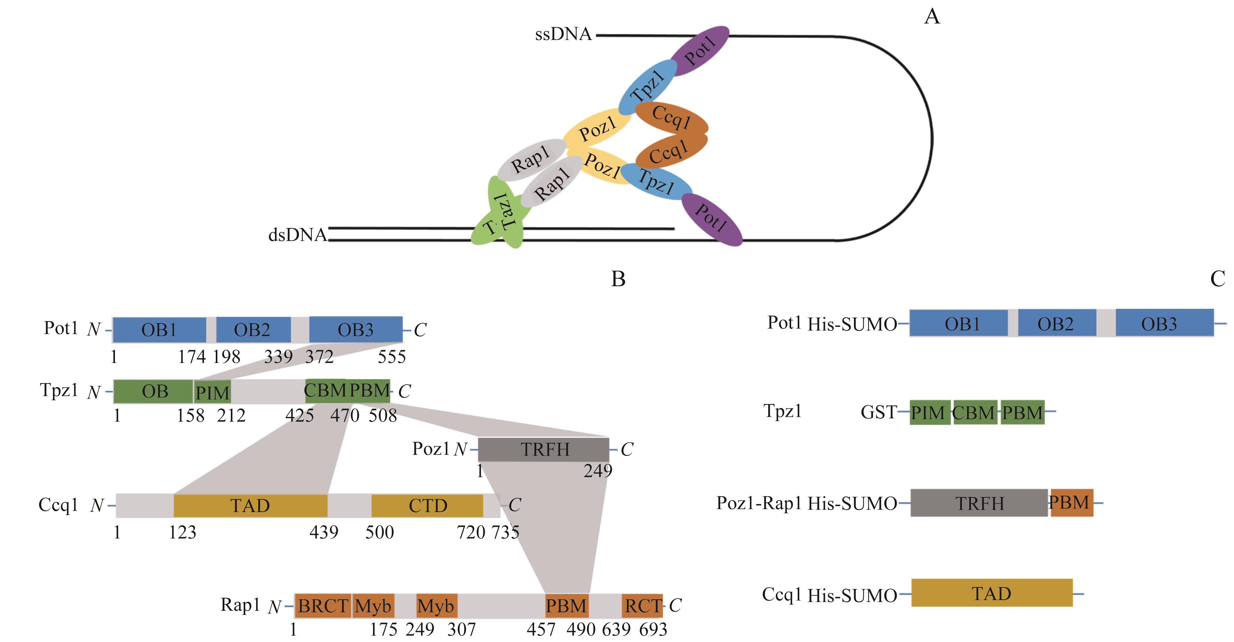
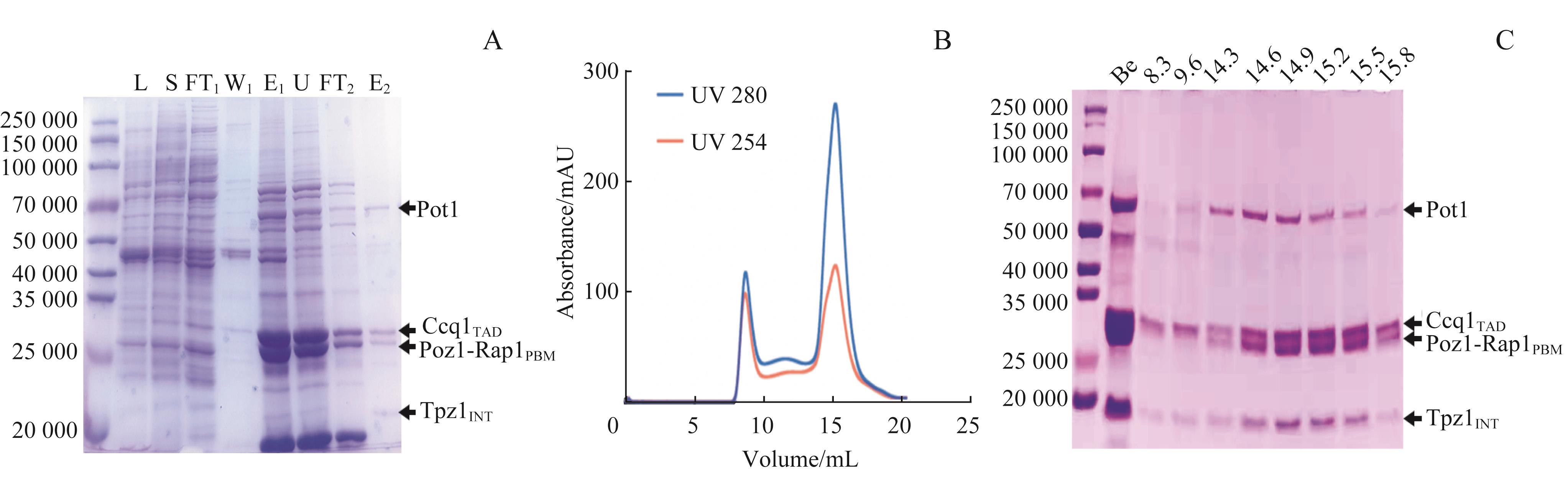
XUE J, CHEN H W, WU J, et al. Structure of the fission yeast S. pombe telomeric Tpz1-Poz1-Rap1 complex[J]. Cell Res, 2017, 27(12): 1503-1520.
|
| 20 |
LEI M, PODELL E R, BAUMANN P, et al. DNA self-recognition in the structure of Pot1 bound to telomeric single-stranded DNA[J]. Nature, 2003, 426(6963): 198-203.
|
| 21 |
KIM J K, LIU J Q, HU X C, et al. Structural basis for shelterin bridge assembly[J]. Mol Cell, 2017, 68(4): 698-714.e5.
|
| 22 |
FAIRALL L, CHAPMAN L, MOSS H, et al. Structure of the TRFH dimerization domain of the human telomeric proteins TRF1 and TRF2[J]. Mol Cell, 2001, 8(2): 351-361.
|
| 23 |
LEI M, PODELL E R, CECH T R. Structure of human POT1 bound to telomeric single-stranded DNA provides a model for chromosome end-protection[J]. Nat Struct Mol Biol, 2004, 11(12): 1223-1229.
|
| 24 |
CHEN C, GU P L, WU J, et al. Structural insights into POT1-TPP1 interaction and POT1 C-terminal mutations in human cancer[J]. Nat Commun, 2017, 8: 14929.
|
| 25 |
KIM S H, DAVALOS A R, HEO S J, et al. Telomere dysfunction and cell survival: roles for distinct TIN2-containing complexes[J]. J Cell Biol, 2008, 181(3): 447-460.
|
| 26 |
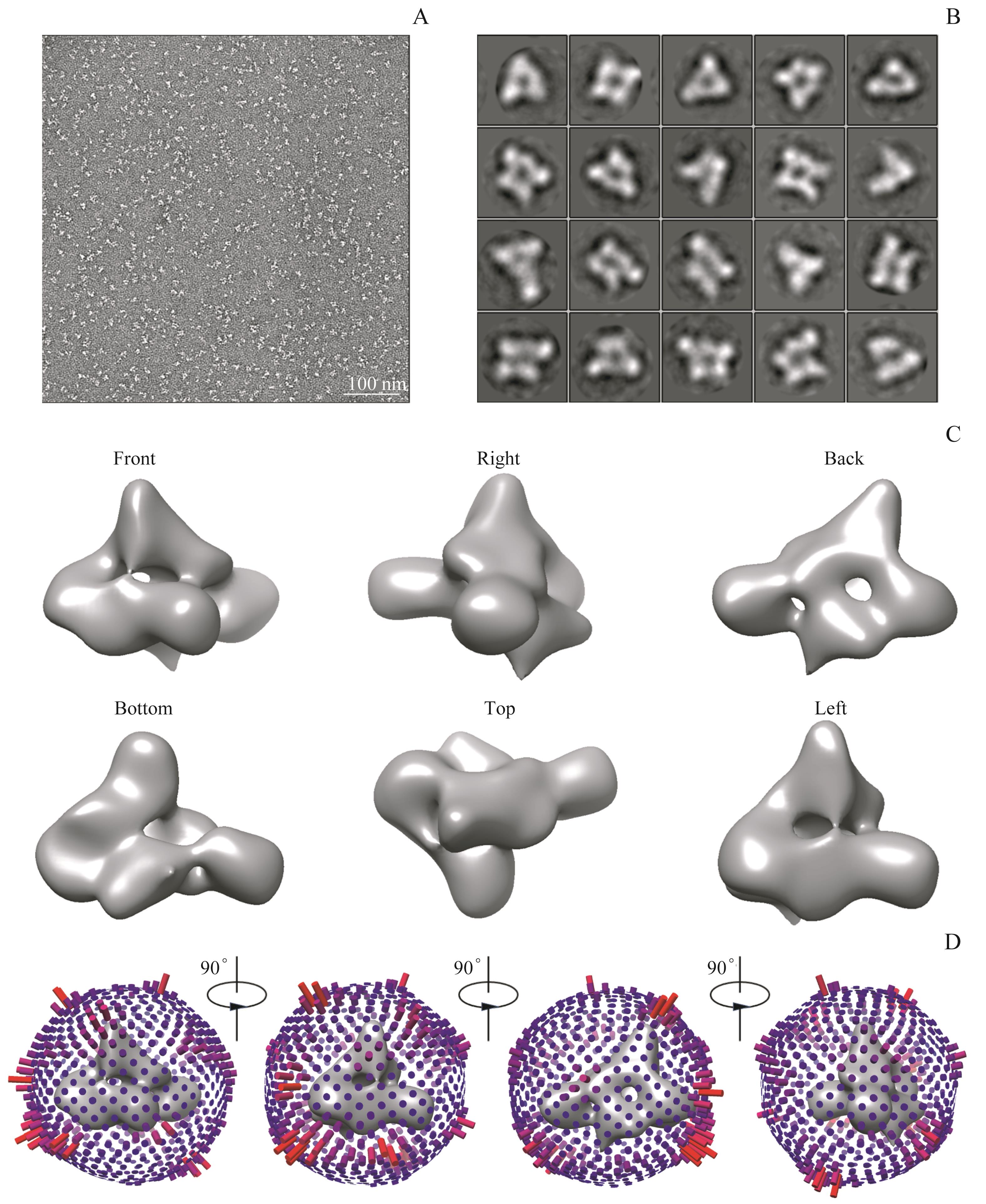
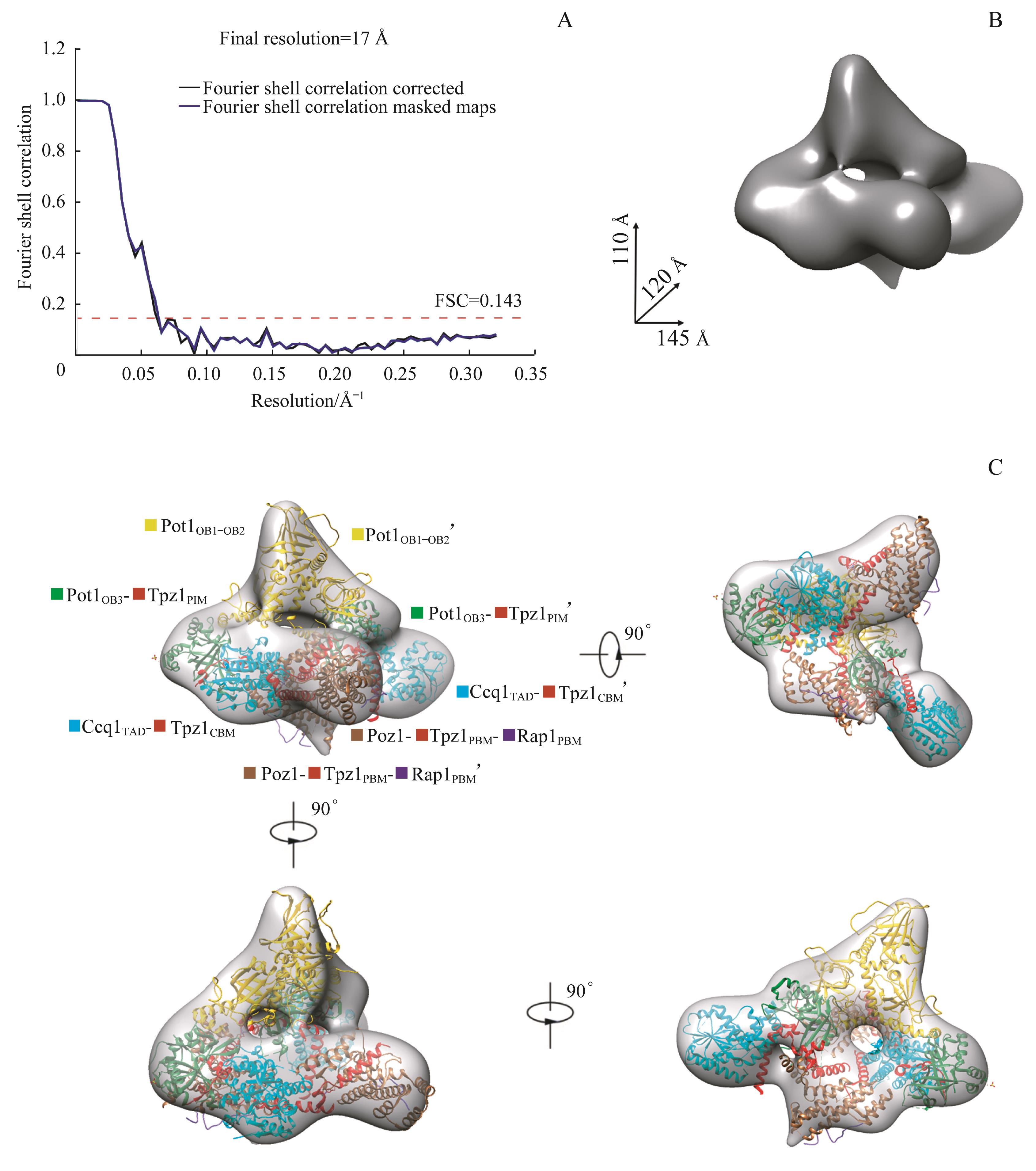
TANG G, PENG L W, BALDWIN P R, et al. EMAN2: an extensible image processing suite for electron microscopy[J]. J Struct Biol, 2007, 157(1): 38-46.
|
| 27 |
SCHERES S H W. RELION: implementation of a Bayesian approach to cryo-EM structure determination[J]. J Struct Biol, 2012, 180(3): 519-530.
|
| 28 |
PETTERSEN E F, GODDARD T D, HUANG C C, et al. UCSF Chimera: a visualization system for exploratory research and analysis[J]. J Comput Chem, 2004, 25(13): 1605-1612.
|
| 29 |
BURLEY S K, BERMAN H M, KLEYWEGT G J, et al. Protein data bank (PDB): the single global macromolecular structure archive[J]. Methods Mol Biol, 2017, 1607: 627-641.
|
| 30 |
SCOTT H, KIM J K, YU C, et al. Spatial organization and molecular interactions of the Schizosaccharomyces pombe Ccq1-Tpz1-Poz1 shelterin complex[J]. J Mol Biol, 2017, 429(19): 2863-2872.
|
| 31 |
SAVAGE S A, GIRI N, BAERLOCHER G M, et al. TINF2, a component of the shelterin telomere protection complex, is mutated in dyskeratosis congenita[J]. Am J Hum Genet, 2008, 82(2): 501-509.
|
| 32 |
MYLER L R, KINZIG C G, SASI N K, et al. The evolution of metazoan shelterin[J]. Genes Dev, 2021, 35(23/24): 1625-1641.
|
| 33 |
WANG J Y, COHEN A L, LETIAN A, et al. The proper connection between shelterin components is required for telomeric heterochromatin assembly[J]. Genes Dev, 2016, 30(7): 827-839.
|
| 34 |
LIU J Q, HU X C, BAO K H, et al. The cooperative assembly of shelterin bridge provides a kinetic gateway that controls telomere length homeostasis[J]. Nucleic Acids Res, 2021, 49(14): 8110-8119.
|
 ), SUN Hong, WU Zhenfang, LEI Ming(