Journal of Shanghai Jiao Tong University (Medical Science) ›› 2023, Vol. 43 ›› Issue (9): 1089-1098.doi: 10.3969/j.issn.1674-8115.2023.09.003
• Basic research • Previous Articles
JIA Junjie( ), XING Haifan, ZHANG Qunzi, LIU Qiye, WANG Niansong, FAN Ying()
Received:2023-04-14
Accepted:2023-08-10
Online:2023-09-28
Published:2023-09-28
Contact:
FAN Ying
E-mail:jekun0610@gmail.com;fanyingsh@126.com
Supported by:CLC Number:
JIA Junjie, XING Haifan, ZHANG Qunzi, LIU Qiye, WANG Niansong, FAN Ying. Renal protective effect and mechanism research of hypoxia inducible factor-1α inhibitor YC-1 in diabetic nephropathy mice[J]. Journal of Shanghai Jiao Tong University (Medical Science), 2023, 43(9): 1089-1098.
| Index | WT group | WT+YC-1 group | DB group | DB+YC-1 group |
|---|---|---|---|---|
| RBG/(mmol·L-1) | 8.10±0.39 | 8.25±0.43 | 28.57±1.18① | 29.15±1.29① |
| BW/g | 27.47±0.58 | 25.98±0.65 | 56.72±1.05① | 54.53±1.02① |
| KW/BW/(mg·g-1) | 9.97±0.23 | 10.19±0.23 | 12.25±0.22① | 11.31±0.14①② |
| Scr/(mg·dL-1) | 0.239±0.010 | 0.263±0.012 | 0.473±0.017① | 0.414±0.016①③ |
| UACR/(μg·mg-1) | 61.68±12.18 | 67.46±10.71 | 1 445.61±63.10① | 663.94±60.19①② |
| uNGAL/(μg·mL-1) | 23.70±1.09 | 23.04±0.95 | 118.66±2.98① | 86.02±3.44①② |
Tab 1 Effect of YC-1 on general physical signs and kidney function indexes in the WT and db/db mice (n=6)
| Index | WT group | WT+YC-1 group | DB group | DB+YC-1 group |
|---|---|---|---|---|
| RBG/(mmol·L-1) | 8.10±0.39 | 8.25±0.43 | 28.57±1.18① | 29.15±1.29① |
| BW/g | 27.47±0.58 | 25.98±0.65 | 56.72±1.05① | 54.53±1.02① |
| KW/BW/(mg·g-1) | 9.97±0.23 | 10.19±0.23 | 12.25±0.22① | 11.31±0.14①② |
| Scr/(mg·dL-1) | 0.239±0.010 | 0.263±0.012 | 0.473±0.017① | 0.414±0.016①③ |
| UACR/(μg·mg-1) | 61.68±12.18 | 67.46±10.71 | 1 445.61±63.10① | 663.94±60.19①② |
| uNGAL/(μg·mL-1) | 23.70±1.09 | 23.04±0.95 | 118.66±2.98① | 86.02±3.44①② |
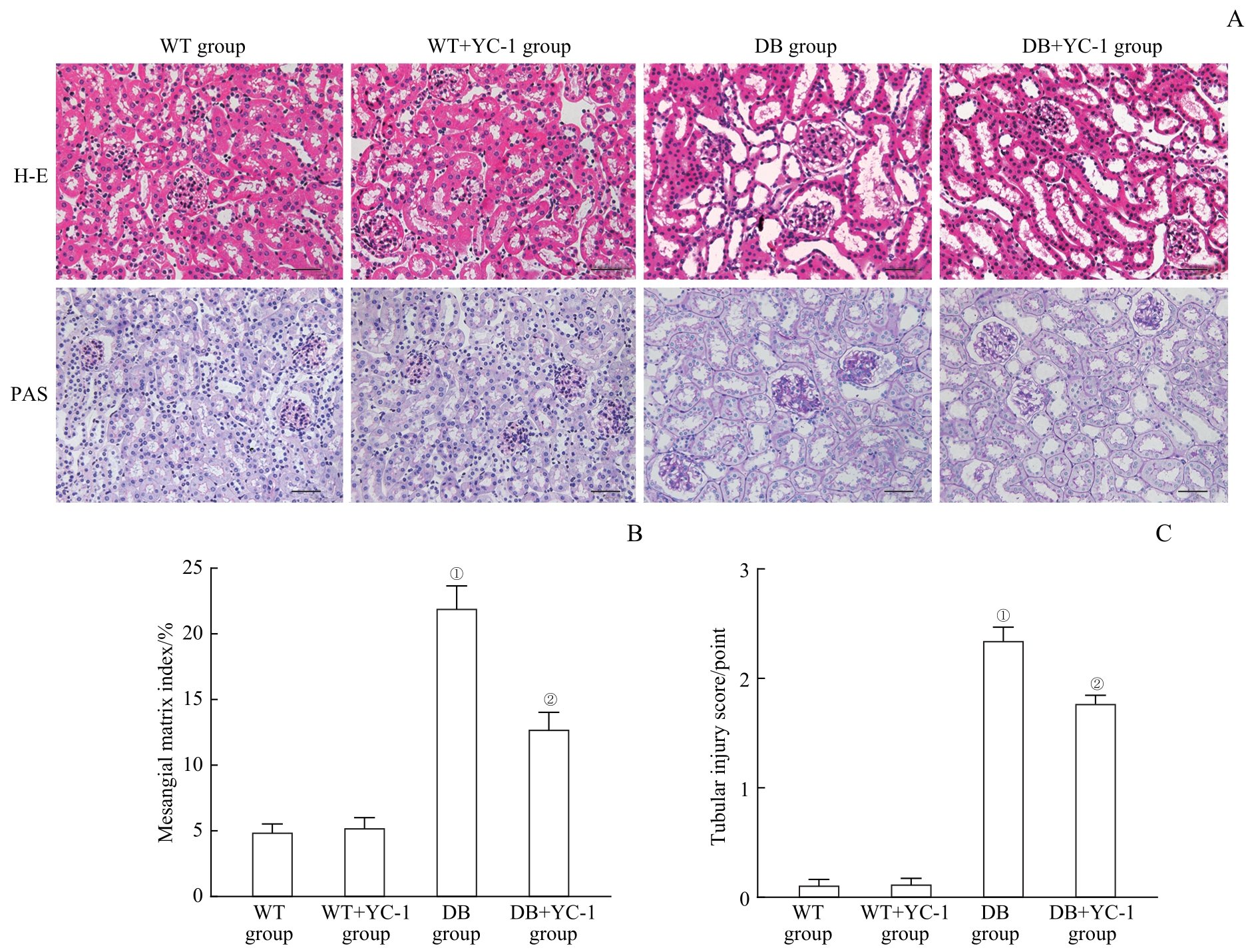
Fig 1 Effect of YC-1 on kidney histopathology injuries in the db/db mice
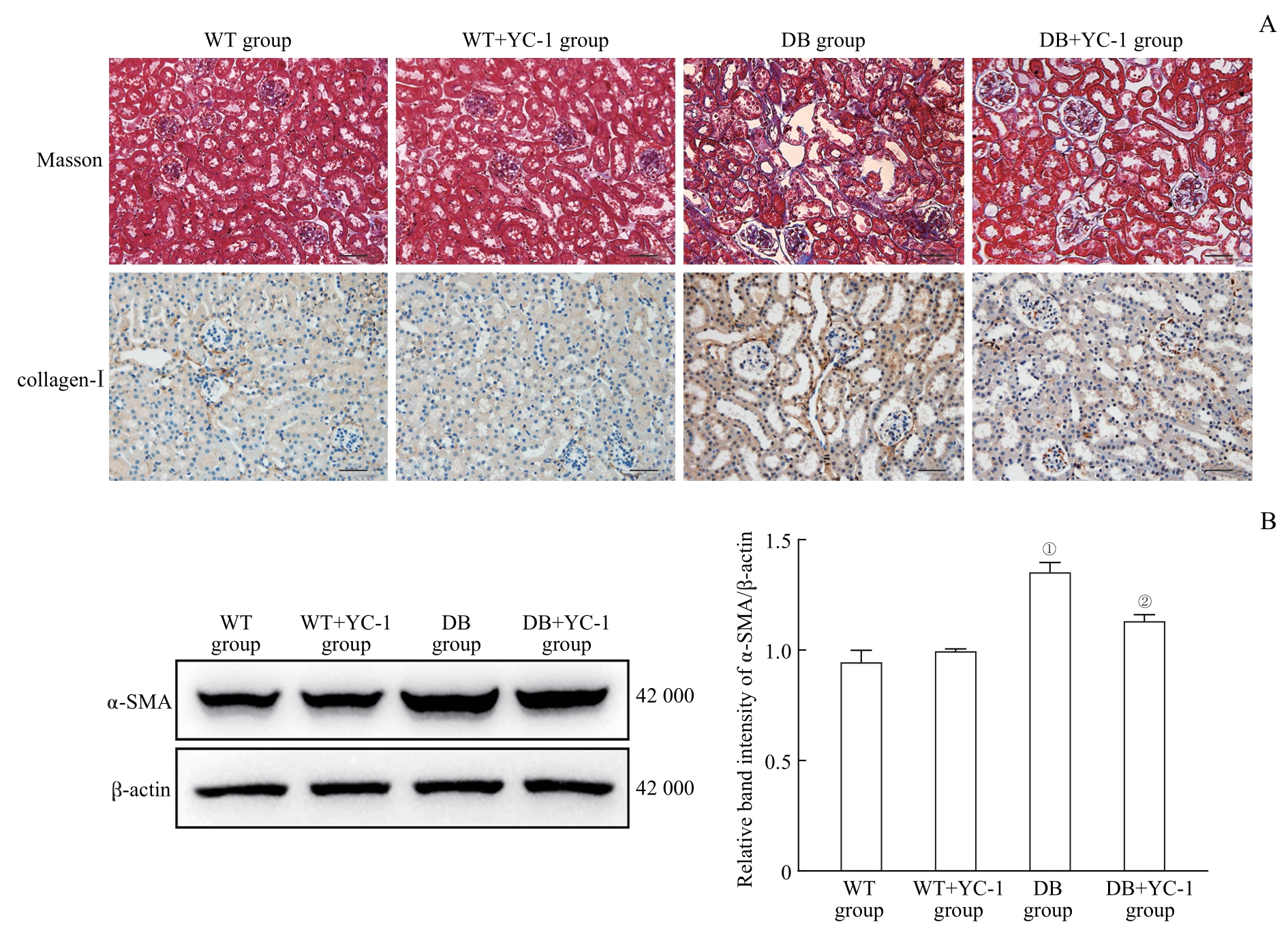
Fig 2 Effect of YC-1 on kidney fibrosis in the db/db mice
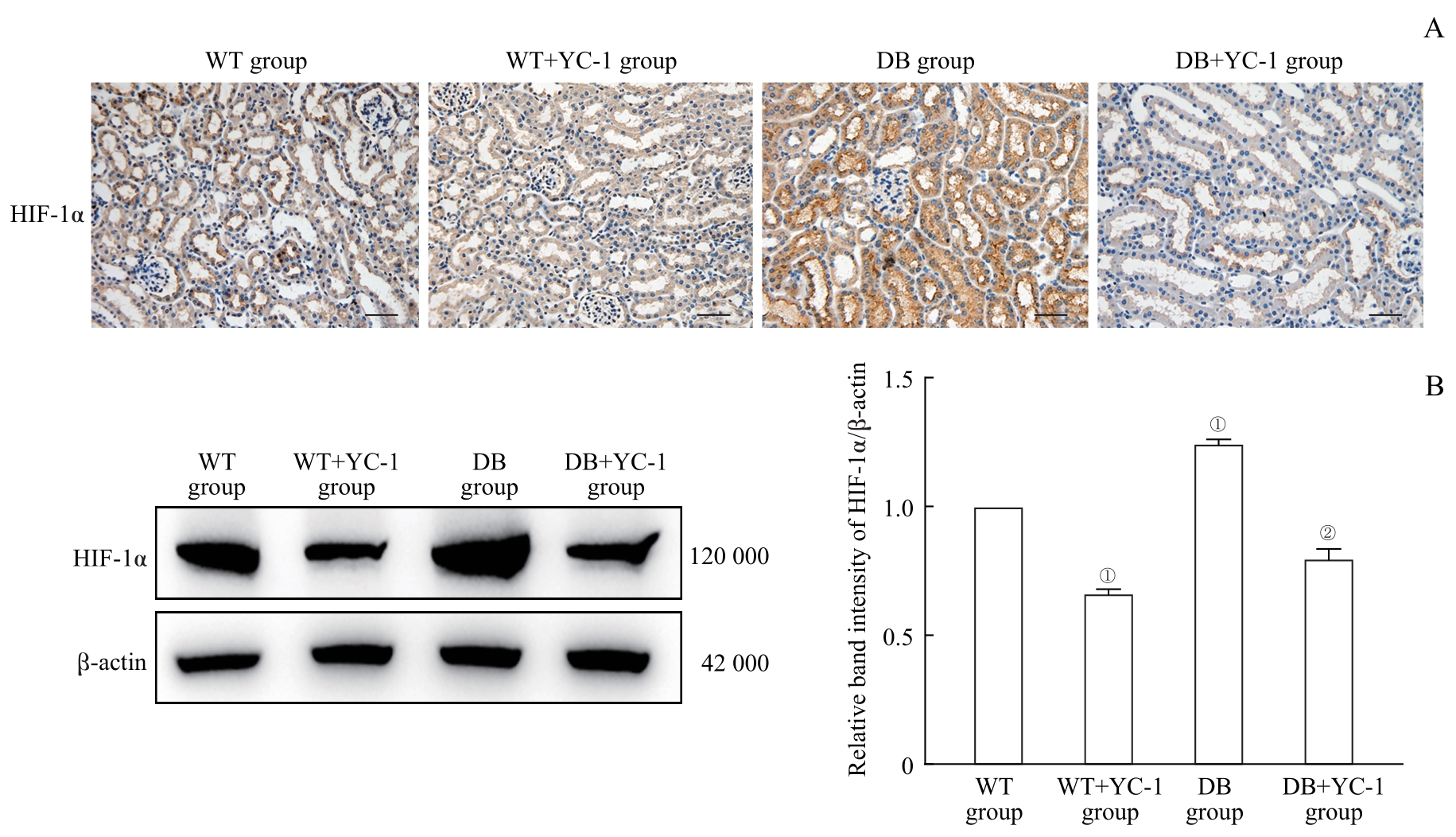
Fig 3 Effect of YC-1 on HIF-1α expression in the kidneys of db/db mice
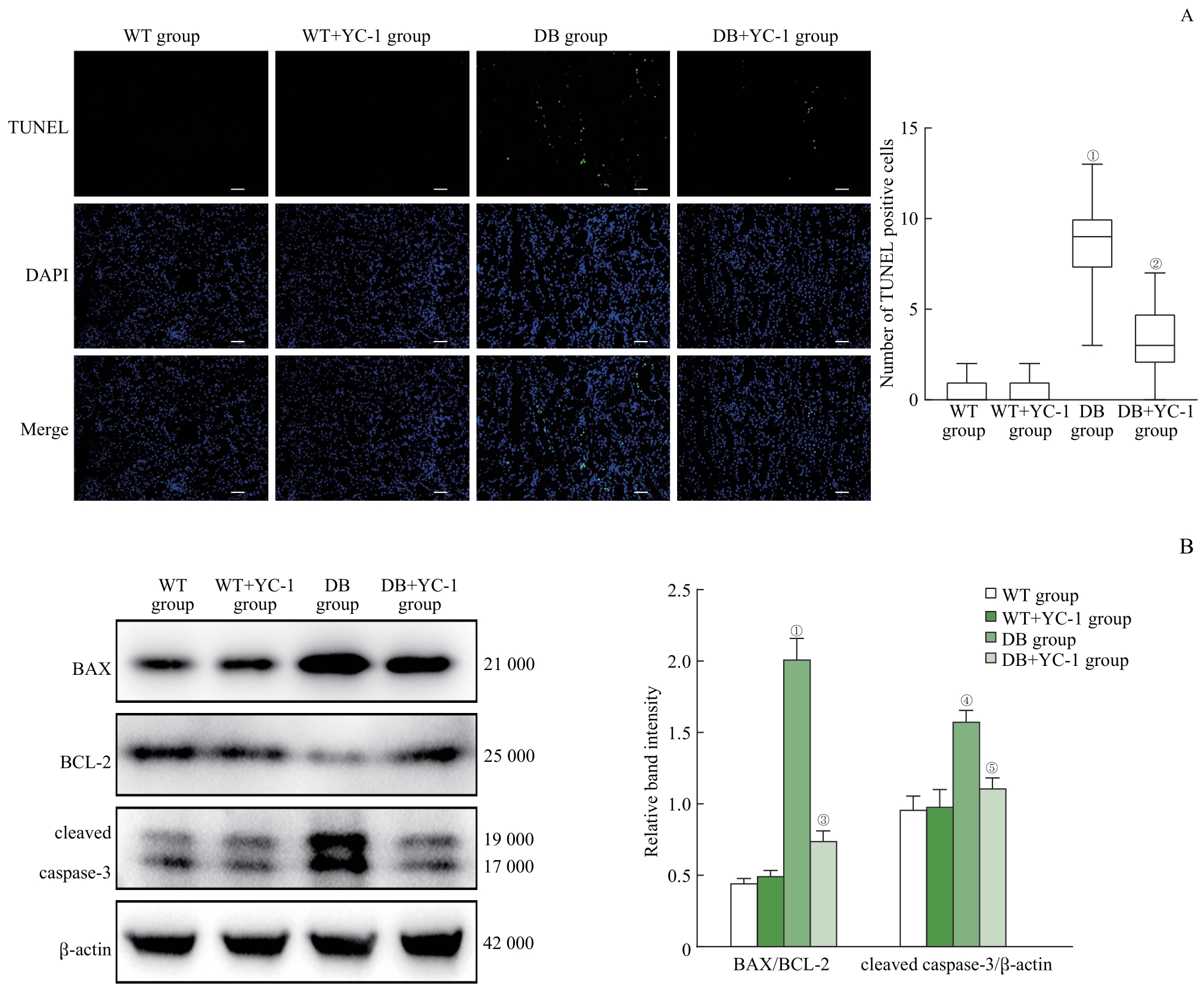
Fig 4 Effect of YC-1 on cell apoptosis in the kidneys of the db/db mice
| Index | WT group | WT+YC-1 group | DB group | DB+YC-1 group |
|---|---|---|---|---|
| MDA level/(nmol·mg-1) | 6.69±0.53 | 5.78±0.82 | 14.05±1.56① | 9.65±0.71② |
| SOD activity/(U·mg-1) | 117.28±11.90 | 115.29±6.72 | 41.71±3.19① | 79.40±2.52③ |
Tab 2 Effect of YC-1 on oxidative stress in the kidneys of db/db mice (n=6)
| Index | WT group | WT+YC-1 group | DB group | DB+YC-1 group |
|---|---|---|---|---|
| MDA level/(nmol·mg-1) | 6.69±0.53 | 5.78±0.82 | 14.05±1.56① | 9.65±0.71② |
| SOD activity/(U·mg-1) | 117.28±11.90 | 115.29±6.72 | 41.71±3.19① | 79.40±2.52③ |

Fig 5 Effect of YC-1 on ERS in the kidneys of db/db mice
| 1 | GUEDES M, PECOITS-FILHO R. Can we cure diabetic kidney disease? Present and future perspectives from a nephrologist′s point of view[J]. J Intern Med, 2022, 291(2): 165-180. |
| 2 | JUNG C Y, YOO T H. Pathophysiologic mechanisms and potential biomarkers in diabetic kidney disease[J]. Diabetes Metab J, 2022, 46(2): 181-197. |
| 3 | HESP A C, SCHAUB J A, PRASAD P V, et al. The role of renal hypoxia in the pathogenesis of diabetic kidney disease: a promising target for newer renoprotective agents including SGLT2 inhibitors?[J]. Kidney Int, 2020, 98(3): 579-589. |
| 4 | SEMENZA G L. Oxygen sensing, homeostasis, and disease[J]. N Engl J Med, 2011, 365(6): 537-547. |
| 5 | LIU H X, LI Y J, XIONG J. The role of hypoxia-inducible factor-1 α in renal disease[J]. Molecules, 2022, 27(21): 7318. |
| 6 | OUYANG C L, ZHANG J, LEI X Y, et al. Advances in antitumor research of HIF-1α inhibitor YC-1 and its derivatives[J]. Bioorg Chem, 2023, 133: 106400. |
| 7 | KHANIJOU V, ZAFARI N, COUGHLAN M T, et al. Review of potential biomarkers of inflammation and kidney injury in diabetic kidney disease[J]. Diabetes Metab Res Rev, 2022, 38(6): e3556. |
| 8 | CHEN P, SHI X Z, XU X J, et al. Liraglutide ameliorates early renal injury by the activation of renal FoxO1 in a type 2 diabetic kidney disease rat model[J]. Diabetes Res Clin Pract, 2018, 137: 173-182. |
| 9 | ZHANG Q Z, HE L, DONG Y, et al. Sitagliptin ameliorates renal tubular injury in diabetic kidney disease via STAT3-dependent mitochondrial homeostasis through SDF-1α/CXCR4 pathway[J]. FASEB J, 2020, 34(6): 7500-7519. |
| 10 | NI L H, YUAN C, WU X Y. Endoplasmic reticulum stress in diabetic nephrology: regulation, pathological role, and therapeutic potential[J]. Oxid Med Cell Longev, 2021, 2021: 7277966. |
| 11 | NANGAKU M. Chronic hypoxia and tubulointerstitial injury: a final common pathway to end-stage renal failure[J]. J Am Soc Nephrol, 2006, 17(1): 17-25. |
| 12 | SHU S Q, WANG Y, ZHENG M L, et al. Hypoxia and hypoxia-inducible factors in kidney injury and repair[J]. Cells, 2019, 8(3): 207. |
| 13 | STANIGUT A M, PANA C, ENCIU M, et al. Hypoxia-inducible factors and diabetic kidney disease-how deep can we go?[J]. Int J Mol Sci, 2022, 23(18): 10413. |
| 14 | ZHANG H, XU R F, WANG Z C. Contribution of oxidative stress to HIF-1-mediated profibrotic changes during the kidney damage[J]. Oxid Med Cell Longev, 2021, 2021: 6114132. |
| 15 | HU J P, WANG W L, ZHANG F, et al. Hypoxia inducible factor-1α mediates the profibrotic effect of albumin in renal tubular cells[J]. Sci Rep, 2017, 7(1): 15878. |
| 16 | MEI S Q, LI L, ZHOU X J, et al. Susceptibility of renal fibrosis in diabetes: role of hypoxia inducible factor-1[J]. FASEB J, 2022, 36(8): e22477. |
| 17 | JIA Y J, CHEN J Q, ZHENG Z K, et al. Tubular epithelial cell-derived extracellular vesicles induce macrophage glycolysis by stabilizing HIF-1α in diabetic kidney disease[J]. Mol Med, 2022, 28(1): 95. |
| 18 | LI X Y, YANG S S, YAN M H, et al. Interstitial HIF1A induces an estimated glomerular filtration rate decline through potentiating renal fibrosis in diabetic nephropathy[J]. Life Sci, 2020, 241: 117109. |
| 19 | NAYAK B K, SHANMUGASUNDARAM K, FRIEDRICHS W E, et al. HIF-1 mediates renal fibrosis in OVE26 type 1 diabetic mice[J]. Diabetes, 2016, 65(5): 1387-1397. |
| 20 | PACKER M. Mechanisms leading to differential hypoxia-inducible factor signaling in the diabetic kidney: modulation by SGLT2 inhibitors and hypoxia mimetics[J]. Am J Kidney Dis, 2021, 77(2): 280-286. |
| 21 | IACOBINI C, VITALE M, HAXHI J, et al. Mutual regulation between redox and hypoxia-inducible factors in cardiovascular and renal complications of diabetes[J]. Antioxidants (Basel), 2022, 11(11): 2183. |
| 22 | DIEBOLD I, PETRY A, HESS J, et al. The NADPH oxidase subunit NOX4 is a new target gene of the hypoxia-inducible factor-1[J]. Mol Biol Cell, 2010, 21(12): 2087-2096. |
| 23 | HETZ C, ZHANG K Z, KAUFMAN R J. Mechanisms, regulation and functions of the unfolded protein response[J]. Nat Rev Mol Cell Biol, 2020, 21(8): 421-438. |
| 24 | MOULIN S, THOMAS A, WAGNER S, et al. Intermittent hypoxia-induced cardiomyocyte death is mediated by HIF-1 dependent MAM disruption[J]. Antioxidants (Basel), 2022, 11(8): 1462. |
| 25 | YANG Y Y, YU H H, JIAO X L, et al. Angiopoietin-like proteins 8 knockout reduces intermittent hypoxia-induced vascular remodeling in a murine model of obstructive sleep apnea[J]. Biochem Pharmacol, 2021, 186: 114502. |
| 26 | DELBREL E, SOUMARE A, NAGUEZ A, et al. HIF-1α triggers ER stress and CHOP-mediated apoptosis in alveolar epithelial cells, a key event in pulmonary fibrosis[J]. Sci Rep, 2018, 8(1): 17939. |
| 27 | SUN F Q, DU J C, LI H B, et al. FABP4 inhibitor BMS309403 protects against hypoxia-induced H9c2 cardiomyocyte apoptosis through attenuating endoplasmic reticulum stress[J]. J Cell Mol Med, 2020, 24(19): 11188-11197. |
| 28 | MOULIN S, THOMAS A, ARNAUD C, et al. Cooperation between hypoxia-inducible factor 1α and activating transcription factor 4 in sleep apnea-mediated myocardial injury[J]. Can J Cardiol, 2020, 36(6): 936-940. |
| [1] | JIN Fangquan, FAN Chenghu, TANG Xiaodong, CHEN Yantong, QI Bingxian. Research progress in the relationship between mitochondrial dysfunction and osteoporosis [J]. Journal of Shanghai Jiao Tong University (Medical Science), 2023, 43(6): 761-767. |
| [2] | WU Jiajin, ZHONG Chen, LI Dawei, CHEN Ruoyang, QU Junwen, ZHANG Ming. Role of methyltransferase like 3 regulating pri-miR-21 methylation in renal fibrosis of diabetes nephropathy [J]. Journal of Shanghai Jiao Tong University (Medical Science), 2023, 43(1): 1-7. |
| [3] | ZHAO Jiuhong, TONG Jiating, SHEN Zhijun, LÜ Yehui. Research progress in the mechanism of interactive regulation between circular RNA and oxidative stress [J]. Journal of Shanghai Jiao Tong University (Medical Science), 2022, 42(3): 393-399. |
| [4] | SUN Jinli, SONG Weiwei, XU Ming, LI Jingquan. Oxidative damage and malignant migration of hepatocellular carcinoma cells LM3 induced by 14 weeks exposure to sodium arsenite [J]. Journal of Shanghai Jiao Tong University (Medical Science), 2022, 42(12): 1677-1684. |
| [5] | Jiu-ang MAO, Zhen WENG, Xiao-yin NIU, Yang HE, Zhen-xin WANG. Role of Tmprss6 gene in radiation-induced intestinal injury of mice [J]. JOURNAL OF SHANGHAI JIAOTONG UNIVERSITY (MEDICAL SCIENCE), 2021, 41(9): 1175-1182. |
| [6] | Sheng CHEN, Zhi-long YAN, Ye-ming WU, Min XU, Song GU, Jing MA. Expression and clinical significance of HIF-1α/SHH signaling pathway in neuroblastoma [J]. JOURNAL OF SHANGHAI JIAOTONG UNIVERSITY (MEDICAL SCIENCE), 2021, 41(8): 1056-1061. |
| [7] | Run-ze YANG, Wen-ning XU, Huo-liang ZHENG, Sheng-dan JIANG. Effects of exosomes derived from human umbilical vein endothelial cells on apoptosis of pre-chondrogenic cells stimulated by inflammatory factors [J]. JOURNAL OF SHANGHAI JIAOTONG UNIVERSITY (MEDICAL SCIENCE), 2021, 41(2): 147-153. |
| [8] | Jing WU, Xue-yi LI, Jing-hong CHEN, Ze-jian WANG. Study on changes of hippocampal bile acid receptors in the depression mouse models [J]. JOURNAL OF SHANGHAI JIAOTONG UNIVERSITY (MEDICAL SCIENCE), 2021, 41(12): 1628-1634. |
| [9] | LU Hai-yang, ZHAO Wei-li. Role of gastrointestinal microbiota in tumorigenesis [J]. , 2019, 39(9): 1083-. |
| [10] | YANG Shuang-shuang1*, GAO Tian-xing2*, HE Xuan1, ZHANG Rui1, ZHANG Yong-fang1. Regulation on brain-derived neurotrophic factor and relevant mechanism of anemarrhena saponin in H2O2-induced SH-SY5Y cells [J]. , 2019, 39(6): 578-. |
| [11] | WANG Hao1*, JIANG Shan1*, GONG Yang-ming2, LIU Yan3, HUA Li1, DENG Xiao-bei1. Atmospheric fine particulate matter causing Alzheimers disease through olfactory bulb pathway: a review of recent studies [J]. , 2019, 39(6): 666-. |
| [12] | WANG Ying-yi,LU Yan-hua,GENG Rui-jie,CHENG Xiao-yan,HUANG Xin-xin,Lü Qin-yu,YING Qi-ang,YI Zheng-hui. Effect of lithium carbonate on oxidative stress in patients with bipolar disorder [J]. , 2019, 39(5): 494-. |
| [13] | LI Qi1, ZHOU Xiang-dong1, ZENG Man1, Victor P. KOLOSOV2, Juliy M. PERELMAN2. Role of inositol-requiring kinase 1α/X-box binding protein 1 in airway mucus secretion inducedneutrophil elastase [J]. , 2019, 39(1): 21-. |
| [14] | ZHANG Jing-jing, ZHOU Yi-jun, HU Yi, SHI Rong, ZHANG Yan, TIAN Ying, GAO Yu. Effects of fenvalerate exposure during puberty on oxidative stress in male rat testis [J]. , 2018, 38(2): 133-. |
| [15] | JI Lin1,RUXIANGULI Aimuzi1, 2, ZHANG Yan1,SHI Rong1,ZHOU Yi-jun1,CHENG Xiao-meng3,WANG Xue-mei3,TIAN Ying1,GAO Yu1. Association between exposure to organophosphate pesticides and levels of oxidative stress in pregnant women with different paraoxonase 1 genotypes [J]. , 2018, 38(2): 174-. |
| Viewed | ||||||
|
Full text |
|
|||||
|
Abstract |
|
|||||