Journal of Shanghai Jiao Tong University (Medical Science) ›› 2025, Vol. 45 ›› Issue (8): 990-1000.doi: 10.3969/j.issn.1674-8115.2025.08.006
• Basic research • Previous Articles Next Articles
ZHOU Xingdie1,2, CHEN Zehao3, LÜ Zhendong2, ZHANG Yuhui2( ), LIU Li1()
Received:2025-01-08
Accepted:2025-04-11
Online:2025-08-28
Published:2025-08-20
Contact:
ZHANG Yuhui, LIU Li
E-mail:zhangyuhui@renji.com;Liuli2002@shu.edu.cn
Supported by:CLC Number:
ZHOU Xingdie, CHEN Zehao, LÜ Zhendong, ZHANG Yuhui, LIU Li. Effect of in situ hypoxia-inducing hydrogelon extracellular matrix secretion in the nucleus pulposus[J]. Journal of Shanghai Jiao Tong University (Medical Science), 2025, 45(8): 990-1000.
Add to citation manager EndNote|Ris|BibTeX
URL: https://xuebao.shsmu.edu.cn/EN/10.3969/j.issn.1674-8115.2025.08.006
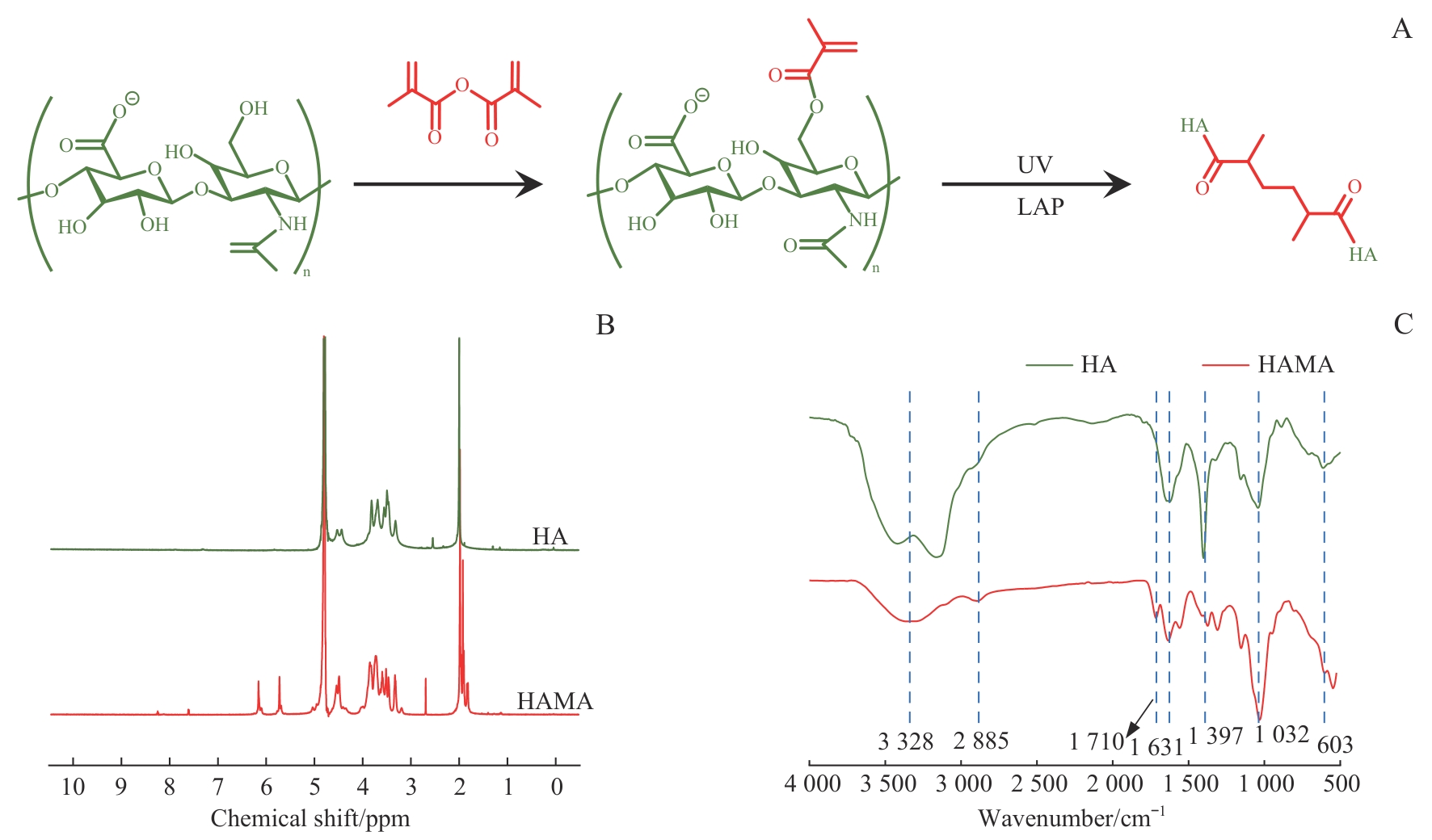
Fig 1 Schematic illustration and structural characterization of HAMA
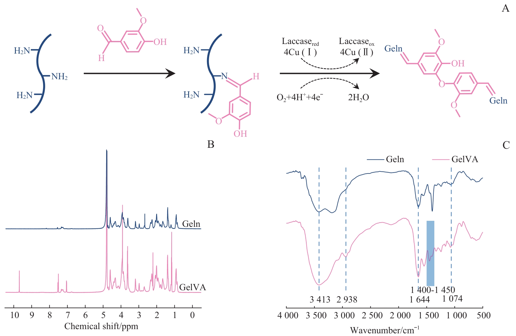
Fig 2 Schematic illustration and structural characterization of GelVA
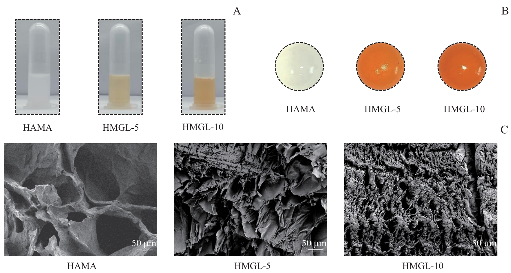
Fig 3 Morphological characterization of different hydrogels
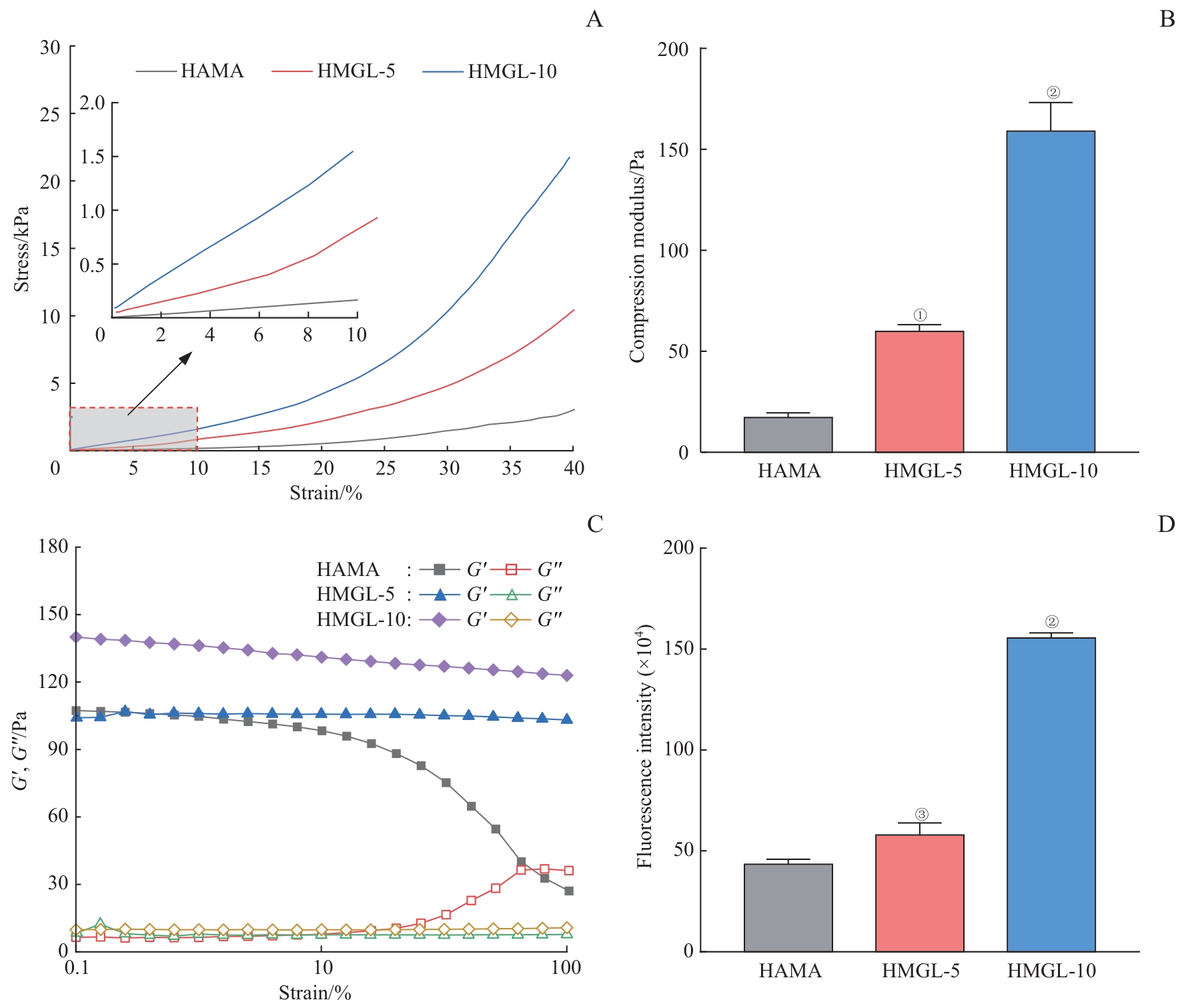
Fig 4 Mechanical properties and hypoxia-inducing behavior of hypoxic hydrogels
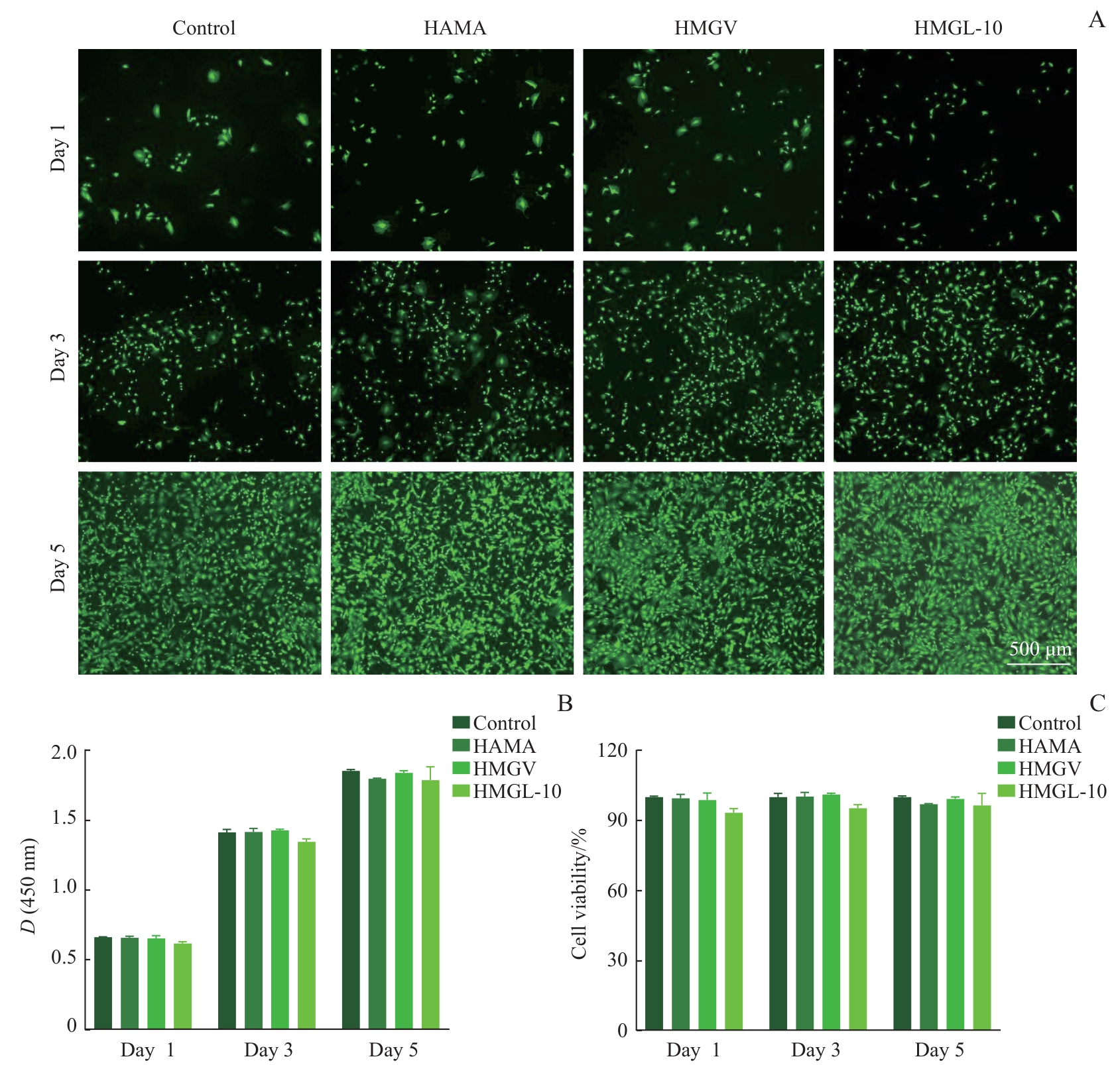
Fig 5 Biocompatibility of hypoxic hydrogels
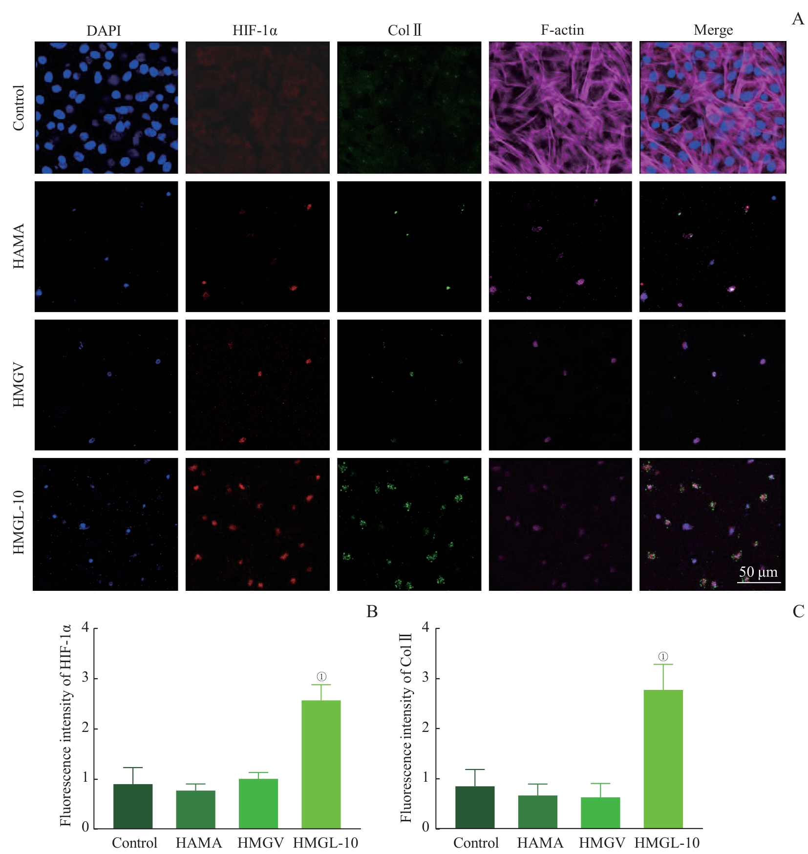
Fig 6 Expression of HIF-1α and Col Ⅱ in nucleus pulposus cells cultured on different hydrogel matrices
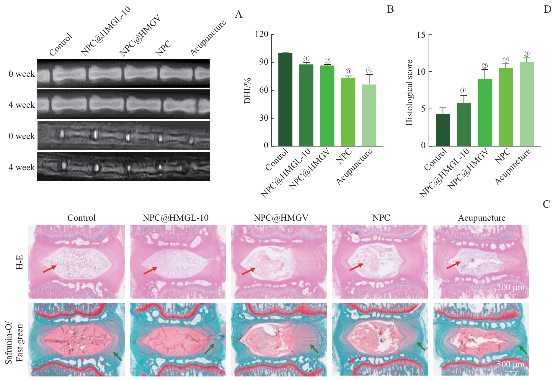
Fig 7 Repair of degenerated intervertebral discs using different hydrogel matrices loaded with nucleus pulposus cells
| [1] | XING H Y, ZHANG Z J, MAO Q J, et al. Injectable exosome-functionalized extracellular matrix hydrogel for metabolism balance and pyroptosis regulation in intervertebral disc degeneration[J]. J Nanobiotechnol, 2021, 19(1): 264. |
| [2] | SAKAI D, GRAD S. Advancing the cellular and molecular therapy for intervertebral disc disease[J]. Adv Drug Deliv Rev, 2015, 84: 159-171. |
| [3] | MERCERON C, MANGIAVINI L, ROBLING A, et al. Loss of HIF-1α in the notochord results in cell death and complete disappearance of the nucleus pulposus[J]. PLoS One, 2014, 9(10): e110768. |
| [4] | BARTELS E M, FAIRBANK J C, WINLOVE C P, et al. Oxygen and lactate concentrations measured in vivo in the intervertebral discs of patients with scoliosis and back pain[J]. Spine (Phila Pa 1976), 1998, 23(1): 1-7;discussion8. |
| [5] | RISBUD M V, SHAPIRO I M. Role of cytokines in intervertebral disc degeneration: pain and disc content[J]. Nat Rev Rheumatol, 2014, 10(1): 44-56. |
| [6] | YANG S, ZHANG F, MA J, et al. Intervertebral disc ageing and degeneration: the antiapoptotic effect of oestrogen[J]. Ageing Res Rev, 2020, 57: 100978. |
| [7] | SEMENZA G L. Hypoxia-inducible factor 1: oxygen homeostasis and disease pathophysiology[J]. Trends Mol Med, 2001, 7(8): 345-350. |
| [8] | PFANDER D, CRAMER T, SCHIPANI E, et al. HIF-1α controls extracellular matrix synthesis by epiphyseal chondrocytes[J]. J Cell Sci, 2003, 116(9): 1819-1826. |
| [9] | PARK K M, BLATCHLEY M R, GERECHT S. The design of dextran-based hypoxia-inducible hydrogels via in situ oxygen-consuming reaction[J]. Macromol Rapid Commun, 2014, 35(22): 1968-1975. |
| [10] | PARK K M, GERECHT S. Hypoxia-inducible hydrogels[J]. Nat Commun, 2014, 5: 4075. |
| [11] | ZHAO R, LIU W, XIA T, et al. Disordered mechanical stress and tissue engineering therapies in intervertebral disc degeneration[J]. Polymers (Basel), 2019, 11(7): E1151. |
| [12] | WANG W T, LIU L, MA W Z, et al. An anti-senescence hydrogel with pH-responsive drug release for mitigating intervertebral disc degeneration and low back pain[J]. Bioact Mater, 2024, 41: 355-370. |
| [13] | HARRINGTON S, WILLIAMS J, RAWAL S, et al. Hyaluronic acid/collagen hydrogel as an alternative to alginate for long-term immunoprotected islet transplantation[J]. Tissue Eng Part A, 2017, 23(19/20): 1088-1099. |
| [14] | A S, ZENG M, JOHNSON M, et al. Green synthetic approach for photo-cross-linkable methacryloyl hyaluronic acid with a tailored substitution degree[J]. Biomacromolecules, 2020, 21(6): 2229-2235. |
| [15] | JIN X, SHANG Y, ZOU Y, et al. Injectable hypoxia-induced conductive hydrogel to promote diabetic wound healing[J]. ACS Appl Mater Interfaces, 2020, 12(51): 56681-56691. |
| [16] | MUNOZ-PINTO D J, JIMENEZ-VERGARA A C, GHARAT T P, et al. Characterization of sequential collagen-poly(ethylene glycol) diacrylate interpenetrating networks and initial assessment of their potential for vascular tissue engineering[J]. Biomaterials, 2015, 40: 32-42. |
| [17] | SI H, XING T, DING Y, et al. 3D bioprinting of the sustained drug release wound dressing with double-crosslinked hyaluronic-acid-based hydrogels[J]. Polymers (Basel), 2019, 11(10): E1584. |
| [18] | VASI A M, POPA M I, BUTNARU M, et al. Chemical functionalization of hyaluronic acid for drug delivery applications[J]. Mater Sci Eng C, 2014, 38: 177-185. |
| [19] | XIA C, CHEN P, MEI S, et al. Photo-crosslinked HAMA hydrogel with cordycepin encapsulated chitosan microspheres for osteoarthritis treatment[J]. Oncotarget, 2017, 8(2): 2835-2849. |
| [20] | XU C H, ZHAN W, TANG X Z, et al. Self-healing chitosan/vanillin hydrogels based on Schiff-base bond/hydrogen bond hybrid linkages[J]. Polym Test, 2018, 66: 155-163. |
| [21] | GÜNGÖR Ö, GÜRKAN P. Synthesis and characterization of higher amino acid Schiff bases, as monosodium salts and neutral forms. Investigation of the intramolecular hydrogen bonding in all Schiff bases, antibacterial and antifungal activities of neutral forms[J]. J Mol Struct, 2014, 1074: 62-70. |
| [22] | CHAO S C, WANG M J, PAI N S, et al. Preparation and characterization of gelatin-hydroxyapatite composite microspheres for hard tissue repair[J]. Mater Sci Eng C, 2015, 57: 113-122. |
| [23] | SONG D, MA L W, PANG B, et al. An active bio-based food packaging material of ZnO@Plant polyphenols/cellulose/polyvinyl alcohol: design, characterization and application[J]. Int J Mol Sci, 2023, 24(2): 1577. |
| [24] | PARK J, NAM J, YUN H, et al. Aquatic polymer-based edible films of fish gelatin crosslinked with alginate dialdehyde having enhanced physicochemical properties[J]. Carbohydr Polym, 2021, 254: 117317. |
| [25] | NACHEMSON A L. Disc pressure measurements[J]. Spine (Phila Pa 1976), 1981, 6(1): 93-97. |
| [26] | LIU H, PAN H, YANG H, et al. LIM mineralization protein-1 suppresses TNF-α induced intervertebral disc degeneration by maintaining nucleus pulposus extracellular matrix production and inhibiting matrix metalloproteinases expression[J]. J Orthop Res, 2015, 33(3): 294-303. |
| [27] | BINCH A L A, FITZGERALD J C, GROWNEY E A, et al. Cell-based strategies for IVD repair: clinical progress and translational obstacles[J]. Nat Rev Rheumatol, 2021, 17(3): 158-175. |
| [28] | LE MAITRE C L, POCKERT A, BUTTLE D J, et al. Matrix synthesis and degradation in human intervertebral disc degeneration[J]. Biochem Soc Trans, 2007, 35(pt 4): 652-655. |
| [29] | BERTRAM H, KROEBER M, WANG H, et al. Matrix-assisted cell transfer for intervertebral disc cell therapy[J]. Biochem Biophys Res Commun, 2005, 331(4): 1185-1192. |
| [30] | WANG P, MENG Q, WANG W, et al. Icariin inhibits the inflammation through down-regulating NF-κB/HIF-2α signal pathways in chondrocytes[J]. Biosci Rep, 2020, 40(11): BSR20203107. |
| [31] | WANG P, ZHU P, LIU R, et al. Baicalin promotes extracellular matrix synthesis in chondrocytes via the activation of hypoxia-inducible factor-1α[J]. Exp Ther Med, 2020, 20(6): 226. |
| [32] | YANG W, JIA C W, LIU L, et al. Hypoxia-inducible factor-1α protects against intervertebral disc degeneration through antagonizing mitochondrial oxidative stress[J]. Inflammation, 2023, 46(1): 270-284. |
| [33] | FENG G, LI L, HONG Y, et al. Hypoxia promotes nucleus pulposus phenotype in 3D scaffolds in vitro and in vivo: laboratory investigation[J]. J Neurosurg Spine, 2014, 21(2): 303-309. |
| [34] | FENG C, ZHANG Y, YANG M, et al. Transcriptome and alternative splicing analysis of nucleus pulposus cells in response to high oxygen tension: involvement of high oxygen tension in the pathogenesis of intervertebral disc degeneration[J]. Int J Mol Med, 2018, 41(6): 3422-3432. |
| [1] | WAN Hongjin, HU Yibin, WANG Xin, ZHANG Kai, QIN An, MA Peixiang, MA Hui, ZHAO Jie. Neferine alleviates intervertebral disc degeneration through KEAP1/NRF2/GPX4 and NF-κB signaling pathways [J]. Journal of Shanghai Jiao Tong University (Medical Science), 2025, 45(3): 261-270. |
| [2] | LIU Linnan, FENG Li, WANG Long, LIU Jiayin, FAN Zhisong. Research progress in the expression of versican in malignant tumors and its biological roles [J]. Journal of Shanghai Jiao Tong University (Medical Science), 2024, 44(4): 525-530. |
| [3] | CHEN Zehao, LÜ Zhendong, ZHANG Zhen, CUI Wenguo, ZHANG Yuhui. Effect of hydrogel stiffness on nucleus pulposus cell phenotypes in vitro and its repairment of intervertebral disc in vivo [J]. Journal of Shanghai Jiao Tong University (Medical Science), 2023, 43(7): 804-813. |
| [4] | TANG Lei, XU Yingchun, ZHANG Fengchun. Review of the role of collagen in tumorigenesis and development [J]. Journal of Shanghai Jiao Tong University (Medical Science), 2023, 43(12): 1577-1584. |
| [5] | Xiao-zhi SUN, Shuang LI, Ying JIN, Bing LIAO. Effect of enzyme digestion into cell clumps on protein levels of OCT4 and SOX2 in human embryonic stem cells [J]. JOURNAL OF SHANGHAI JIAOTONG UNIVERSITY (MEDICAL SCIENCE), 2021, 41(4): 413-420. |
| [6] | LIANG Zhi-hao, CHEN Zhi-qian, CHEN Chen, ZHOU Yi-fan, YANG Xiao, ZHAO Jie. Effect and mechanism of ginsenoside Re on intervertebral disc degeneration [J]. JOURNAL OF SHANGHAI JIAOTONG UNIVERSITY (MEDICAL SCIENCE), 2020, 40(10): 1347-1353. |
| [7] | JI An-qi, DENG Guo-ying, WANG Qiu-gen, WANG Qian . Research advances in the mechanism of osteoarthritis caused by the mechanical instability [J]. , 2017, 37(4): 561-. |
| [8] | SONG Fei, LIU Ying-kai, WANG Xi-qiao. Study on dynamic changes in microvessels and partial pressure of oxygen during the occurrence and development of hypertrophic scars [J]. , 2016, 36(11): 1553-. |
| [9] | BAO Jing, XIE Ming, JIAO Ting. Effect of tensile force on expression of extracellular matrix metalloproteinase inducer in human periodontal ligament fibroblasts and study on relevant signal pathways [J]. , 2015, 35(9): 1280-. |
| [10] | HUANG Li, XIE Ming. Functional role of EMMPRIN in morphogenesis of tooth germ and formation of hard tissues in mouse mandibular first molars [J]. , 2014, 34(1): 23-. |
| [11] | FENG Jie, WU Qing-kai, WU Deng-long, et al. Features of extracellular matrix of pelvic floor muscles in pregnant women with stress urinary incontinence [J]. , 2013, 33(7): 981-. |
| [12] | XU Yan-chun, YAO Xiao-hong, ZHU Ming-jie. Research progress of breast atypical ductal hyperplasia [J]. , 2013, 33(4): 516-. |
| [13] | KANG Yu-jun, WANG Chuan, JIANG Zheng, et al. Effect of recombinant plasmid pEGFP-N1-EMMPRIN on invasion and migration of gastric cancer SGC-7901 cells [J]. , 2013, 33(2): 155-. |
| [14] | KANG Yu-jun, WANG Chuan, JIANG Zheng, et al. Influence of short hairpin RNA of EMMPRIN on invasion and migration of SGC-7901 cells [J]. , 2013, 33(1): 12-. |
| [15] | GUO Shan-mai, ZHONG Fang, ZHOU Qiao, et al. Renal protective effect of Cordyceps sinensis on 5/6 nephrectomy-induced renal fibrosis in rats [J]. , 2012, 32(1): 1-. |
| Viewed | ||||||
|
Full text |
|
|||||
|
Abstract |
|
|||||