JOURNAL OF SHANGHAI JIAOTONG UNIVERSITY (MEDICAL SCIENCE) ›› 2022, Vol. 42 ›› Issue (1): 70-76.doi: 10.3969/j.issn.1674-8115.2022.01.010
• Clinical research • Previous Articles Next Articles
Cui CHEN( ), Ye JIN, Lin WANG, Hongli LI, Caifeng WAN(), Lixin JIANG()
Received:2021-07-23
Online:2022-01-28
Published:2022-01-28
Contact:
Caifeng WAN,Lixin JIANG
E-mail:sjtuchencui1989@163.com;wancaifengky@sina.com;jinger_28@sina.com
Supported by:CLC Number:
Cui CHEN, Ye JIN, Lin WANG, Hongli LI, Caifeng WAN, Lixin JIANG. Comparative analysis of 30 cases of metaplastic carcinoma of the breast[J]. JOURNAL OF SHANGHAI JIAOTONG UNIVERSITY (MEDICAL SCIENCE), 2022, 42(1): 70-76.
Add to citation manager EndNote|Ris|BibTeX
URL: https://xuebao.shsmu.edu.cn/EN/10.3969/j.issn.1674-8115.2022.01.010
| Item | Squamous cell carcinoma (n=18) | Carcinoma with mesenchymal differentiation (n=6) | Spindle cell carcinoma (n=2) | Myoepithelial carcinoma (n=1) | Mixed metaplastic carcinoma (n=3) | Total/n(%) |
|---|---|---|---|---|---|---|
| Shape | ||||||
| Regular | 2 | 3 | 0 | 1 | 1 | 7 (23.3) |
| Irregular | 16 | 3 | 2 | 0 | 2 | 23 (76.7) |
| Margin | ||||||
| Clear | 7 | 5 | 1 | 1 | 2 | 16 (53.3) |
| Not clear | 11 | 1 | 1 | 0 | 1 | 14 (46.7) |
| Echo | ||||||
| Hypoechoic | 14 | 3 | 1 | 0 | 1 | 19 (63.3) |
| Hybrid echo | 4 | 3 | 1 | 1 | 2 | 11 (36.7) |
| Calcification | ||||||
| Tiny | 5 | 2 | 0 | 0 | 0 | 7 (23.3) |
| Coarse | 0 | 0 | 0 | 0 | 0 | 0 (0) |
| None | 13 | 4 | 2 | 1 | 3 | 23 (76.7) |
| Axillary lymph node | ||||||
| Swollen | 5 | 0 | 0 | 0 | 0 | 5 (16.7) |
| Normal | 13 | 6 | 2 | 1 | 3 | 25 (83.3) |
Tab 1 Ultrasound features of MBC subtypes
| Item | Squamous cell carcinoma (n=18) | Carcinoma with mesenchymal differentiation (n=6) | Spindle cell carcinoma (n=2) | Myoepithelial carcinoma (n=1) | Mixed metaplastic carcinoma (n=3) | Total/n(%) |
|---|---|---|---|---|---|---|
| Shape | ||||||
| Regular | 2 | 3 | 0 | 1 | 1 | 7 (23.3) |
| Irregular | 16 | 3 | 2 | 0 | 2 | 23 (76.7) |
| Margin | ||||||
| Clear | 7 | 5 | 1 | 1 | 2 | 16 (53.3) |
| Not clear | 11 | 1 | 1 | 0 | 1 | 14 (46.7) |
| Echo | ||||||
| Hypoechoic | 14 | 3 | 1 | 0 | 1 | 19 (63.3) |
| Hybrid echo | 4 | 3 | 1 | 1 | 2 | 11 (36.7) |
| Calcification | ||||||
| Tiny | 5 | 2 | 0 | 0 | 0 | 7 (23.3) |
| Coarse | 0 | 0 | 0 | 0 | 0 | 0 (0) |
| None | 13 | 4 | 2 | 1 | 3 | 23 (76.7) |
| Axillary lymph node | ||||||
| Swollen | 5 | 0 | 0 | 0 | 0 | 5 (16.7) |
| Normal | 13 | 6 | 2 | 1 | 3 | 25 (83.3) |
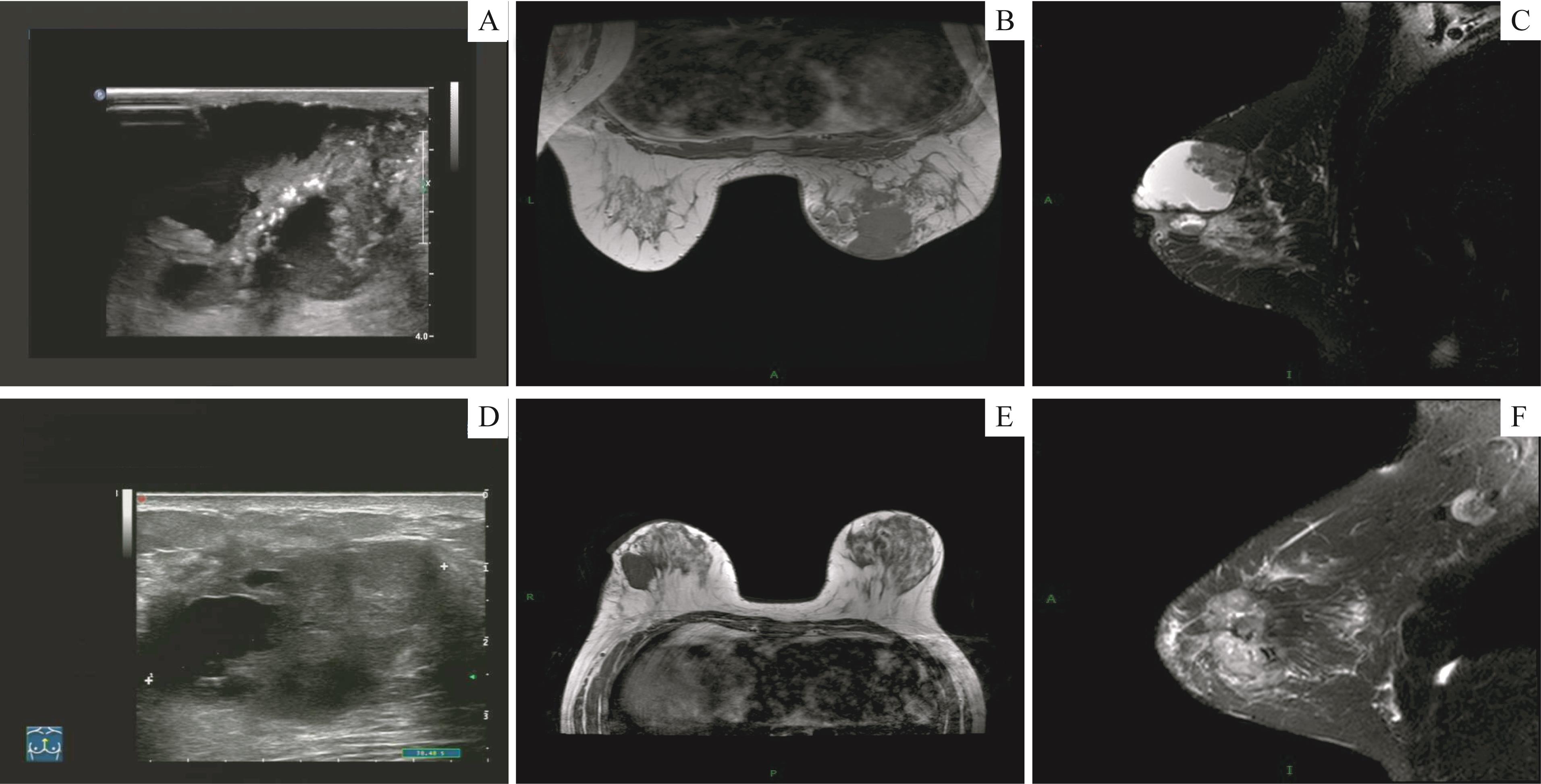
Fig 1 Ultrasound and MRI imaging features about two cases of squamous cell carcinoma
| Subtype | Shape, signal and echo | Margin | Calcification | BI-RADS category | Swollen axillary lymph node | ||||||||||
|---|---|---|---|---|---|---|---|---|---|---|---|---|---|---|---|
| CR | MRI | Ultrasound | CR | MRI | Ultrasound | CR | MRI | Ultrasound | CR | MRI | Ultrasound | CR | MRI | Ultrasound | |
| Squamous cell carcinoma | Irregularly high density | Cyst-like mass with thick wall, low T1WI and T2WI signals; multiregional non-mass-like enhancement | Irregular shape, hybrid echoic | Not clear | Not clear | Not clear | Multiple and tiny | None | Multiple and tiny | 5 | 5 | 5 | Both sides | Both sides | Right side |
| Squamous cell carcinoma | Irregularly high density | Module-like, low T1WI and T2WI signals; unevenly enhancement | Irregular shape, hypoechoic | Not clear | Not clear | Not clear | None | None | None | 4c | 5 | 5 | None | None | None |
| Squamous cell carcinoma | Irregularly high density | Module-like, low T1WI and T2WI signals; early enhancement | Irregular shape, hypoechoic | Not clear | Not clear | Not clear | None | None | None | 0 | 5 | 5 | None | None | None |
| Spindle cell carcinoma | Irregularly high density | Module-like, low T1WI and T2WI signals; unevenly enhancement | Irregular shape, hypoechoic | Not clear | Not clear | Not clear | Tiny | None | Multiple and tiny | 4c | 5 | 4a | None | None | None |
Tab 2 CR, MRI and ultrasound features of four MBC cases
| Subtype | Shape, signal and echo | Margin | Calcification | BI-RADS category | Swollen axillary lymph node | ||||||||||
|---|---|---|---|---|---|---|---|---|---|---|---|---|---|---|---|
| CR | MRI | Ultrasound | CR | MRI | Ultrasound | CR | MRI | Ultrasound | CR | MRI | Ultrasound | CR | MRI | Ultrasound | |
| Squamous cell carcinoma | Irregularly high density | Cyst-like mass with thick wall, low T1WI and T2WI signals; multiregional non-mass-like enhancement | Irregular shape, hybrid echoic | Not clear | Not clear | Not clear | Multiple and tiny | None | Multiple and tiny | 5 | 5 | 5 | Both sides | Both sides | Right side |
| Squamous cell carcinoma | Irregularly high density | Module-like, low T1WI and T2WI signals; unevenly enhancement | Irregular shape, hypoechoic | Not clear | Not clear | Not clear | None | None | None | 4c | 5 | 5 | None | None | None |
| Squamous cell carcinoma | Irregularly high density | Module-like, low T1WI and T2WI signals; early enhancement | Irregular shape, hypoechoic | Not clear | Not clear | Not clear | None | None | None | 0 | 5 | 5 | None | None | None |
| Spindle cell carcinoma | Irregularly high density | Module-like, low T1WI and T2WI signals; unevenly enhancement | Irregular shape, hypoechoic | Not clear | Not clear | Not clear | Tiny | None | Multiple and tiny | 4c | 5 | 4a | None | None | None |
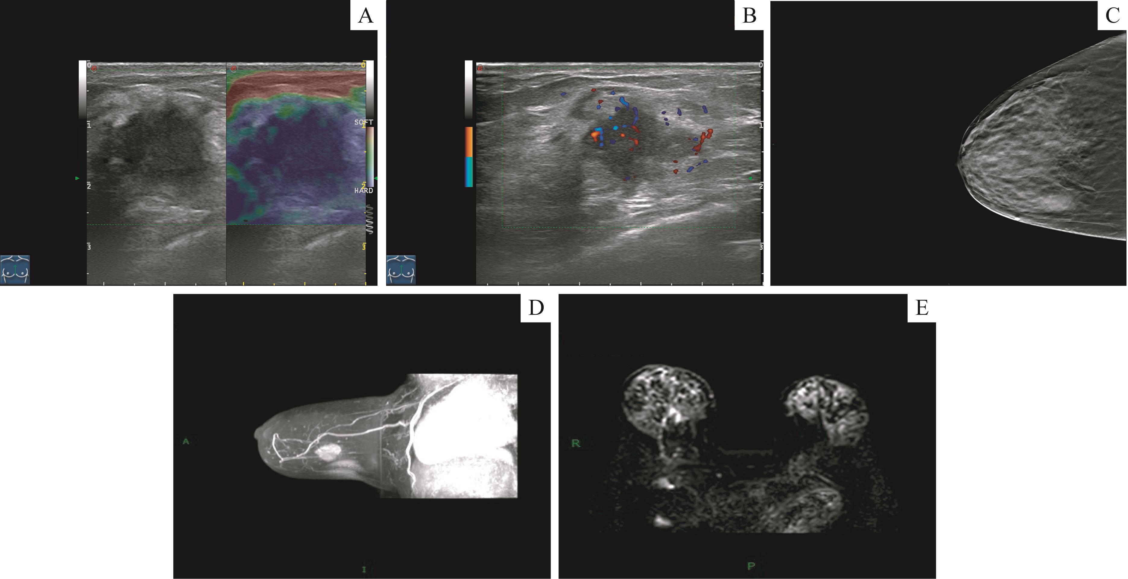
Fig 2 Ultrasound, CR and MRI imaging features of one squamous cell carcinoma case
| 1 | ZEIN DEI, HUGHES M, KUMAR S, et al. Metaplastic carcinoma of the breast is more aggressive than triple-negative breast cancer: a study from a single institution and review of literature[J]. Clin Breast Cancer, 2017, 17(5): 382-391. |
| 2 | LAKHANI S R. WHO classification of tumours of the breast[M]. 4th. Lyon: International Agency for Research on Cancer, 2012. |
| 3 | ONG C T, CAMPBELL B M, THOMAS S M, et al. Metaplastic breast cancer treatment and outcomes in 2500 patients: a retrospective analysis of a national oncology database[J]. Ann Surg Oncol, 2018, 25(8): 2249-2260. |
| 4 | 颜红菊, 许晓静, 谭艳娟, 等. 化生性乳腺癌的多模态超声特征[J]. 医学影像学杂志, 2018, 28(6): 945-949. |
| 5 | ADLER D D, CARSON P L, RUBIN J M, et al. Doppler ultrasound color flow imaging in the study of breast cancer: preliminary findings[J]. Ultrasound Med Biol, 1990, 16(6): 553-559. |
| 6 | 中国抗癌协会乳腺癌专业委员会. 中国抗癌协会乳腺癌诊治指南与规范(2019年版)[J]. 中国癌症杂志, 2019, 29(8): 609-680. |
| 7 | HUVOS A G, LUCAS J C JR, FOOTE F W JR. Metaplastic breast carcinoma. Rare form of mammary cancer[J]. N Y State J Med, 1973, 73(9): 1078-1082. |
| 8 | HARDY B M, CORTINA C S, JAVIDIPARSIJANI S, et al. Hypercalcemia in metaplastic squamous cell carcinoma of the breast[J]. Am J Case Rep, 2019, 20: 366-369. |
| 9 | GREENBERG D, MCINTYRE H, BIERRE T. Metaplastic breast cancer[J]. Australas Radiol, 2004, 48(2): 243-247. |
| 10 | LANGLANDS F, CORNFORD E, RAKHA E, et al. Imaging overview of metaplastic carcinomas of the breast: a large study of 71 cases[J]. Br J Radiol, 2016, 89(1064): 20140644. |
| 11 | CHOI B B, SHU K S. Metaplastic carcinoma of the breast: multimodality imaging and histopathologic assessment[J]. Acta Radiol Stock Swed, 2012, 53(1): 5-11. |
| 12 | 姜珊珊, 张乃千, 佟凌霞. 乳腺化生性癌的超声表现与临床病理特点[J]. 中国实验诊断学, 2020, 24(10): 1637-1639. |
| 13 | BAE S Y, LEE S K, KOO M Y, et al. The prognoses of metaplastic breast cancer patients compared to those of triple-negative breast cancer patients[J]. Breast Cancer Res Treat, 2011, 126(2): 471-478. |
| 14 | 贾懿, 詹维伟, 朱樱. 化生性乳腺癌的影像学诊断研究[J]. 医学影像学杂志, 2019, 29(5): 779-782. |
| 15 | RAKHA E A, TAN P H, VARGA Z, et al. Prognostic factors in metaplastic carcinoma of the breast: a multi-institutional study[J]. Br J Cancer, 2015, 112(2): 283-289. |
| 16 | LUINI A, AGUILAR M, GATTI G, et al. Metaplastic carcinoma of the breast, an unusual disease with worse prognosis: the experience of the European Institute of Oncology and review of the literature[J]. Breast Cancer Res Treat, 2007, 101(3): 349-353. |
| 17 | ZHANG Y Q, LV F, YANG Y L, et al. Clinicopathological features and prognosis of metaplastic breast carcinoma: experience of a major Chinese Cancer Center[J]. PLoS One, 2015, 10(6): e0131409. |
| 18 | TZANNINIS I G, KOTTEAS E A, NTANASIS-STATHOPOULOS I, et al. Management and outcomes in metaplastic breast cancer[J]. Clin Breast Cancer, 2016, 16(6): 437-443. |
| 19 | SALEMIS NS. Metaplastic carcinoma of the breast with mesenchymal differentiation (carcinosarcoma). A unique presentation of an aggressive malignancy and literature review[J]. Breast Dis, 2018, 37(3): 169-175. |
| 20 | HU Q, CHEN W X, ZHONG S L, et al. Current progress in the treatment of metaplastic breast carcinoma[J]. Asian Pac J Cancer Prev, 2013, 14(11): 6221-6225. |
| 21 | KRINGS G, CHEN Y Y. Genomic profiling of metaplastic breast carcinomas reveals genetic heterogeneity and relationship to ductal carcinoma[J]. Mod Pathol, 2018, 31(11): 1661-1674. |
| [1] | SUN Lei, DAI Shifeng, CHEN Yuhua, XU Xinyi, JIANG Kele, LI Xiaowen, LI Chengjing, WU Tingting. Quantitative analysis of the distance between articular disc and condyle in patients with temporomandibular disorders [J]. Journal of Shanghai Jiao Tong University (Medical Science), 2025, 45(6): 684-692. |
| [2] | LI Zhuohang, YU Xindi, REN Jingya, SHEN Jia, DONG Suzhen, WANG Wei. Postoperative neurodevelopmental outcomes of end-to-side anastomosis for coarctation of the aorta [J]. Journal of Shanghai Jiao Tong University (Medical Science), 2025, 45(6): 753-759. |
| [3] | LUO Rui, YANG Gongxin, SHI Huimin, HAN Yongshun, HE Yining, TIAN Zhen, WU Yingwei. Study of imaging characteristics of Kimura disease in the head and neck [J]. Journal of Shanghai Jiao Tong University (Medical Science), 2024, 44(9): 1182-1189. |
| [4] | HUANG Qin, HUANG Ying, LI Wen. Timing of ultrasonography in the diagnosis of fallopian tubal heterotopic pregnancy [J]. Journal of Shanghai Jiao Tong University (Medical Science), 2024, 44(12): 1545-1551. |
| [5] | LU Xiaobing, YUE Jiang, HE Shengyun, DONG Ying, LU Qing, MA Jing. Effect of intramuscular adipose tissue in the skeletal muscle of thigh on glucose metabolism in male patients with obesity [J]. Journal of Shanghai Jiao Tong University (Medical Science), 2023, 43(9): 1169-1174. |
| [6] | CHEN Jiaye, ZHANG Huifeng, LU Wenxian, PENG Daihui. Research progress of brain magnetic resonance imaging related to suicide in bipolar disorder patients [J]. Journal of Shanghai Jiao Tong University (Medical Science), 2023, 43(7): 939-944. |
| [7] | LIU Siyu, WU Bing, LI Xiaomin, ZHAO Lulu, CHEN Jun, AI Songtao. Preliminary exploration of diffusion-weighted imaging in pre-surgical planning of dermatofibrosarcoma protuberans [J]. Journal of Shanghai Jiao Tong University (Medical Science), 2022, 42(8): 1095-1102. |
| [8] | CHEN Liqi, XUE Zhuowei, WU Qingkai. Review of MRI-based three-dimensional digital model reconstruction of female pelvic floor organs [J]. Journal of Shanghai Jiao Tong University (Medical Science), 2022, 42(3): 381-386. |
| [9] | Xuehong WANG, Xuzhuo CHEN, Yi MAO, Da SHEN, Shanyong ZHANG. Difference in recurrence rates after temporomandibular joint disc repositioning surgery with miniscrew anchor at different developmental stages in adolescents [J]. JOURNAL OF SHANGHAI JIAOTONG UNIVERSITY (MEDICAL SCIENCE), 2022, 42(2): 173-177. |
| [10] | WEI Yifan, ZHU Yueniu, KONG Xiangmei, XU Yaya, ZHU Xiaodong. Effects of early mechanical ventilation on the morphology and function of the diaphragm in children [J]. Journal of Shanghai Jiao Tong University (Medical Science), 2022, 42(12): 1712-1719. |
| [11] | Yihuan WANG, Ruokun LI, Huanhuan CHONG, Fuhua YAN. Research progress of Gd-EOB-DTPA-enhanced magnetic resonance imaging in the evaluation of biological behavior of hepatocellular carcinoma [J]. JOURNAL OF SHANGHAI JIAOTONG UNIVERSITY (MEDICAL SCIENCE), 2022, 42(1): 130-134. |
| [12] | Yan-jie JI, Hao LUO, Hai-yan CAI, Xin-yu LIU, Shi-jia JIN, Shen-yue SU, Han-zhang XU, Hu LEI, Ying-li WU. Inhibition of CDDO-ME on ubiquitin-specific protease 2a activity and cell proliferation in triple negative breast cancer cells [J]. JOURNAL OF SHANGHAI JIAOTONG UNIVERSITY (MEDICAL SCIENCE), 2021, 41(8): 1025-1032. |
| [13] | JI Ying-ying, XUE Bin, HUANG Yue, ZHANG Jian-wei. Efficacy and safety of oral midazolam in combination with intranasal dexmedetomidine for paediatric magnetic resonance imaging sedation [J]. JOURNAL OF SHANGHAI JIAOTONG UNIVERSITY (MEDICAL SCIENCE), 2020, 40(8): 1098-1102. |
| [14] | YANG Tao, CHEN Jun, FANG Yi-ru. Advances in magnetic resonance imaging study of bipolar Ⅰdisorder [J]. JOURNAL OF SHANGHAI JIAOTONG UNIVERSITY (MEDICAL SCIENCE), 2020, 40(12): 1660-1664. |
| [15] | YUE Xiu-hui, KONG Wei-dan, REN Ji-liang, YUAN Ying#, TAO Xiao-feng#. Value of 3.0-T MR diffusion-weighted imaging combined with dynamic contrast-enhanced imaging in differentiating benign and malignant thyroid nodules [J]. JOURNAL OF SHANGHAI JIAOTONG UNIVERSITY (MEDICAL SCIENCE), 2020, 40(10): 1393-1397. |
| Viewed | ||||||
|
Full text |
|
|||||
|
Abstract |
|
|||||