上海交通大学学报(医学版), 2023, 43(1): 52-60 doi: 10.3969/j.issn.1674-8115.2023.01.007

论著 · 临床研究

丙型病毒性肝炎肝硬化失代偿期患者发生细菌感染的列线图预测模型构建及评价

薛淋淋,1, 李秉翰1, 常丽仙2, 李卫昆2, 刘春云2, 刘立,2

1.大理大学公共卫生学院,大理 671000

2.云南省昆明市第三人民医院肝病免疫科,昆明 650041

Construction and evaluation of a nomogram prediction model for bacterial infection in patients with decompensated hepatitis C cirrhosis

XUE Linlin,1, LI Binghan1, CHANG Lixian2, LI Weikun2, LIU Chunyun2, LIU Li,2

1.School of Public Health, Dali University, Dali 671000, China

2.Department of Liver Diseases and Immunology, The Third People's Hospital of Kunming, Yunnan Province, Kunming 650041, China

通讯作者: 刘 立,电子信箱:liuli197210@163.com

编委: 包玲

收稿日期: 2022-09-01   接受日期: 2022-12-09   网络出版日期: 2023-01-28

基金资助: 佑安专科联盟科研专项基金.  LM202014

Corresponding authors: LIU Li, E-mail:liuli197210@163.com.

Received: 2022-09-01   Accepted: 2022-12-09   Online: 2023-01-28

作者简介 About authors

薛淋淋(1993—),女,硕士生;电子信箱:18860230076@163.com。 E-mail:18860230076@163.com

摘要

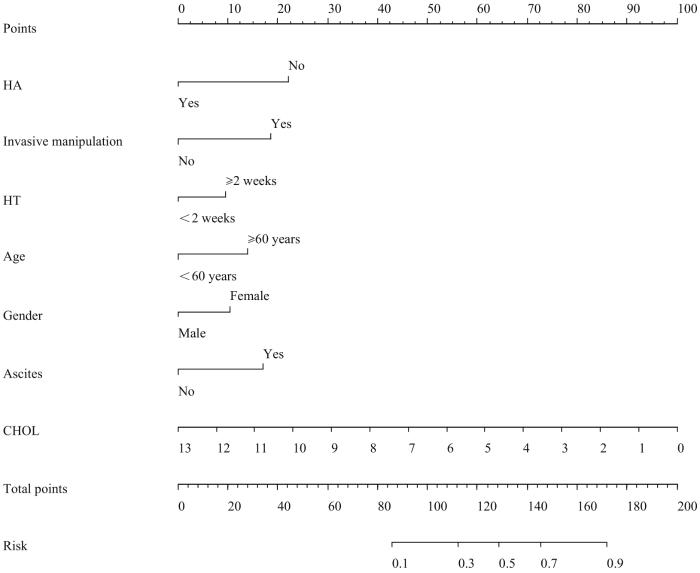
目的·探讨丙型病毒性肝炎(丙肝)肝硬化失代偿期患者发生细菌感染的影响因素,建立列线图预测模型并进行评价。方法·回顾分析昆明市第三人民医院肝病科2020年1月—2021年12月因丙肝肝硬化住院的失代偿期患者574例,以是否发生细菌感染分为细菌感染组和非细菌感染组。收集患者的一般资料、入院合并症及实验室指标。经单因素分析、最小绝对收缩和选择算子(least absolute shrinkage and selection operator,LASSO)回归筛选变量,采用多因素Logistic回归分析影响因素,据此构建列线图模型并进行验证。采用决策曲线及临床影响曲线(clinical impact curve,CIC)评估模型的临床实际应用价值。结果·纳入患者中28.4%(163/574)的患者发生细菌感染,共191个部位,以自发性细菌性腹膜炎(86/191)和肺部细菌感染(79/191)为主;共分离培养出病原菌78株,以肺炎克雷伯菌(15/78)和大肠埃希菌(15/78)为主。多因素Logistic回归分析显示年龄≥60岁[比值比(odds ratio,OR)=2.054,95%置信区间(confidence interval,CI) 1.104~3.822,P=0.023]、女性(OR=1.701,95%CI 1.112~2.602,P=0.014)、腹水(OR=2.386,95%CI 1.601~3.557,P=0.000)、近2周有创操作史(OR=2.605,95%CI 1.368~4.960,P=0.004)、住院时间≥2周(OR=1.629,95%CI 1.098~2.416,P=0.015)是丙肝肝硬化失代偿期患者发生细菌感染的独立危险因素;输注人血白蛋白(OR=0.324,95%CI 0.194~0.542,P=0.000)和高总胆固醇(total cholesterol,CHOL;OR=0.675,95%CI 0.549~0.830,P=0.000)水平是其保护因素。用以上7个影响因素构建列线图模型,采用受试者工作特征曲线(receiver operator characteristic curve,ROC曲线)分析显示曲线下面积(area under the curve,AUC)为0.736,敏感度80.4%,特异度65.1%。Hosmer-lemeshow检验显示,模型具有较好的拟合度(χ2=9.030,P=0.340)。使用Bootstrap法内部重复抽样1 000次进行验证,平均绝对误差0.010,校正曲线和理想曲线基本拟合,预测值和实际值一致性较好。决策曲线显示列线图模型在高风险阈值(0.040~0.715)范围时,有着一定的临床实用性。CIC显示该列线图模型可进行高风险人群分层预测。结论·研究所构建的列线图模型具有较好的预测性、一致性和临床实用性,可为临床医师初步判断丙肝肝硬化失代偿期患者发生细菌感染的风险提供依据。

关键词: 丙型病毒性肝炎 ; 细菌感染 ; 肝硬化失代偿期 ; 危险因素 ; 列线图

Abstract

Objective ·To explore the influencing factors of bacterial infection in decompensated stage of hepatitis C cirrhosis, and establish a risk prediction model of nomogram. Methods ·A total of 574 patients with decompensated hepatitis C cirrhosis were retrospectively collected from The Third People′s Hospital of Kunming between January 2020 and December 2021, and divided into non-infected and infected groups according to whether bacterial infection occurred. The general information, complications, and laboratory indicators were collected. The variables were screened by univariate analysis, and least absolute shrinkage and selection operator (LASSO) regression, and the nomogram model were constructed and verified by multivariate Logistic regression analysis of influencing factors. The decision curve and clinical impact curve (CIC) were used to evaluate the clinical application value of the model. Results ·Bacterial infections occurred in 28.4% (163/574) of the patients, with a total of 191 sites, mainly including spontaneous bacterial peritonitis (86/191) and pulmonary bacterial infections (79/191). Totally 78 strains of pathogens were isolated and cultured, mainly including Klebsiella pneumoniae (15/78) and Escherichia coli (15/78). Multivariate Logistic regression analysis showed that age ≥60 years [odds ratio (OR)=2.054, 95% confidence interval (CI) 1.104‒3.822, P=0.023], female (OR=1.701, 95%CI 1.112‒2.602, P=0.014), ascites (OR=2.386, 95%CI 1.601‒3.557, P=0.000), history of invasive procedures in the last two weeks (OR=2.605, 95%CI 1.368‒4.960, P=0.004), and hospitalization time≥2 weeks (OR=1.629, 95%CI 1.098‒2.416, P=0.015) were independent risk factors for bacterial infection in decompensated hepatitis C cirrhosis patients, while infusing human serum albumin (OR=0.324, 95%CI 0.194‒0.542, P=0.000) and high level of total cholesterol (OR=0.675, 95%CI 0.549‒0.830, P=0.000) were protective factors. The nomogram model was constructed with the above seven influencing factors. Receiver operator characteristic (ROC) curve analysis showed that the area under the curve (AUC) was 0.736 and the sensitivity was 80.4%; and the specificity was 65.1%. Hosmer-lemeshow test showed that the model had a good degree of fit (χ2=9.030, P=0.340). The bootstrap method was used for internal repeated sampling for 1 000 times, the average absolute error was 0.010, the calibration curve and the ideal curve were basically fitted, and the predicted values were in good agreement with the actual values. The decision curve showed that the nomogram model had certain clinical practicability in the high risk threshold range (0.040‒0.715). CIC showed that the nomogram model can be used to forecast the high-risk population in different levels. Conclusion ·The nomogram risk prediction model constructed in this study has good predictability, consistency and clinical practicability, and can provide evidence for clinicians to preliminary judge the risk of bacterial infection in patients with decompensated hepatitis C cirrhosis.

Keywords: viral hepatitis C ; bacterial infection ; decompensated cirrhosis ; risk factor ; nomogram

PDF (2418KB) 元数据 多维度评价 相关文章 导出 EndNote| Ris| Bibtex  收藏本文

本文引用格式

薛淋淋, 李秉翰, 常丽仙, 李卫昆, 刘春云, 刘立. 丙型病毒性肝炎肝硬化失代偿期患者发生细菌感染的列线图预测模型构建及评价. 上海交通大学学报(医学版)[J], 2023, 43(1): 52-60 doi:10.3969/j.issn.1674-8115.2023.01.007

XUE Linlin, LI Binghan, CHANG Lixian, LI Weikun, LIU Chunyun, LIU Li. Construction and evaluation of a nomogram prediction model for bacterial infection in patients with decompensated hepatitis C cirrhosis. Journal of Shanghai Jiao Tong University (Medical Science)[J], 2023, 43(1): 52-60 doi:10.3969/j.issn.1674-8115.2023.01.007

丙型病毒性肝炎(丙肝)是由丙型肝炎病毒(hepatitis C virus,HCV)引起的以肝损伤为主的传染性疾病。研究发现,HCV急性感染者有75%~85%会发展为慢性HCV感染1,可导致慢性肝炎、肝纤维化,部分患者可发展为肝硬化甚至肝细胞癌2。大量研究3-4表明,细菌感染作为丙肝肝硬化失代偿期患者常见的并发症,伴有高致残率和病死率,预后差,是临床治疗的重点和难点。有文献显示,39.7%的肝硬化患者可并发各种细菌感染5,常起病隐匿6,症状缺乏特异性,约1/3患者可无症状,其临床表现多样、病情轻重不一,且致病病原体种类繁多,腹水培养、血培养阳性率低或有其他疾病常见的症状和体征,容易漏诊或误诊,使得早期临床诊断十分困难。实验室细菌感染指标,如C反应蛋白、降钙素原和白介素-6(interleukin-6,IL-6)有时在细菌感染发生后变化并不明显7-8,在炎症早期波动幅度不大。列线图模型可较为准确地预测某些疾病在未来的发生率与复发率9。因此,本研究拟建立基于实验室指标的细菌感染预测模型10-11,采用受试者工作特征曲线(receiver operator characteristic curve,ROC曲线)、校准曲线(calibration curve)、决策曲线(decision curve analysis,DCA)和临床影响曲线(clinical impact curve,CIC)对模型的预测效能、准确性、实用性进行验证,旨在帮助预判丙肝肝硬化失代偿期患者发生细菌感染的风险,从而尽早进行预防性治疗。

1 对象与方法

1.1 研究对象及样本量计算

纳入2020年1月—2021年12月于昆明市第三人民医院肝病科574例首次住院就诊的慢性HCV患者作为研究对象。纳入标准:① 年龄大于18周岁。② 临床资料完整可靠。③ 丙型肝炎及肝硬化失代偿期诊断标准参考《丙型肝炎防治指南(2019年版)》12。④ 细菌感染标准参考《终末期肝病合并感染诊治专家共识(2021年版)》6。排除标准:① 入院不足24 h死亡。② 妊娠期妇女。③ 非丙肝引起的肝硬化失代偿。④ 获得性免疫缺陷综合征。⑤ 继发性腹膜炎。根据是否发生细菌感染,将患者分为细菌感染组与非细菌感染组。

本研究采用横断面研究样本量计算公式计算样本量:

n=p(1-p)z-α/2+z-βp-p02

查阅文献,肝硬化发生感染的患病率p约为36%,设双侧α=0.05,效力1-β为0.8,p为0.36,p0 为0.3,使用软件计算样本量为502例,考虑失访率10%,因此至少需要调查550例丙肝肝硬化失代偿期的患者。

1.2 资料收集

收集丙肝肝硬化失代偿期患者的年龄、性别、入院时是否有合并症(糖尿病、高血压、上消化道出血、腹水、肝肾综合征、肝性脑病、门脉高压性胃肠病、高氨血症)、近2周有创操作史(中心静脉置管、胃镜检查、肠镜检查、腹水穿刺引流、脾动脉栓塞术)、输注人血白蛋白史;收集患者入院时实验室检查结果,包括白细胞计数(white blood cell count,WBC)、红细胞计数(red blood cell count,RBC)、血小板计数(platelet count,PLT)、中性粒细胞计数(neutrophil count,NEUT),血红蛋白(hemoglobin,HGB)、总蛋白(total protein,TP)、白蛋白(albumin,ALB)、前白蛋白(prealbumin,PA)、谷丙转氨酶(glutamic-pyruvic transaminase,GPT)、谷草转氨酶(glutamic-oxaloacetic transaminase,GOT)、总胆红素(total bilirubin,TBIL)、三酰甘油(triaclyglycerol,TAG)、总胆固醇(total cholesterol,CHOL)、高密度脂蛋白胆固醇(high-density lipoprotein,HDL-C)、低密度脂蛋白胆固醇(low-density lipoprotein,LDL-C)、尿素(urea,UR)、肌酐(creatinine,CREA)、尿酸(uric acid,UA)、乳酸脱氢酶(lactic dehydrogenase,LDH)、甲胎蛋白(alpha fetoprotein,AFP)、碱性磷酸酶(alkaline phosphatase,ALP)、IL-6、补体1q(complement 1q,C1q)、超敏C反应蛋白(high sensitivity C-reactive protein,hs-CRP)水平,以及凝血酶原时间(prothrombin time,PT)、凝血酶原活动度(prothrombin time activity,PTA)、国际标准化比值(international normalized ratio,INR)、住院时间(≥2周和<2周)等临床资料。

1.3 标本采集

丙肝肝硬化失代偿期患者在住院48 h内采集静脉血送检;有腹水者,采集腹水送细菌培养;怀疑呼吸道细菌感染患者,采集其咽拭子或者痰液送细菌培养;怀疑泌尿道细菌感染患者,采集其尿液送细菌培养。所有标本在送检中严格遵循无菌、密封、低温原则,时间不超过30 min。

1.4 检测仪器及试剂

临床生化检测使用日本奥林巴斯AU400全自动生化仪。外周血检测采用Sysmex-XT 4000i全自动血细胞分析仪检测(日本Sysmex公司)。使用仪器VITEK2 Compact(法国梅里埃公司)全自动细菌鉴定及药敏分析仪进行细菌鉴定。

1.5 统计学分析

采用SPSS 26.0软件和R 4.04软件进行统计学分析。非正态分布的定量资料用MQ1Q3)进行描述,2组间比较采用Mann-Whitney U检验。定性资料用频数(百分比)表示,2组间比较采用χ2检验。采用LASSO回归的10折交叉验证(10-fold cross validation)法筛选预测变量,纳入多因素Logistic回归分析,并建立列线图模型。采用ROC曲线评价模型的预测效能;采用bootstrap自抽样法重复抽样1 000次进行内部验证,绘制校准曲线评价模型的准确度;采用DCA评价模型的临床实用性;采用CIC进行高风险人群分层预测,分析预测模型的高风险例数与实际发生细菌感染(结局事件)例数的关系,评估模型的临床实际应用价值。P<0.05表示差异具有统计学意义。

2 结果

2.1 一般基线特征比较

共纳入574例丙肝肝硬化失代偿期患者,其中男性409例,女性165例,年龄30~80岁,平均年龄(50.5±7.8)岁。根据是否发生细菌感染分为细菌感染组(163例)与非细菌感染组(411例),细菌感染的患病率为28.4%。2组患者一般基线资料比较如表1所示,患者在入院时的合并症(糖尿病、高血压、上消化道出血、肝肾综合征、肝性脑病、门脉高压性胃肠病等)、RBC、PLT、HGB、GPT、GOT、TAG、LDL-C、UR、CREA、UA、LDH、PT、PTA、AFP、ALP、C1q、hs-CRP方面的差异无统计学意义。

表1   2组丙肝肝硬化失代偿期患者基线资料比较

Tab 1  Baseline data comparison between the two groups of patients with decompensated hepatitis C cirrhosis

ItemNon-bacterial infection group (n=411)

Bacterial infection group

n=163)

χ2 /Z valueP value
Age≥60 years/n (%)32 (7.8)24 (14.7)6.2830.012
Gender/n (%)6.3810.012
Male304 (74.0)105 (64.4)
Female107 (26.0)58 (35.6)
Complication on admission/n (%)
Diabetes74 (16.9)20 (12.0)2.8030.094
Hypertension107 (24.4)5 (3.0)1.9260.588
Upper gastrointestinal Hemorrhage19 (4.3)4 (2.4)1.4270.232
Ascites113 (25.7)81 (48.5)28.0730.000
Hepatorenal syndrome7 (1.6)5 (3.0)1.0610.303
Hepatic encephalopathy21 (4.8)10 (6.0)0.2400.624
Portal hypertensive Gastroenteropathy74 (16.9)24 (14.4)0.6730.412
Hyperammonemia24 (5.5)18 (10.8)4.6600.031
History of invasive procedures in the last two weeks/n (%)29 (7.1)23 (14.1)7.0500.008
Infusing human serum albumin/n (%)134 (32.6)24(14.7)18.7030.000
Laboratory index
WBC/(×109·L-14.0 (3.0, 5.2)4.6 (3.6, 6.2)-3.9200.000
RBC/(×109·L-14.0 (3.2, 4.8)3.8 (3.0, 4.5)-1.8580.063
PLT/(×109·L-176.0 (52.0, 110.0)71.0 (47.0, 104.0)-0.9170.359
NEUT/(×109·L-12.3 (1.6, 3.3)3.0 (2.1, 4.4)-4.7300.000
HGB/(g·L-1124.0 (101.0, 145.0)121.3 (90.0, 145.0)-1.8170.069
TP/(g·L-168.0 (60.8, 72.8)66.3 (58.2, 70.5)-3.1070.002
ALB/(g·L-132.9 (28.0, 38.7)30.5 (24.9, 35.0)-4.2800.000
PA/(mg·L-1114.9 (93.1, 134.7)114.9 (76.6, 115.0)-2.8700.004
GPT/(U·L-139.0 (24.0, 64.0)36.0 (23.0, 59.0)-1.5090.131
GOT/(U·L-159.0 (36.0, 94.0)59.0 (36.0, 94.0)-0.6080.543
TBIL/(μmol·L-128.3 (18.0, 45.5)36.0 (19.9, 69.4)-2.7280.006
TAG/(mmol·L-11.2 (0.7, 4.1)1.1 (0.7, 4.1)-0.4960.620
CHOL/(mmol·L-13.3 (2.7, 3.9)3.2 (2.4, 3.4)-4.0810.000
HDL-C/(mmol·L-11.4 (1.0, 1.9)1.3 (0.9, 1.4)-3.5810.000
LDL-C/(mmol·L-11.3 (0.8, 1.6)1.3 (0.8, 1.6)-0.8720.383
UR/(mmol·L-14.7 (3.4, 6.3)5.3 (3.6, 7.4)-1.5900.112
CREA/(mmol·L-165.0 (54.0, 79.0)71.0 (55.0, 90.0)-3.1000.002
UA/(μmol·L-1354.0 (276.0, 443.0)366.0 (303.0, 447.0)-0.9040.366
LDH/(U·L-1210.1 (156.0, 231.0)210.1 (159.0, 252.0)-1.7320.083
PT/s15.1 (13.1, 16.8)15.1 (10.3, 17.8)-0.3930.694
PTA/%58.0 (42.0, 71.0)54.7 (20.3, 68.0)-1.9370.053
INR1.3 (1.2, 1.5)1.3 (1.2, 1.6)-2.7080.007
AFP/(ng·mL-110.2 (5.2, 26.3)8.7 (3.8, 1 476.3)-0.0730.942
ALP/(U·L-1133.0 (99.0, 200.6)143.0 (98.0, 200.6)-0.8060.420
IL-6/(pg·mL-121.0 (8.4, 64.5)62.7 (23.1, 64.5)-5.9080.000
C1q/(mg·L-1170.0 (142.0, 190.0)164.7 (140.0, 177.0)-1.8780.060
hs-CRP/(mg·L-15.3 (0.8, 11.8)4.2 (0.8, 11.8)-0.6390.523
Hospitalization time ≥2 weeks/n (%)177 (43.1)95 (58.3)10.8390.001

Note:Invasive procedures included central vein catheterization, gastroscopy/colonoscopy, ascites puncture drainage, and splenic artery embolization.

新窗口打开| 下载CSV


2.2 细菌感染分布及病原学培养结果

163例患者共发生191个部位的感染,分别为自发性腹膜炎86个(45.0%)、肺部感染79个(41.4%)、泌尿系统感染13个(6.8%)和急性上呼吸道感染13个(6.8%);共分离培养出病原菌78株,分别为肺炎克雷伯菌15株(19.2%)、大肠埃希菌15株(19.2%)、金黄色葡萄球菌14株(17.9%)、表皮葡萄球菌12株(15.4%)、屎肠球菌10株(12.8%)、微球菌9株(11.5%)和羊莫拉菌3株(3.8%)。

2.3 单因素Logistic回归和LASSO回归筛选变量

单因素Logistic回归分析(表2)发现,年龄≥60岁、女性、腹水、高氨血症、近2周有创操作史、输注人血白蛋白、WBC、NEUT、TP、ALB、PA、TBIL、CHOL、HDL-C、CREA、IL-6、C1q和住院时间≥2周是丙肝肝硬化失代偿期患者发生细菌感染的影响因素,结果有统计学意义(均P<0.05)。将单因素分析结果有统计学意义的变量纳入LASSO回归模型(图1)筛选预测变量,通过10折交叉验证,最终筛选出的变量是7个,分别为年龄≥60岁、女性、腹水、输注人血白蛋白、近2周有创操作史、CHOL和住院时间≥2周。

表2   丙肝肝硬化失代偿期患者发生细菌感染的单因素及多因素Logistic回归分析结果

Tab 2  Results of univariate and multivariate Logistic regression analysis of bacterial infection in patients with decompensated hepatitis C cirrhosis

ItemUnivariate analysisMultivariate analysis
OR (95%CIP valueOR (95%CIP value
Age≥60 years2.045 (1.164‒3.594)0.0132.054 (1.104‒3.822)0.023
Gender (female)1.569 (1.064‒2.316)0.0231.701 (1.112‒2.602)0.014
Diabetes1.567 (0.921‒2.666)0.098
Hypertension1.167 (0.709‒1.918)0.544
Hyperammonemia2.002 (1.055‒3.797)0.034
Ascites2.605 (1.789‒3.793)0.0002.386 (1.601‒3.557)0.000
History of invasive procedures in the last two weeks2.164 (1.211‒3.867)0.0092.605 (1.386‒4.960)0.004
Infusing human serum albumin0.357 (0.221‒0.577)0.0000.324 (0.194‒0.542)0.000
Laboratory index
WBC1.187 (1.099‒1.282)0.000
NEUT1.239 (1.131‒1.359)0.000
HGB0.995 (0.990‒1.000)0.068
TP0.980 (0.964‒0.997)0.018
ALB0.952 (0.930‒0.975)0.000
PA0.992 (0.988‒0.997)0.000
TBIL1.007 (1.003‒1.010)0.000
CHOL0.680 (0.559‒0.827)0.0000.675 (0.549‒0.830)0.000
HDL-C0.644 (0.485‒0.854)0.002
LDL-C1.001 (1.000‒1.003)0.071
CREA1.005 (1.001‒1.009)0.007
IL-61.001 (1.000‒1.002)0.027
C1q0.995 (0.992‒0.999)0.018
LDH1.001 (1.000‒1.003)0.071
INR1.015 (0.932‒1.107)0.727
Hospitalization time ≥2 weeks1.847 (1.279‒2.677)0.0011.629 (1.098‒2.416)0.015

Note:Invasive procedures included central vein catheterization, gastroscopy/colonoscopy, ascites puncture drainage, and splenic artery embolization.OR—odds ratio; CI—confidence interval.

新窗口打开| 下载CSV


图1

图1   丙肝肝硬化失代偿期患者发生细菌感染的LASSO回归模型

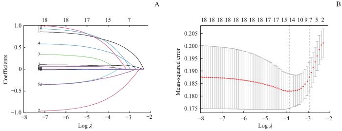
Note: A. Path diagram of regression coefficient. The upper abscissas was the number of variables with non-zero coefficients in the model at this time, the lower abscissas was the logarithm of the penalty coefficient (λ), and the ordinate was the value of the coefficient. B. Cross-verification curve of LASSO regression. The upper and lower abscissas were the same as Fig A, and the ordinate was likelihood bias. The dotted line on the left of Fig B indicates the number of variables corresponding to the minimum λ (when the model has the highest fitting effect), and the number of variables was 14. The dotted line on the right indicates one standard error of the least λ (when the model has better fitting effect, fewer and simpler variables are included), and the number of variables was 7.

Fig 1   LASSO regression model of bacterial infection in patients with decompensated hepatitis C cirrhosis


2.4 列线图预测模型的构建

将LASSO回归筛选出的7个变量纳入多因素Logistic回归,校正混杂因素(高氨血症、WBC、NEUT、TP、ALB、PA、TBIL、HDL-C、CREA、IL-6、C1q)后,结果如表2所示。根据多因素Logistic回归模型结果构建丙肝肝硬化失代偿期患者发生细菌感染的列线图(图2)。Hosmer-lemeshow检验显示,模型具有较好的拟合度(χ2=9.030,P=0.340)。

图2

图2   丙肝肝硬化失代偿期患者发生细菌感染多因素Logistic回归分析的列线图

Note: HA—human serum albumin was infused; HT—hospitalization time.

Fig 2   Multivariate Logistic regression analysis of nomogram of bacterial infection in patients with decompensated hepatitis C cirrhosis
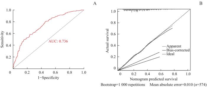
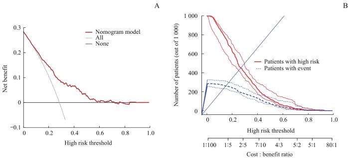

2.5 列线图预测模型的评价

采用ROC曲线分析(图3A)显示,曲线下面积(area under the curve,AUC)为0.736(95%CI 0.692~0.781,P=0.000),敏感度80.4%,特异度65.1%。校准曲线(图3B)显示平均绝对误差0.010,表明细菌感染发生的实际值和预测值之间有较好的一致性。DCA(图4A)显示,阈值为0.040~0.715时,列线图模型有一定的临床实用性。使用CIC进行列线图模型预测1 000人的风险分层,显示“损失∶受益”坐标轴,赋以8个刻度,显示置信区间,如图4B所示:红色曲线表示,在各个阈概率下,被列线图模型划分为阳性(高风险)的人数;蓝色曲线为各个阈概率下真阳性的人数,随着阈值增大,预测的阳性例数减少,其中真阳性的例数也逐渐减少。

图3

图3   列线图模型的ROC曲线(A)和校准曲线(B

Fig 3   ROC curve (A) and calibration plot (B) of the prognostic model


图4

图4   列线图预测模型的DCAA)和CICB

Fig 4   Decision curve analysis (A) and clinical impact curve (B) of the prognostic model


3 讨论

细菌感染是导致肝硬化失代偿期患者全因死亡率升高主要的可预防的危险因素之一3。肝脏功能明显减退、肝脏微循环障碍、肝脏局部以及全身性炎症反应、免疫麻痹及缺陷、肠道菌群异位及微生态失衡等均为细菌感染的危险因素13-14。本研究回顾性分析574例丙肝肝硬化失代偿期患者,其中163例发生细菌感染,患病率28.4%,提示丙肝肝硬化失代偿期患者是合并细菌感染的高危人群。因此,探讨丙肝肝硬化失代偿期患者发生细菌感染的影响因素以及构建相关模型预测其风险至关重要。

本研究尽可能纳入全面广泛的变量,较以往变量筛选增加LASSO回归,可避免各变量之间可能存在的共线性问题,筛选出与研究目的相关的变量。研究建立并验证了列线图预测模型,结果显示该模型具有较高的预测效能、较好的实用性、可观的临床净获益,能够实现对丙肝肝硬化失代偿期患者发生细菌感染的风险预测。

本研究结果表明,年龄≥60岁、女性、腹水、近2周有创操作史及住院时间≥2周是丙肝肝硬化失代偿期患者发生细菌感染的独立危险因素;输注人血白蛋白和高CHOL是其独立的保护因素。与既往研究揭示性别与肝硬化患者发生细菌感染之间无相关性15不同,本研究结果显示性别(女性)与丙肝肝硬化失代偿期患者发生细菌感染有关。分析可能原因为雌激素与氧化应激、肝星状细胞的活化、细胞外基质的堆积、免疫调节的关系密切,当体内雌激素水平降低时,机体免疫力随之降低16。另外,女性患者随着年龄增加,雌激素水平下降,基础疾病增多,机体组织和脏器功能不断发生退变,其储备能力和代偿能力下降,而肝纤维化的严重程度与雌激素的缺乏状态成正比17。本研究中丙肝肝硬化失代偿期患者常需进行胃镜、肠镜、导尿、腹腔穿刺引流、中心静脉穿刺置管、脾动脉栓塞术等有创操作,这些操作均会增加患者细菌感染的可能性18-19。研究20提示,住院时间越长,患者交叉感染细菌的机会越多。本研究结果表明,住院时间≥2周是患者发生细菌感染的独立危险因素,相比较既往研究21发现肝硬化合并上消化道出血患者住院>4周更易发生感染的时间更短,表明丙肝肝硬化失代偿期患者病情可能较其他肝硬化患者更重。细菌感染可加剧肝功能损害,并且肝功能受损后易发生细菌感染,两者之间相互影响,形成恶性循环22

CHOL常被用来作为肝功能受损程度的评价指标。本研究结果显示,未发生细菌感染组的CHOL水平高于感染组,并且正常范围内高水平的CHOL是丙肝肝硬化失代偿期患者发生细菌感染的独立保护因素。NING等23在脂多糖诱导的急性肝衰竭小鼠实验中发现,CHOL在体内炎症反应、细胞凋亡和细胞存活中发挥重要作用。高鹏等24研究也得出相似结论。国外已有学者25提出,充足的CHOL供应对肝脏再生及肝细胞、星状细胞和库普弗细胞的中间代谢发挥着重要作用。ALB、TP水平反映了机体的营养状况,ALB水平下降提示出现肝硬化或重症肝炎26-27。DESCHÊNES等28的研究也支持这一结论。白蛋白在维持血浆胶体渗透压和免疫调节方面起到重要作用,及时输注人血白蛋白对预防和降低丙肝肝硬化失代偿期患者合并细菌感染有着积极意义。本研究结果表明,丙肝肝硬化失代偿期患者补充人血白蛋白在纠正低蛋白血症的同时,达到了预防细菌感染的作用。有趣的是,C1q在感染组与非感染组的基线分析中差异无统计学意义,而在单因素Logistic分析中显示与患者发生细菌感染有关。既往研究29提出,C1q与某些自身抗体的分布模式、自身免疫性疾病(autoimmune disease,AID)患者的病情相关,表明C1q在AID中具有一定的临床意义。因此,本研究中将C1q作为混杂因素纳入多因素Logistic回归。

丙肝肝硬化失代偿期患者与乙肝肝硬化患者相比较,两者发生感染的相同之处为:革兰阳性菌均以金黄色葡萄球菌多见,革兰阴性菌均以大肠埃希菌、肺炎克雷伯菌多见30-31。不同之处在于:本研究年龄≥60岁、女性是丙肝肝硬化患者发生细菌感染的独立危险因素,而乙肝肝硬化合并感染与性别无相关性15;ALB在本研究中不是独立影响因素,而在乙肝肝硬化合并感染中为独立影响因素21;输注人血白蛋白在本研究中是独立的保护因素,但也有研究32表明,输注白蛋白并不会降低肝硬化失代偿期患者新发感染和死亡等风险。

本研究存在一定局限性。研究样本来自单中心,样本量小,代表性不足;并且构建的列线图风险预测模型只进行了内部验证,待今后开展多中心、大样本外部验证。

本研究对丙肝肝硬化失代偿期患者发生细菌感染进行列线图预测模型构建及验证,该列线图预测模型具有良好的临床实用价值,能够帮助临床医师更加直观地预判丙肝肝硬化失代偿期患者发生细菌感染的风险。

作者贡献声明

李秉翰、刘春云、李卫昆参与研究设计,刘立指导论文修改,薛淋淋、常丽仙负责数据收集与分析、论文撰写与修改。

The study was designed by LI Binghan, LIU Chunyun, and LI Weikun. The manuscript of revision was directed by LIU Li. XUE Linlin and CHANG Lixian were responsible for data collection and analysis as well as paper writing and modification.

利益冲突声明

所有作者声明不存在利益冲突。

All authors disclose no relevant conflict of interests.

参考文献

中华人民共和国国家卫生健康委. 中国病毒性肝炎防治规划(2017—2020年)[EB/OL]. (2017-11-10)[2022-09-01]. http://www.nhc.gov.cn/ewebeditor/uploadfile/2017/11/20171113134002475.pdf.

[本文引用: 1]

National Health Commission of the People′s Republic of China. Prevention and control plan of viral hepatitis in China (2017‒2020)[EB/OL]. (2017-11-10)[2022-09-01]. http://www.nhc.gov.cn/ewebeditor/uploadfile/2017/11/20171113134002475.pdf.

[本文引用: 1]

HEI F X, YE S D, DING G W, et al. Epidemiological analysis on reported hepatitis C cases in China from 2012 to 2016[J]. Biomed Environ Sci, 2018, 31(10): 773-776.

[本文引用: 1]

PIANO S, SINGH V, CARACENI P, et al. Epidemiology and effects of bacterial infections in patients with cirrhosis worldwide[J]. Gastroenterology, 2019, 156(5): 1368-1380.e10.

[本文引用: 2]

PLEGUEZUELO M, BENITEZ J M, JURADO J, et al. Diagnosis and management of bacterial infections in decompensated cirrhosis[J]. World J Hepatol, 2013, 5(1): 16-25.

[本文引用: 1]

FERNÁNDEZ J, PRADO V, TREBICKA J, et al. Multidrug-resistant bacterial infections in patients with decompensated cirrhosis and with acute-on-chronic liver failure in Europe[J]. J Hepatol, 2019, 70(3): 398-411.

[本文引用: 1]

中华医学会感染病学分会. 终末期肝病合并感染诊治专家共识(2021年版)[J]. 中华肝脏病杂志, 2022, 30(2): 147-158.

[本文引用: 2]

Chinese Society of Infectious Diseases, Chinese Medical Association. Expert consensus on diagnosis and treatment of end-stage liver disease complicated infection (2021 version)[J]. Chinese Journal of Hepatology, 2022, 30(2): 147-158.

[本文引用: 2]

MIKUŁA T, SAPUŁA M, JABŁOŃSKA J, et al. Significance of heparin-binding protein and D-dimers in the early diagnosis of spontaneous bacterial peritonitis[J]. Mediators Inflamm, 2018, 2018: 1969108.

[本文引用: 1]

SPAHR L, MORARD I, HADENGUE A, et al. Procalcitonin is not an accurate marker of spontaneous bacterial peritonitis in patients with cirrhosis[J]. Hepato gastroenterology, 2001, 48(38): 502-505.

[本文引用: 1]

ZHU Y, CHENG H, MIN R, et al. Computed tomography images under the nomogram mathematical prediction model in the treatment of cerebral infarction complicated with nonvalvular atrial fibrillation and the impacts of virus infection[J]. Contrast Media Mol Imaging, 2022, 2022: 3950641.

[本文引用: 1]

LI S Y, YIN C H, CHEN J S, et al. A nomogram for predicting the development of serious bacterial infections in febrile term neonates: a single medical center experience in southern Taiwan[J]. Pediatr Neonatol, 2022, 63(6): 605-612.

[本文引用: 1]

XU X F, LI H W, SHENG Y J, et al. Nomogram for prediction of bronchial mucus plugs in children with Mycoplasma pneumoniae pneumonia[J]. Sci Rep, 2020, 10(1): 4579.

[本文引用: 1]

中华医学会肝病学分会, 中华医学会感染病学分会. 丙型肝炎防治指南(2019年版)[J]. 实用肝脏病杂志, 2020, 23(1): S33-S52.

[本文引用: 1]

Chinese Society of Hepatology and Chinese Society of Infectious Diseases, Chinese Medical Association. Guidelines for prevention and treatment of hepatitis C (2019 edition)[J]. Journal of Practical Hepatology, 2020, 23(1): S33-S52.

[本文引用: 1]

PIANO S, TONON M, ANGELI P. Changes in the epidemiology and management of bacterial infections in cirrhosis[J]. Clin Mol Hepatol, 2021, 27(3): 437-445.

[本文引用: 1]

ALBILLOS A, DE GOTTARDI A, RESCIGNO M. The gut-liver axis in liver disease: pathophysiological basis for therapy[J]. J Hepatol, 2020, 72(3): 558-577.

[本文引用: 1]

吴柳, 洪灏, 李维正, 等. 肝硬化患者合并细菌感染的临床特征分析[J]. 中国感染与化疗杂志, 2020, 20(6): 601-606.

[本文引用: 2]

WU L, HONG H, LI W Z, et al. Clinical characteristics of bacterial infections in patients with liver cirrhosis[J]. Chinese Journal of Infection and Chemotherapy, 2020, 20(6): 601-606.

[本文引用: 2]

徐文倩, 吴瑶麒, 张近远, 等. 慢性肝病女性患者绝经后雌激素抗肝纤维化的作用机制[J]. 临床肝胆病杂志, 2021, 37(10): 2425-2428.

[本文引用: 1]

XU W Q, WU Y Q, ZHANG J Y, et al. Mechanism of estrogen against liver fibrosis in postmenopausal women with chronic liver disease[J]. Journal of Clinical Hepatology, 2021, 37(10): 2425-2428.

[本文引用: 1]

KLAIR J S, YANG J D, ABDELMALEK M F, et al. A longer duration of estrogen deficiency increases fibrosis risk among postmenopausal women with nonalcoholic fatty liver disease[J]. Hepatology, 2016, 64(1): 85-91.

[本文引用: 1]

MARTIN MATEOS R, ALBILLOS A. Sepsis in patients with cirrhosis awaiting liver transplantation: new trends and management[J]. Liver Transpl, 2019, 25(11): 1700-1709.

[本文引用: 1]

MARTÍNEZ J, HERNÁNDEZ-GEA V, RODRÍGUEZ-DE-SANTIAGO E, et al. Bacterial infections in patients with acute variceal bleeding in the era of antibiotic prophylaxis[J]. J Hepatol, 2021, 75(2): 342-350.

[本文引用: 1]

宫能凯, 全斌, 鲁俊, 等. 回归分析失代偿期肝硬化患者医院感染的危险因素[J]. 齐齐哈尔医学院学报, 2021, 42(9): 772-775.

[本文引用: 1]

GONG N K, QUAN B, LU J, et al. Analysis of the risk factors of nosocomial infection among patients with decompensated liver cirrhosis inpatients based on regression analysis[J]. Journal of Qiqihar Medical University, 2021, 42(9): 772-775.

[本文引用: 1]

徐升, 徐芳, 应丽园, 等. 肝硬化合并上消化道出血患者医院感染的病原学特点及影响因素研究[J]. 中华医院感染学杂志, 2019, 29(1): 71-74.

[本文引用: 2]

XU S, XU F, YING L Y, et al. Etiological charateristics and influencing factor for nosocomial infection in liver cirrhosis patients complicated with upper gastrointestinal hemorrhage[J]. Chinese Journal of Nosocomiology, 2019, 29(1): 71-74.

[本文引用: 2]

ARROYO V, ANGELI P, MOREAU R, et al. The systemic inflammation hypothesis: towards a new paradigm of acute decompensation and multiorgan failure in cirrhosis[J]. J Hepatol, 2021, 74(3): 670-685.

[本文引用: 1]

NING Y X, KIM J K, MIN H K, et al. Cholesterol metabolites alleviate injured liver function and decrease mortality in an LPS-induced mouse model[J]. Metabolism, 2017, 71: 83-93.

[本文引用: 1]

高鹏, 肖萍, 陈青锋, 等. 慢性乙型肝炎患者血脂水平的影响因素分析[J]. 第二军医大学学报, 2011, 32(12): 1375-1377.

[本文引用: 1]

GAO P, XIAO P, CHEN Q F, et al. Analysis of factors influencing serum lipids of patients with chronic hepatitis B infection[J]. Academic Journal of Second Military Medical University, 2011, 32(12): 1375-1377.

[本文引用: 1]

DELGADO-COELLO B, BRIONES-ORTA M A, MACÍAS-SILVA M, et al. Cholesterol: recapitulation of its active role during liver regeneration[J]. Liver Int, 2011, 31(9): 1271-1284.

[本文引用: 1]

薛永举, 杨丽, 朱玉, 等. 血清白蛋白、胆碱酯酶及凝血酶原活动度对病毒性肝炎肝硬化的诊断价值[J]. 蚌埠医学院学报, 2019, 44(3): 306-308, 313.

[本文引用: 1]

XUE Y J, YANG L, ZHU Y, et al. Value of the serum albumin, cholinesterase and prothrombin activity in the diagnosis of viral hepatitis cirrhosis[J]. Journal of Bengbu Medical College, 2019, 44(3): 306-308, 313.

[本文引用: 1]

文关良. 肝炎肝硬化患者血清CHE、ALB、CHO水平检测在肝功能评估中的临床应用价值[J]. 检验医学与临床, 2017, 14(18): 2741-2742.

[本文引用: 1]

GUAN W L. Clinical application value of serum CHE, ALB and CHO levels in liver function assessment of patients with hepatitis cirrhosis[J]. Laboratory Medicine and Clinic, 2017, 14(18): 2741-2742.

[本文引用: 1]

DESCHÊNES M, VILLENEUVE J P. Risk factors for the development of bacterial infections in hospitalized patients with cirrhosis[J]. Am J Gastroenterol, 1999, 94(8): 2193-2197.

[本文引用: 1]

谭立明, 丁耀东, 陈娟娟, 等. C1q在自身免疫性疾病中的临床意义[J]. 检验医学, 2017, 32(8): 686-690.

[本文引用: 1]

TAN L M, DING Y D, CHEN J J, et al. Complement 1q determination for autoimmune diseases[J]. Laboratory Medicine, 2017, 32(8): 686-690.

[本文引用: 1]

ZHANG Q, SHI B X, WU L. Characteristics and risk factors of infections in patients with HBV-related acute-on-chronic liver failure: a retrospective study[J]. PeerJ, 2022, 10: e13519.

[本文引用: 1]

杨慧玲, 刘小静, 何英利, 等. 失代偿期乙型肝炎肝硬化患者发生医院感染临床特点及危险因素分析[J]. 实用肝脏病杂志, 2020, 23(1): 78-81.

[本文引用: 1]

YANG H L, LIU X J, HE Y L, et al.Clinical characteristics of and risk factors for nosocomial infections in patients with hospitalized decompensated hepatitis B liver cirrhosis[J]. Journal of Practical Hepatology, 2020, 23(1): 78-81.

[本文引用: 1]

CHINA L, FREEMANTLE N, FORREST E, et al. A randomized trial of albumin infusions in hospitalized patients with cirrhosis[J]. N Engl J Med, 2021, 384(9): 808-817.

[本文引用: 1]

/