上海交通大学学报(医学版), 2023, 43(11): 1339-1347 doi: 10.3969/j.issn.1674-8115.2023.11.001

创新团队成果专栏

PTEN对FoxM1可变剪接的调控及该过程在肿瘤细胞迁移中的作用

王晓玲,1, 葛梦凯2, 沈少明,2

1.上海交通大学医学院基础医学公共技术平台,上海 200025

2.上海交通大学基础医学院细胞病理生理系,分化与凋亡教育部重点实验室,上海 200025

PTEN-regulated alternative splicing of FoxM1 affects tumor cell migration

WANG Xiaoling,1, GE Mengkai2, SHEN Shaoming,2

1.Common Technology Platform, Shanghai Jiao Tong University College of Basic Medical Sciences, Shanghai 200025, China

2.Department of Pathophysiology, Key Laboratory of Cell Differentiation and Apoptosis of Chinese Ministry of Education, Shanghai Jiao Tong University College of Basic Medical Sciences, Shanghai 200025, China

通讯作者: 沈少明,电子信箱:smshen@shsmu.edu.cn

编委: 崔黎明

收稿日期: 2023-06-29   接受日期: 2023-09-15   网络出版日期: 2023-11-28

基金资助: 上海市高水平地方高校创新团队.  SHSMU-ZDCX20211800

Corresponding authors: SHEN Shaoming, E-mail:smshen@shsmu.edu.cn.

Received: 2023-06-29   Accepted: 2023-09-15   Online: 2023-11-28

作者简介 About authors

王晓玲(1984—),女,助理实验师,本科;电子信箱:wxl1984.1234@aliyun.com。 E-mail:wxl1984.1234@aliyun.com

摘要

目的·探讨10号染色体磷酸酶和张力同源蛋白(phosphatase and tensin homolog,PTEN)对叉头框转录因子M1(forkhead box M1,FoxM1)可变剪接的调控,以及该调控作用对肿瘤细胞迁移的影响。方法·在人胚肾293T细胞,人前列腺癌DU145细胞,人结肠腺癌RKO细胞,人结肠癌SW480、SW620细胞中用短发夹RNA(short hairpin RNA,shRNA)敲低PTEN的mRNA水平。设计针对FoxM1及其亚型FoxM1BFoxM1C的特异性引物,用实时荧光定量PCR(quantitative real-time PCR,qRT-PCR)检测FoxM1BFoxM1C的mRNA表达水平在PTEN敲低前后的差异。在DU145细胞中分别过表达FoxM1B和FoxM1C,利用Transwell细胞迁移实验和划痕实验检测两者对肿瘤细胞迁移的影响。通过免疫荧光和双荧光素酶报告基因检测实验,探索FoxM1B和FoxM1C对肿瘤细胞迁移调控差异的潜在机制。结果·① 将293T、DU145、RKO、SW480和SW620细胞内的PTEN敲低后,qRT-PCR检测发现,与对照细胞相比,在PTEN敲低的细胞中FoxM1B的mRNA均显著增多,而FoxM1C的mRNA表现为减少或不变。PTEN敲低不影响FoxM1的转录水平,而影响FoxM1的可变剪接,促进了FoxM1B亚型的生成。② Transwell细胞迁移实验结果显示,与对照细胞相比,FoxM1B过表达组DU145细胞迁移数量增多(P=0.024),FoxM1C过表达组DU145细胞迁移数量减少(P=0.000)。细胞划痕实验结果显示,过表达FoxM1B的DU145细胞愈合能力显著增强(P=0.001),过表达FoxM1C的DU145细胞愈合能力减弱(P=0.021)。FoxM1B和FoxM1C对肿瘤细胞迁移具有相反的调节作用:FoxM1B促进肿瘤细胞迁移,而FoxM1C则抑制肿瘤细胞迁移。③ FoxM1B和FoxM1C过表达,均未引起β连环蛋白入核。双荧光素酶报告基因检测实验发现FoxM1BFoxM1C的转录活性没有差别,FoxM1B和FoxM1C在调控肿瘤转移方面的差异不是由β连环蛋白转位介导的。结论·PTEN敲低影响肿瘤细胞内FoxM1的可变剪接,导致促迁移的FoxM1B表达增加,从而促进肿瘤细胞迁移。

关键词: 磷酸酶和张力同源蛋白 ; 叉头框转录因子M1 ; 可变剪接 ; 肿瘤细胞 ; 迁移

Abstract

Objective ·To study the effect of phosphatase and tensin homolog on chromosome 10 (PTEN) on alternative splicing of forkhead box M1 (FoxM1), and its impact on tumor cell migration. Methods ·PTEN was knocked down by short hairpin RNA (shRNA) in human embryonic kidney 293T cells, human prostate cancer DU145 cells, human colorectal adenocarcinoma RKO cells, and human colon cancer SW480 and SW620 cells. Specific primers were designed for FoxM1 and its subtypes FoxM1B and FoxM1C, and the mRNA expression levels of FoxM1B and FoxM1C were detected by quantitative real-time PCR (qRT-PCR). FoxM1B and FoxM1C were overexpressed in DU145 cells, and their effects on tumor cell migration were tested by Transwell assay and wound healing assay. Immunofluorescence and dual luciferase reporter gene assay were used to explore the potential mechanism of differential regulation of tumor cell migration by FoxM1B and FoxM1C. Results ·① PTEN was knocked down in 293T, DU145, RKO, SW480, and SW620 cell lines. qRT-PCR results showed that compared with the control cells, the mRNA expression level of FoxM1B significantly increased in PTEN knockdown cells, while the mRNA expression level of FoxM1C decreased or remained unchanged. Knockdown of PTEN did not affect the transcription level of FoxM1, but caused the variable splicing of FoxM1 and promoted the generation of FoxM1B. ② Compared with the control cells, the number of DU145 cells migrating to the below chamber increased in the FoxM1B overexpression group (P=0.024), while the number of migrating DU145 cells in the FoxM1C overexpression group was lower (P=0.000). The healing ability of DU145 cells was significantly enhanced in the FoxM1B overexpression group (P=0.001), while the healing ability of DU145 cells was weakened in the FoxM1C overexpression group (P=0.021). Overall, FoxM1B and FoxM1C had opposite effects on tumor cell migration. FoxM1B promoted tumor cell migration, while FoxM1C inhibited tumor cell migration. ③ Neither FoxM1B nor FoxM1C overexpression could induce β-catenin to enter the nucleus. Dual luciferase reporter gene assay showed no difference in the transcriptional activity of FoxM1B and FoxM1C. The difference between FoxM1B and FoxM1C in the regulation of tumor metastasis was also not mediated by β-catenin translocation. Conclusion ·Knockdown of PTEN regulates the alternative splicing of FoxM1, leading to increasing expression of transcript FoxM1B, which plays a positive role in tumor cell migration.

Keywords: phosphatase and tensin homolog (PTEN) ; forkhead box M1 (FoxM1) ; alternative splicing ; tumor cell ; migration

PDF (3622KB) 元数据 多维度评价 相关文章 导出 EndNote| Ris| Bibtex  收藏本文

本文引用格式

王晓玲, 葛梦凯, 沈少明. PTEN对FoxM1可变剪接的调控及该过程在肿瘤细胞迁移中的作用. 上海交通大学学报(医学版)[J], 2023, 43(11): 1339-1347 doi:10.3969/j.issn.1674-8115.2023.11.001

WANG Xiaoling, GE Mengkai, SHEN Shaoming. PTEN-regulated alternative splicing of FoxM1 affects tumor cell migration. Journal of Shanghai Jiao Tong University (Medical Science)[J], 2023, 43(11): 1339-1347 doi:10.3969/j.issn.1674-8115.2023.11.001

10号染色体磷酸酶和张力同源蛋白(phosphatase and tensin homolog,PTEN)是一种重要的抑癌因子1-2,参与调控细胞的生长、增殖、代谢等多种重要生命活动3。PTEN既可作为脂质磷酸酶,也可作为蛋白磷酸酶,主要依赖其双重特异磷酸酶活性起作用。其作为脂质磷酸酶可以通过使底物磷脂酰肌醇3-激酶(phosphatidylinositide 3-kinases,PIP3)去磷酸化,抑制丝氨酸/苏氨酸激酶(serine/threonine kinase,Akt)信号通路,从而发挥抑癌功能4-5。此外,PTEN也可以进入细胞核发挥“脚手架”功能,调节细胞周期6。近年来,PTEN还被报道与前体mRNA的可变剪接有关7

前体mRNA的可变剪接是真核系统调控蛋白表达的重要机制。它能使同一基因编码产生多种转录本,丰富蛋白的表达形式8。可变剪接不但在细胞表达调控、生物体发育中发挥重要作用,而且与肿瘤等系统性疾病的发生密切相关。在肿瘤细胞中,一些重要基因通过可变剪接产生不同于正常细胞的剪接异构体,并直接导致肿瘤的发生和发展9。此前,我们发现PTEN敲低会引起一系列基因前体mRNA的可变剪接异常,其中就包括叉头框转录因子M1(forkhead box M1,FoxM1)。

FoxM1属于Forkhead家族,与细胞的增殖、氧化压力保护以及染色体稳定性有关。在许多类型的肿瘤中均检测到FoxM1的高表达10FoxM1前体mRNA通过可变剪接可产生3种经典亚型。其中,FoxM1A在人正常细胞中的丰度最低,且为失活状态11。目前有关FoxM1B的报道相对较多。FoxM1B参与调控细胞周期12,其表达上调会引发基因组不稳定并诱导肿瘤产生13;FoxM1B又可以促进血管生成和肿瘤转移14。FoxM1C被报道能拮抗高压氧诱导的衰老,并维持癌细胞干性15;在胰腺癌中,FoxM1C与上皮间质转换(epithelial-mesenchymal transition,EMT)和肿瘤转移有关16。根据文献报道,FoxM1B和FoxM1C的功能比较接近,因此传统观点认为不同的亚型只是作为FoxM1的备份,以便在某一种亚型功能失常时由其他亚型代偿其功能。

我们在之前关于PTEN与前体mRNA可变剪接的研究中,通过二代测序发现FoxM1的可变剪接是PTEN所调控的可变剪接事件之一17。本研究拟利用实时荧光定量PCR(quantitative real-time PCR,qRT-PCR)在PTEN敲低细胞中对这一现象进行验证,并探究PTEN敲低对FoxM1BFoxM1C mRNA产生的影响,从而进一步探讨FoxM1可变剪接对肿瘤细胞迁移的影响及其潜在机制。

1 材料和方法

1.1 实验材料

1.1.1 细胞系

人胚肾293T细胞,人前列腺癌DU145细胞,人结肠腺癌RKO细胞,人结肠癌SW480、SW620细胞,均购自中国科学院细胞库。使用添加10%胎牛血清的高糖DMEM培养液,于37 °C、含5% CO2的细胞培养箱中培养。

1.1.2 试剂和仪器

胎牛血清购自德国Sigma,TRIzol购自美国Invitrogen,反转录随机引物购自日本Takara,M-MLV反转录酶购自德国Promega,SYBR BR Green PCR Master Mix购自美国Applied Biosystems,硝化纤维素膜购自德国Bio-Rad,辣根过氧化物酶标记的二抗购自美国Cell Signaling Technology,ECL化学发光试剂盒购自德国Merck Millipore,荧光素酶检测试剂盒购自美国Promega,4%多聚甲醛、磷酸盐缓冲液购自上海碧云天生物技术有限公司,PTEN兔单克隆抗体(货号9188)购自美国Cell Signaling Technology,Flag-HRP抗体(货号A8592)购自德国Sigma,β-actin兔单克隆抗体(货号GB15003)购自武汉赛维尔生物科技有限公司。生物安全柜、CO2细胞培养箱、qPCR仪来自美国Thermo,荧光显微镜、Leica TCS Sp8STED共聚焦显微镜来自德国Leica。

1.2 实验方法

1.2.1 慢病毒包装

293T细胞生长密度达80%时进行慢病毒包装。在1.5 mL无菌EP管中加入470 μL减血清培养基和30 μL聚乙烯亚胺(polyethylenimine,PEI),室温静置5 min。将Δ8.9、PMDG、目的质粒按照6∶3∶9的比例加至1.5 mL无菌EP管中,再加入500 μL静置后的PEI悬液。振荡5 s后室温静置10 min,旋转缓慢滴入预先铺好293T细胞的培养皿内,于37 ℃、含5% CO2的细胞培养箱中继续培养。24 h后换成新鲜培养基,收集慢病毒感染24 h、48 h的细胞。对于过表达慢病毒包装,目的质粒为PLVX-EV-Flag-ZSG(对照)和PLVX-FoxM1B/C-Flag-ZSG;对于敲减慢病毒包装,目的质粒为PGIPZ-shEV(空载)和PGIPZ-shPTEN#1/#2。对照及针对PTEN的短发夹RNA(short hairpin RNA,shRNA)序列见表1

表1   PTEN 敲减序列

Tab 1  PTEN knockdown sequences

shRNASequences (5′→3′)
shEVCCTAAGGTTAAGTCGCCCTCGCTCGAGCGAGGGCGACTTAACCTTAGG
shPTEN#1GATCTTGACCAATGGCTAAGT
shPTEN#2CGGGAAGACAAGTTCATGTACTT

Note: shEV—knockdown control.

新窗口打开| 下载CSV


1.2.2 构建FoxM1B/C过表达细胞系和PTEN敲减细胞系

收集过表达和敲减慢病毒液,添加至293T、DU145、RKO、SW480和SW620细胞中。培养48 h后在荧光显微镜下通过绿色荧光强度观察病毒感染效率;使用qRT-PCR和蛋白印迹法(Western blotting)验证PTEN敲减效率,随后进行后续实验。

1.2.3 qRT-PCR

用TRIzol裂解293T、DU145、RKO、SW480和SW620细胞,每毫升TRIzol加267 μL三氯甲烷,抽提mRNA。定容后用反转录随机引物和M-MLV反转录酶反转为互补DNA(complementary DNA,cDNA)。将cDNA稀释20倍。扩增需要的引物序列如表2所示。扩增体系为:5 μL 2×SYBR,1 μL模板,0.2 μL上、下游引物,3.8 μL无菌水。将上述液体充分混合,加至384孔板中,每孔10 μL,每组设置3个复孔。设置如下反应程序:95 ℃,30 s;95 ℃,5 s;60 ℃,20 s,72 ℃,30 s;60 ℃,1 min。循环数为40。使用2-ΔΔCT方法分析目的基因的表达量。

表2   qRT-PCR引物序列

Tab 2  Primer sequences for qRT-PCR

GenePrimer sequence (5′→3′)
FoxM1b-FAAGCCAGGCTGGAAGAAC
FoxM1b-RGTTTCTGCTGCTTAAACACC
FoxM1c-FGAAGAACTCCATCCGCCACA
FoxM1c-RTGATTCCAAGTGCTCGGGCA
FoxM1-FGAGCAGAAACGGGAGACCTG
FoxM1-RACCTTAACCTGTCGCTGCTC

Note: F—forward; R—reverse.

新窗口打开| 下载CSV


1.2.4 Western blotting

细胞用含溴酚蓝的1×SDS裂解,105 ℃加热10 min,冰上静置5 min,再次105 ℃加热10 min。细胞裂解液用聚丙烯酰胺凝胶电泳(SDS-PAGE)分离后,将凝胶转移到硝化纤维素膜。用1×磷酸盐缓冲液配制5%的脱脂奶粉,室温封闭1 h,用一抗PTEN抗体(1∶100)、Flag-HRP抗体(1∶1 000)、β-actin抗体(1∶1 000)4 ℃孵育过夜。第2日用辣根过氧化物酶标记的二抗(1∶2 000)在室温孵育1 h。使用辣根过氧化物酶底物试剂盒进行显影检测。

1.2.5 Transwell细胞迁移实验

FoxM1B/C过表达DU145细胞提前饥饿24 h,取5×10⁴个细胞加入Transwell小室的上室。下室加入500 µL含胎牛血清的培养基,常规培养6~8 h。取两室中间隔膜,风干,用0.1%结晶紫染色。去离子水清洗浮色,使用扫描仪扫描细胞图像,使用Image J软件统计细胞数量。

1.2.6 细胞划痕实验

用记号笔在6孔板背面,以直尺为参照,每隔0.5 cm均匀地划横线。在孔中加入约5×10⁵个FoxM1B/C过表达DU145细胞,使其过夜能铺满孔板。第2日用枪头尽量垂直于横线划痕。用磷酸盐缓冲液洗去划下的细胞,加入无血清培养基。放入37 ℃含5% CO2的细胞培养箱培养。培养0、6、12、24 h后取出6孔板,在解剖镜下拍照,使用Image J软件对细胞的迁移距离进行分析。

1.2.7 免疫荧光实验

将2×10⁵个瞬时转染0.5 μg Plvx-FoxM1B-Flag和Plvx-FoxM1C-Flag的293T细胞,培养在含载玻片的6孔板内。第2日取出载玻片,用4%多聚甲醛固定,用0.3%的Triton-X透化15 min,再用2%的牛血清白蛋白室温封闭1 h。4 ℃一抗β连环蛋白(β-catenin)抗体和Flag-HRP抗体孵育过夜,第2日洗涤后使用异硫氰酸荧光素(FITC)标记的山羊抗兔荧光二抗和四甲基异硫氰酸罗丹明(TRITC)标记的山羊抗鼠荧光二抗在湿盒中避光孵育1 h,漂洗后加入带淬灭剂的DAPI,封片。使用共聚焦荧光显微镜观察并拍摄荧光图片。

1.2.8 荧光素酶报告基因检测转录因子活性

取FoxM1B、FoxM1C过表达及空载293T稳转细胞系,共转染Firefly和Renilla的双荧光素酶质粒,培养24 h后收集样品。加入裂解液,室温裂解细胞15 min。向40 µL的LAR Ⅱ中加入10 µL细胞裂解液,吹打混匀后,检测读数,即为萤火虫荧光素酶(Firefly luciferase)的活性。加入40 µL Stop&Glo试剂,再次读数,即为海肾荧光素酶(Renilla luciferase)的活性。计算每管Firefly luciferase/Renilla luciferase活性的比值(Luc/Ren)。再将空载组的比值定为单位1,即可得到不同处理组相对空载组的luciferase活性,也就是该处理组基因转录的调控活性。

1.3 统计学分析

数据采用GraphPad Prism 9.0软件进行统计学分析。定量资料以x±s表示,组间比较采用独立样本t检验。P<0.05表示差异具有统计学意义。

2 结果

2.1 PTEN 敲低对 FoxM1 的可变剪接以及FoxM1B生成的影响

为了验证PTEN敲低可能引起FoxM1的A1外显子逃逸,导致转录本FoxM1CFoxM1B转换,首先我们分别构建FoxM1BFoxM1C过表达及对照293T细胞系,并有针对性地设计3对qRT-PCR引物,分别检测FoxM1BFoxM1C以及总体FoxM1的mRNA水平。发现所设计的引物确实能分别特异性识别目标mRNA(图1A、B)。

图1

图1   PTEN 敲低细胞内 FoxM1 mRNA亚型的变化

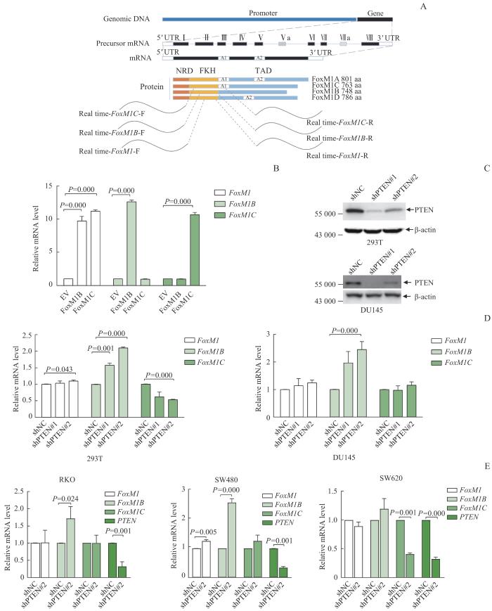
Note: A. Schematic diagram of primer design strategy for mRNA of FoxM1B, FoxM1C and total FoxM1. B. The specificity of the primers was verified by using qRT-PCR in FoxM1B/FoxM1C overexpressing 293T cells. C. Western blotting results showed that PTEN was efficiently knocked down by two pairs of shRNA both in 293T and DU145 cells. D. qRT-PCR results showed that with PTEN knockdown, the expression of FoxM1B mRNA was upregulated in 293T (left) and DU145 (right) cells, whilethe expression of FoxM1C mRNA was downregulated in 293T cells but did not change in DU145 cells. E. qRT-PCR results showed upregulation of FoxM1B mRNA expression, but no significant change in FoxM1C mRNA expression after PTEN being knocked down in RKO (left), SW480 (middle) and SW460 (right) cells. NRD—N-terminal repressor domain; FKH—forkhead/winged-helix domain; TAD—transcativation domain; EV—empty vector.

Fig 1   FoxM1 mRNA isoforms changed in PTEN knockdown cells


用2条针对PTEN的shRNA,敲低293T、DU145、RKO、SW480和SW620细胞内的PTEN。Western blotting结果显示,2条shRNA均可有效降低293T和DU145细胞内的PTEN蛋白的表达水平(图1C)。qRT-PCR检测发现,在293T和DU145细胞中,与对照(shNC)细胞相比,PTEN敲低后的细胞中的FoxM1B形式的mRNA均显著增多,而FoxM1C形式的mRNA表现为减少或不变,FoxM1的mRNA总体水平没有显著变化(图1D)。在RKO、SW480和DU145细胞中,与对照细胞相比,PTEN敲低后的细胞中FoxM1B形式的mRNA也显著增多,而FoxM1C形式的mRNA同样表现为减少或不变,FoxM1的mRNA总体水平依然没有显著变化(图1E)。这些结果说明在PTEN敲低后,FoxM1在转录水平并没有发生变化,但其可变剪接发生了改变:其他形式的FoxM1FoxM1B转换。因此,细胞内PTEN敲低影响FoxM1的可变剪接。

2.2 FoxM1B/C对肿瘤细胞迁移的相反作用

DU145是一种PTEN单拷贝缺失的前列腺癌细胞系;PTEN表达量较低,可以很好地模拟PTEN缺失状态下肿瘤细胞的生理特性18。利用DU145细胞构建FoxM1B和FoxM1C稳定过表达的细胞系。Western blotting结果显示,对应亚型蛋白的表达上调,表明FoxM1B和FoxM1C过表达成功(图2A)。通过Transwell细胞迁移实验发现:相较于空载(EV)组,FoxM1B过表达组DU145细胞迁移数量更多(P=0.024),迁移能力增强;有趣的是,与空载组相比FoxM1C过表达组细胞迁移数量减少(P=0.000),迁移能力减弱(图2B)。

图2

图2   FoxM1BFoxM1CDU145细胞迁移的影响

Note: A. Western blotting was used to detect the overexpression level of FoxM1B and FoxM1C in DU145 cells. B. Transwell migration analysis results of DU145 cells overexpressing FoxM1B and FoxM1C. Left—typical crystal violet staining photos. Bar=100 μm. Right—statistical analysis of cell migration numbers. C. Wound healing assay of DU145 cells transfected with FoxM1B and FoxM1C overexpression lentivirus. Left—photos of cells incubated for 0 and 24 h in the scratch experiment. Bar=100 μm. Right—statistical analysis of the wound healing rate.

Fig 2   Effects of FoxM1B and FoxM1C on DU145 migration


接下来我们又进行了细胞划痕实验,发现过表达FoxM1B组的细胞与空载组相比,其愈合能力显著增强(P=0.001),而过表达FoxM1C的细胞愈合能力则比空载组弱(P=0.021)(图2C)。这说明FoxM1B能促进DU145细胞愈合,增强DU145细胞的迁移能力;而FoxM1C则会抑制DU145细胞愈合,减弱DU145细胞的迁移能力。FoxM1B/C对于肿瘤细胞的迁移起着截然相反的作用:FoxM1B发挥促进肿瘤细胞迁移的功能,而FoxM1C则表现为抑制肿瘤细胞迁移。

2.3 FoxM1B/C转录活性以及对β-catenin转位的影响

文献18报道,FoxM1可引起β-catenin入细胞核。但我们通过免疫荧光实验发现,无论是FoxM1B还是FoxM1C过表达,都没能引起β-catenin入细胞核(图3A)。FoxM1B和FoxM1C作为转录因子,对肿瘤细胞转移调控的差异是否是两者转录活性不同导致的呢?用荧光素酶报告基因系统可以检测转录因子的转录能力(图3B)。我们首先构建了FoxM1B或FoxM1C过表达的293T稳转细胞系,然后共转染Firefly和Renilla的双荧光素酶质粒。Western blotting检测到FoxM1B和FoxM1C成功表达(图3C)。但荧光素底物检测发现FoxM1B或FoxM1C过表达细胞内报告基因的表达差异没有统计学意义(P>0.05,图3D),这说明FoxM1B和FoxM1C的转录活性没有差别。因此FoxM1B和FoxM1C在调控肿瘤转移方面的差异也不是由β-catenin转位到细胞核介导的。其具体机制还需要利用其他实验进一步探究。

图3

图3   FoxM1B/C转录活性和β-catenin定位鉴定

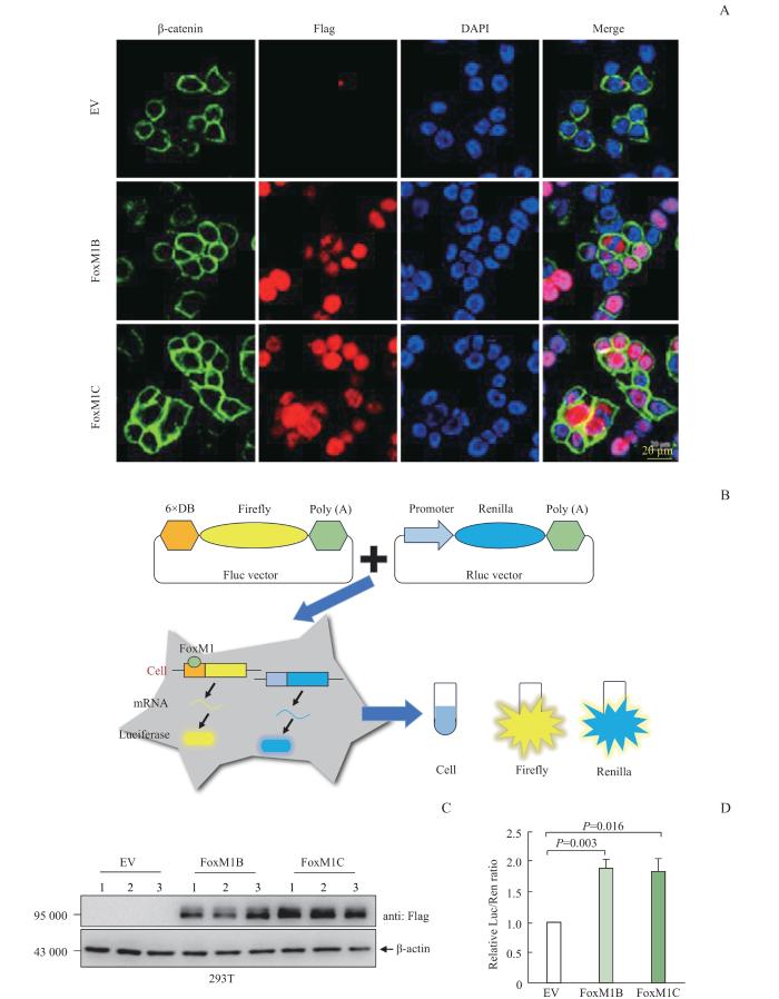
Note: A. Immunofluorescence of 293T cells transfected with Plvx-FOXM1B-Flag and Plvx-FoxM1C-Flag. DAPI represents nucleus, green represents β-catenin, and red represents Flag. Neither FoxM1B nor FoxM1C could cause β-catenin to enter the nucleus. B. Schematic diagram of double luciferase reporter expression system. C. Protein expression of FoxM1B and FoxM1C in FoxM1B/C overexpressing 293T cells transfected with Firefly luciferase and Renilla luciferase reporter gene plasmids. D. Transcriptional activity of FoxM1B and FoxM1C. There was no significant difference in the relative Luc/Ren ratio between FoxM1B and FoxM1C overexpressing 293T cells.

Fig 3   FoxM1B /C transcriptional activities and β-catenin localization


3 讨论

本研究发现PTEN可以调控FoxM1的可变剪接;证实在PTEN敲低细胞中,存在FoxM1的其他亚型通过可变剪接向FoxM1B亚型转换,从而增加肿瘤细胞迁移能力。人类FoxM1基因有10个外显子;通过外显子的可变剪接,主要产生3个变体:FoxM1A、FoxM1C和FoxM1B。FoxM1B和FoxM1C是转录激活因子,而FoxM1A则没有转录活性19。此外,有研究20-21报道,在肿瘤中FoxM1B或FoxM1C的表达水平较高,而FoxM1A的表达水平较低。故本研究侧重对FoxM1B和FoxM1C亚型进行研究。

既往报道22多集中于FoxM1的促癌效应,研究最多的是FoxM1B。其对肿瘤转移的功能早有报道,并发现其与肿瘤细胞增殖、血管生成密切相关23-24。FoxM1B在口腔鳞状细胞癌早期异常上调,通过诱导遗传不稳定性发挥致瘤作用25。同样,FoxM1B被发现在76.9%的宫颈鳞状细胞癌中过表达,提示其对宫颈癌的发展至关重要26。关于FoxM1C,一些研究报道了其促肿瘤的功能22。FoxM1C过表达与胰腺癌的上皮间质化和转移有关27,与卵巢癌的细胞增殖、锚定性生长、迁移和侵袭有关28。此外,FoxM1C通过转录调控干扰素调节因子1的表达,促进食管癌转移29。目前大量研究侧重于FoxM1B和FoxM1C在肿瘤转移、迁移中的作用,故本文也主要研究其对肿瘤转移而非增殖方面的影响。有研究把不同的FoxM1亚型归结为功能相互代偿的备份体。然而本研究发现FoxM1B和FoxM1C在肿瘤转移过程中存在相反的效应。在人类前列腺癌中,PTEN突变和缺失发生频率很高,有40%的病例发生PTEN基因失活,且与较差的预后和较高的转移能力相关30。本研究在PTEN敲低细胞中发现FoxM1的其他亚型通过可变剪接向FoxM1B转换;在PTEN单拷贝缺失的前列腺癌细胞DU145中发现FoxM1B起着促进肿瘤迁移的功能,而FoxM1C则表现为抑制肿瘤迁移的功能。

这对关于FoxM1变异体的传统认知是一种挑战。但是FoxM1B和FoxM1C的转录活性并没有明显差别,在肿瘤迁移中截然相反的效应似乎与它们的转录活性没有关系。不过FoxM1B和FoxM1C之间存在15个氨基酸的差异。这15个氨基酸的差别是否会使它们各自调控特定基因的转录呢?另外这种差异对其与其他蛋白的相互作用是否有影响?这些都可能是引起两者在肿瘤迁移中差异作用的因素,这背后具体的联系和机制仍有待探索。

FoxM1与多种肿瘤具有关联性,在肿瘤中的重要地位使其具备成为一个靶点的潜力。对FoxM1不同变异体差异性功能的解读有助于我们更深入理解FoxM1和肿瘤的关系。本研究发现,FoxM1B发挥促进肿瘤迁移的功能,而FoxM1C则表现为抑制肿瘤迁移。FoxM1B/C对于肿瘤细胞的迁移起着截然相反的作用,通过增加FoxM1C的表达水平或抑制FoxM1B的表达可以帮助我们在针对FoxM1的肿瘤治疗上寻找新思路。

作者贡献声明

沈少明负责课题设计及论文的修改。葛梦凯、王晓玲负责实验操作、数据分析和论文写作。所有作者均阅读并同意了最终稿件的提交。

AUTHOR's CONTRIBUTIONS

SHEN Shaoming was responsible for project design and thesis revision. GE Mengkai and WANG Xiaoling were responsible for experimental operation, data analysis and thesis writing. All authors have read the final manuscript and agreed to the submission.

利益冲突声明

所有作者声明不存在利益冲突。

COMPETING INTERESTS

All authors disclose no relevant conflict of interests.

参考文献

NUSSINOV R, TSAI C J, JANG H. Anticancer drug resistance: an update and perspective[J]. Drug Resist Updat, 2021, 59: 100796.

[本文引用: 1]

LIN Y X, WANG Y, DING J X, et al. Reactivation of the tumor suppressor PTEN by mRNA nanoparticles enhances antitumor immunity in preclinical models[J]. Sci Transl Med, 2021, 13(599): eaba9772.

[本文引用: 1]

GAO P, HAO J L, XIE Q W, et al. PELO facilitates PLK1-induced the ubiquitination and degradation of Smad4 and promotes the progression of prostate cancer[J]. Oncogene, 2022, 41(21): 2945-2957.

[本文引用: 1]

LIU W W, XU L, WANG X, et al. PRDX1 activates autophagy via the PTEN-AKT signaling pathway to protect against cisplatin-induced spiral ganglion neuron damage[J]. Autophagy, 2021, 17(12): 4159-4181.

[本文引用: 1]

ÁLVAREZ-GARCIA V, TAWIL Y, WISE H M, et al. Mechanisms of PTEN loss in cancer: it′s all about diversity[J]. Semin Cancer Biol, 2019, 59: 66-79.

[本文引用: 1]

BYRNES K, BLESSINGER S, BAILEY N T, et al. Therapeutic regulation of autophagy in hepatic metabolism[J]. Acta Pharm Sin B, 2022, 12(1): 33-49.

[本文引用: 1]

FENG J W, DANG Y P, ZHANG W Q, et al. PTEN arginine methylation by PRMT6 suppresses PI3K-AKT signaling and modulates pre-mRNA splicing[J]. Proc Natl Acad Sci U S A, 2019, 116(14): 6868-6877.

[本文引用: 1]

PENG Q, ZHOU Y J, OYANG L, et al. Impacts and mechanisms of alternative mRNA splicing in cancer metabolism, immune response, and therapeutics[J]. Mol Ther, 2022, 30(3): 1018-1035.

[本文引用: 1]

FRANKIW L, BALTIMORE D, LI G D. Alternative mRNA splicing in cancer immunotherapy[J]. Nat Rev Immunol, 2019, 19(11): 675-687.

[本文引用: 1]

WANG K, DAI X Y, YU A, et al. Peptide-based PROTAC degrader of FOXM1 suppresses cancer and decreases GLUT1 and PD-L1 expression[J]. J Exp Clin Cancer Res, 2022, 41(1): 289.

[本文引用: 1]

VANGENDEREN C, HARKNESS T A A, ARNASON T G. The role of anaphase promoting complex activation, inhibition and substrates in cancer development and progression[J]. Aging, 2020, 12(15): 15818-15855.

[本文引用: 1]

HU G H, YAN Z W, ZHANG C, et al. FOXM1 promotes hepatocellular carcinoma progression by regulating KIF4A expression[J]. J Exp Clin Cancer Res, 2019, 38(1): 188.

[本文引用: 1]

NAKAMURA S, HIRANO I, OKINAKA K, et al. The FOXM1 transcriptional factor promotes the proliferation of leukemia cells through modulation of cell cycle progression in acute myeloid leukemia[J]. Carcinogenesis, 2010, 31(11): 2012-2021.

[本文引用: 1]

RATHER T B, PARVEIZ I, BHAT G A, et al. Evaluation of forkhead box M1 (FOXM1) gene expression in colorectal cancer[J]. Clin Exp Med, 2023, 23(6): 2385-2405.

[本文引用: 1]

LI S K M, SMITH D K, LEUNG W Y, et al. FoxM1c counteracts oxidative stress-induced senescence and stimulates Bmi-1 expression[J]. J Biol Chem, 2008, 283(24): 16545-16553.

[本文引用: 1]

NILSSON M B, SUN H Y, ROBICHAUX J, et al. A YAP/FOXM1 axis mediates EMT-associated EGFR inhibitor resistance and increased expression of spindle assembly checkpoint components[J]. Sci Transl Med, 2020, 12(559): eaaz4589.

[本文引用: 1]

SHEN S M, JI Y, ZHANG C, et al. Nuclear PTEN safeguards pre-mRNA splicing to link Golgi apparatus for its tumor suppressive role[J]. Nat Commun, 2018, 9(1): 2392.

[本文引用: 1]

MATSUSHITA M, FUJITA K, HAYASHI T, et al. Gut microbiota-derived short-chain fatty acids promote prostate cancer growth via IGF1 signaling[J]. Cancer Res, 2021, 81(15): 4014-4026.

[本文引用: 2]

KORVER W, ROOSE J, CLEVERS H. The winged-helix transcription factor Trident is expressed in cycling cells[J]. Nucleic Acids Res, 1997, 25(9): 1715-1719.

[本文引用: 1]

BARGER C J, ZHANG W, HILLMAN J, et al. Genetic determinants of FOXM1 overexpression in epithelial ovarian cancer and functional contribution to cell cycle progression[J]. Oncotarget, 2015, 6(29): 27613-27627.

[本文引用: 1]

TASSI R A, TODESCHINI P, SIEGEL E R, et al. FOXM1 expression is significantly associated with chemotherapy resistance and adverse prognosis in non-serous epithelial ovarian cancer patients[J]. J Exp Clin Cancer Res, 2017, 36(1): 63.

[本文引用: 1]

BU H T, TANG S S, LIU G T, et al. In silico, in vitro and in vivo studies: dibutyl phthalate promotes prostate cancer cell proliferation by activating Forkhead Box M1 and remission after Natura-α pretreatment[J]. Toxicology, 2023, 488: 153465.

[本文引用: 2]

MADHI H, LEE J S, CHOI Y E, et al. FOXM1 inhibition enhances the therapeutic outcome of lung cancer immunotherapy by modulating PD-L1 expression and cell proliferation[J]. Adv Sci (Weinh), 2022, 9(29): e2202702.

[本文引用: 1]

SHER G, MASOODI T, PATIL K, et al. Dysregulated FOXM1 signaling in the regulation of cancer stem cells[J]. Semin Cancer Biol, 2022, 86(Pt 3): 107-121.

[本文引用: 1]

KELLEHER F C, O′SULLIVAN H. FOXM1 in sarcoma: role in cell cycle, pluripotency genes and stem cell pathways[J]. Oncotarget, 2016, 7(27): 42792-42804.

[本文引用: 1]

CHEN W, SHIMANE T, KAWANO S, et al. Human papillomavirus 16 E6 induces FoxM1B in oral keratinocytes through GRHL2[J]. J Dent Res, 2018, 97(7): 795-802.

[本文引用: 1]

HUANG C, XIE D C, CUI J J, et al. FOXM1c promotes pancreatic cancer epithelial-to-mesenchymal transition and metastasis via upregulation of expression of the urokinase plasminogen activator system[J]. Clin Cancer Res, 2014, 20(6): 1477-1488.

[本文引用: 1]

LOK G T M, CHAN D W, LIU V W S, et al. Aberrant activation of ERK/FOXM1 signaling cascade triggers the cell migration/invasion in ovarian cancer cells[J]. PLoS One, 2011, 6(8): e23790.

[本文引用: 1]

ZHOU Y Z, WANG Q, CHU L, et al. FOXM1c promotes oesophageal cancer metastasis by transcriptionally regulating IRF1 expression[J]. Cell Prolif, 2019, 52(2): e12553.

[本文引用: 1]

TAYLOR B S, SCHULTZ N, HIERONYMUS H, et al. Integrative genomic profiling of human prostate cancer[J]. Cancer Cell, 2010, 18(1): 11-22.

[本文引用: 1]

/