

上颌矢状向发育不足及横向发育不足是口腔正畸临床中常见的骨性错
1 对象与方法
1.1 研究对象
本研究回顾性分析了2023年7月—8月在上海交通大学医学院附属第九人民医院口腔颅颌面科就诊患者的CBCT影像资料。研究对象排除了唇腭裂、颅面部综合征以及有正畸正颌治疗史或颌面部外伤史的患者,最终纳入134例(男性67例、女性67例)。样本年龄范围为6~25岁,平均年龄为(15.0±5.3)岁。依据年龄特点,将样本划分为6组,如表1所示,覆盖了从童年期至成年期的范围。
表1 研究对象的分组(N=134)
Tab 1
| Group | Age/year | Male (n=67) | Female (n=67) |
|---|---|---|---|
| 1 | 6≤Age<9 | 11 | 11 |
| 2 | 9≤Age<12 | 11 | 11 |
| 3 | 12≤Age<15 | 11 | 11 |
| 4 | 15≤Age<18 | 12 | 12 |
| 5 | 18≤Age<21 | 11 | 11 |
| 6 | 21≤Age<25 | 11 | 11 |
1.2 方法
1.2.1 测量方法
患者采取直立位,头部维持自然状态,通过颏托及外耳道杆及逆行固定,确保咬合稳定于牙尖交错位。调整影像资料中头部位置,确保正中矢状面精确通过前鼻棘点(anterior nasal spine,ANS)、后鼻棘点(posterior nasal spine,PNS)和鼻根点(nasion,N)。横断面设定为经过ANS点与PNS点,且与正中矢状面保持垂直,以下称为腭平面。冠状面沿矢状向定位,以双侧腭大孔后缘连线的中点为基准,同时保持与正中矢状面及腭平面的垂直关系(图1)。
图1
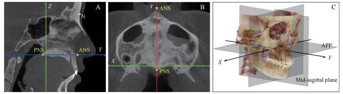
图1
翼腭缝宏观解剖的标准观测头位
Note: A. Mid-sagittal plane. B. Axial palatal plane. C. Head position in space. APP—axial palatal plane.
Fig 1
Standard observational head position of macroscopic anatomy of the pterygopalatine suture
1.2.2 仪器
采用KaVo OP3D Vision系统(KaVo公司,德国)获取CBCT影像。扫描参数设定为管电压120 kV,管电流5 mA,体素层厚0.3 mm,总扫描时间8.9 s,其扫描视野直径为23 cm,高度为17 cm。对于CBCT DICOM数据的三维重建、头位调整及标志点定位,则通过Dolphin Imaging软件(版本11.9,美国)实现。所有参数的计算处理均基于Windows系统,通过PyCharm集成的Python环境(版本3.8.12)完成。
1.2.3 观察指标
腭平面与上颌扩弓及前牵引治疗之间存在密切的关联性,该平面成为本研究的关键观测基准。在此研究中,PPS被腭平面划分为内外2个部分,腭骨锥突嵌入蝶骨翼切迹的结构简称为“锥突嵌体”。对于每侧PPS区域,本研究标定了4个特定的标志点,分别为锥突嵌体的最外侧后点、最外侧前点、最内侧后点、最内侧前点,见图2A~C。
图2
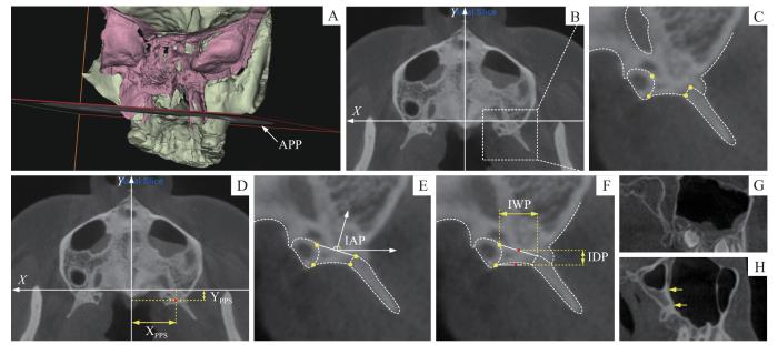
图2
翼腭缝宏观解剖结构的标志点与观测指标
Note: A. Positional relationship between the axial palatal plane and pterygopalatine sutures. Pink block represents the sphenoid bone, and green block represents the rest. B. Axial palatal plane. C. Landmarks of the pterygopalatine suture. D. Transverse and sagittal position of the PPS (XPPS and YPPS). E. Insertion angle of the pyramidal process (IAP). F. Insertion width and depth of the pyramidal process (IWP and IDP). G. Pterygomaxillary junction-noncontacted (PMJ-N). H. Pterygomaxillary junction-contacted (PMJ-C); the segment between yellow arrows represents the contacting segment.
Fig 2
Landmarks and observation indexes of the macroscopic anatomical structure of pterygopalatine suture
表2 参数计算方法
Tab 2
| Measurement | Calculation |
|---|---|
| XPPS | |Xb+Xd|/2 |
| YPPS | (Yb+Yd)/2 |
| IAP | cos-1[(Yb-Yd)/|bd|] |
| IWP | |Xd-Xb|/2 |
| IDP | (Yb+Yd-Ya-Yc)/2 |
| PMJ | Noncontact/contact (N/C) |
定量指标如下。①PPS横向位置(transverse position of PPS,XPPS)即锥突嵌体前缘中点至ANS-PNS连线的水平距离。②PPS矢状向位置(sagittal position of PPS,YPPS)即锥突嵌体前缘中点至腭大孔后缘(X轴)的矢状向距离。③锥突嵌入角(insertion angle of pyramidal process,IAP)定义为锥突嵌体前缘的前向垂线与水平外侧线之间的夹角。④锥突嵌入宽度(insertion width of pyramidal process,IWP)指锥突嵌体前缘的横向延伸宽度。⑤锥突嵌入深度(insertion depth of pyramidal process,IDP)即锥突嵌体前缘中点至后缘中点的矢状向距离。⑥翼上颌联合类型:非连接型(pterygomaxillary junction-noncontact type,PMJ-N),即上颌骨后壁未与蝶骨翼突直接相连;连接型(pterygomaxillary junction-contact type,PMJ-C),即上颌骨后壁与蝶骨翼突直接连接。
1.3 统计学方法
由单一研究者对所有影像资料进行头位校正、标志点标定及翼上颌联合情况的判定。2周后,随机抽取20%的样本,由2名研究者独立并行地重复相同流程。组内相关系数(intraclass correlation coefficient,ICC)及Kappa系数用于评价组间及组内的一致性。
使用PASS 15软件进行样本量估算,确定每组所需的最小样本量为21人,本研究的样本量符合标准。使用SPSS 26.0软件进行统计学分析。PMJ-N被指定为0,PMJ-C被指定为1。通过逐步线性回归分析及二元逻辑回归,探究定量和定性参数与年龄、性别的整体相关性。进一步开展多重比较分析,以确定这些参数快速变化和稳定的年龄段。定量参数的双侧差异采用配对t检验进行分析,定性参数的双侧差异则采用配对χ2检验。P<0.05表示差异具有统计学意义。
2 结果
5项定量参数的组内ICC值为0.830~0.934,组间ICC值为0.822~0.920,表明观测结果可信度高。翼上颌联合类型判定的组内及组间Kappa系数分别为0.982和0.957,表明结果高度一致。
2.1 观察指标与年龄及性别的相关性
观察指标与年龄及性别的相关性分析结果见表3。XPPS与年龄呈正相关,且男性的测量值显著高于女性;YPPS与年龄、性别均未显示出显著的相关性。就内部嵌合形态而言,IAP及IWP与年龄增长呈正相关,与性别无显著相关性;IDP与年龄和性别均无显著相关性。此外,翼上颌连接型的形成与年龄的增加呈现显著相关性。
表3 翼腭缝位置及形态与年龄及性别的相关性(N=268)
Tab 3
| Measurement | Age | Gender | |||
|---|---|---|---|---|---|
| r | P | r | P | ||
| XPPS | 0.098 | <0.001 | 0.560 | 0.008 | |
| YPPS | 0.011 | 0.565 | 0.054 | 0.181 | |
| IAP | 0.835 | <0.001 | 1.098 | 0.517 | |
| IWP | 0.079 | 0.001 | 0.371 | 0.133 | |
| IDP | -0.009 | 0.218 | 0.078 | 0.309 | |
| PMJ/(N/C) | 0.304 | <0.001 | -0.653 | 0.060 | |
2.2 发育节点比较
为了确定发育节点,本研究在样本按年龄分组的基础上进行了进一步分析。多重比较的结果如图3所示,在翼腭缝的横向位置上,第1组和第2组之间均显示出统计学差异(女性,P=0.006;男性,P=0.004),而其余相邻组间的差异不显著。其余相邻组间的差异无统计学意义。IAP(锥突嵌入角)在组2和组3之间的比较显示出统计学差异(P=0.042),其他相邻组间的差异不显著。IAP的数值范围为3.6°~167.2°,但各年龄组多数个体的IAP均处于4°~90°范围内(图4)。IWP(锥突嵌入宽度)在组1和组3之间的比较显示出统计学差异(P=0.016),相邻组间的差异不显著。翼上颌联合类型在第1组和第2组之间(P<0.001)、组2和组3之间(P=0.037)的分布显示出统计学差异,其他相邻组间的差异不显著。在本研究纳入的样本中,翼上颌连接型的比例为77.6%(208/268),而在纳入的成年样本中,该比例为95.5%(84/88)。
图3
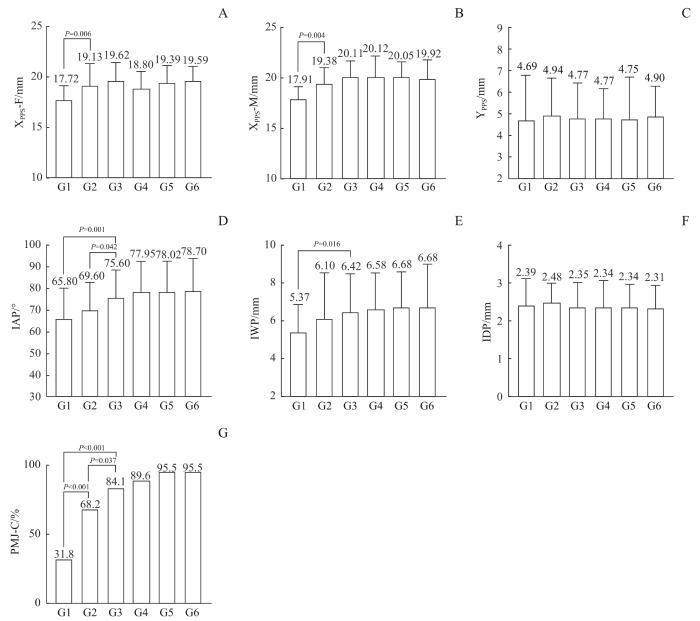
图3
翼腭缝位置及形态参数的年龄组间比较
Note: A. Transverse position of PPS-Female (XPPS-F). B. Transverse position of PPS -Male (XPPS-M). C. Sagittal position of PPS (YPPS). D. Insertion angle of the pyramidal process (IAP). E. Insertion width of the pyramidal process (IWP). F. Insertion depth of the pyramidal process (IDP). G. The proportion of pterygomaxillary junction-Contact (PMJ-C). G1‒6 represent Group 1‒6.
Fig 3
Comparison of the parameters of pterygopalatine suture morphology and position between age groups
图4
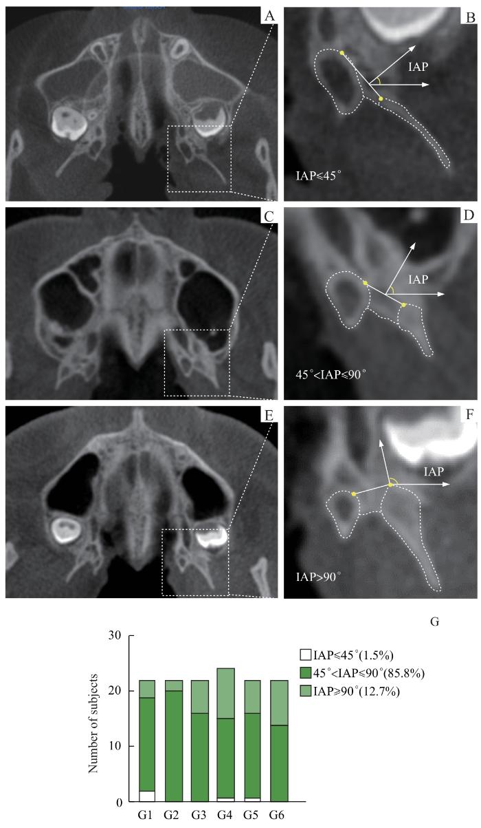
图4
不同角度范围的IAP
Note: A/B. Insertion angle of the pyramidal process (IAP) less than 45º. C/D. IAP larger than 45º and less than 90º. E/F. IAP larger than 90º. G. Proportion of 3 types of IAP in each age group.
Fig 4
IAP for different angular ranges
2.3 双侧结构对称性比较
样本间双侧PPS横向及矢状向位置,锥突嵌入角、嵌入宽度、嵌入深度、翼上颌连接情况的差异均无统计学意义(P>0.05)。
3 讨论
6岁以后的成长期间,翼腭缝相对于腭大孔的矢状向位置稳定。但随着上颌第二(第三)磨牙的萌出,上颌结节区域的骨沉积持续向后推进,使得上颌结节与蝶骨翼突的间距逐渐缩短[2],并倾向于在翼腭缝的上方外侧形成翼上颌联合结构。成人人群中,翼上颌联合的发生率高于90%。此联合的形成及其连接面积的增加,可能会引起翼颌区域骨性阻力的增强。此外,在翼腭缝的嵌合形态方面,IAP在发育过程中逐步扩大,其嵌入方向亦逐步趋向矢状向。
因此,除了业界广泛接受的扩弓治疗通过松解上颌周缘骨缝以增强前牵引疗效的作用机制外[6],上颌前牵引治疗具备通过促进翼腭缝分离以进一步增强扩弓效果的可能性。因此针对上颌横向生长不足的骨性Ⅲ类错
本研究创新性地应用CBCT技术对中国人群的翼腭缝宏观形态进行了探讨,但仍存在一定局限性。首先,对翼腭缝形态的分析仅限于腭平面,尚未涵盖立体三维空间的全面评估。此外,本研究采用回顾性横断面设计,且样本量相对有限,只是翼腭缝宏观解剖的初步探索,未来将开展基于更大数据样本的纵向研究。
作者贡献声明
章文益负责研究方案设计、资料收集、数据分析及初稿撰写;郑美里参与研究方案设计、资料收集、论文撰写及修改;谢羽番参与资料收集及数据分析;江凌勇负责研究指导及最终审核。所有作者均阅读并同意了最终稿件的提交。
AUTHOR's CONTRIBUTIONS
ZHANG Wenyi was responsible for the research protocol design, data collection, statistical analysis, and writing original draft. CHUNG Miri contributed to the research protocol design, data collection, writing review and editing. XIE Yufan contributed to the data collection and statistical analysis. JIANG Lingyong contributed to the research supervision and final review. All authors have read the final manuscript and approved for submission.
利益冲突声明
所有作者声明不存在利益冲突。
COMPETING INTERESTS
All authors disclose no relevant conflict of interests.
参考文献
