上海交通大学学报(医学版) ›› 2021, Vol. 41 ›› Issue (7): 876-883.doi: 10.3969/j.issn.1674-8115.2021.07.005

• 论著 · 基础研究 • 上一篇    

6-OHDA诱导的帕金森病小鼠表现出以p16Ink4a上调和星形胶质细胞衰老为特征的衰老表型

袁笑(), 田野野, 薛峥()   

  1. 华中科技大学同济医学院附属同济医院神经内科,武汉 430030
  • 出版日期:2021-07-28 发布日期:2021-08-03
  • 通讯作者: 薛峥 E-mail:xiaoyuan07@outlook.com;xuezheng@tjh.tjmu.edu.cn
  • 作者简介:袁 笑(1994—),女,硕士生;电子信箱:xiaoyuan07@outlook.com
  • 基金资助:
    国家自然科学基金(91849121)

6-OHDA-induced Parkinson disease mice exhibit senescent phenotypes characterized by upregulation of p16Ink4a and astrocyte senescence

Xiao YUAN(), Ye-ye TIAN, Zheng XUE()   

  1. Department of Neurology, Tongji Hospital, Tongji Medical College, Huazhong University of Science and Technology, Wuhan 430030, China
  • Online:2021-07-28 Published:2021-08-03
  • Contact: Zheng XUE E-mail:xiaoyuan07@outlook.com;xuezheng@tjh.tjmu.edu.cn
  • Supported by:
    Funding Information National Natural Science Foundation of China(91849121)

摘要:

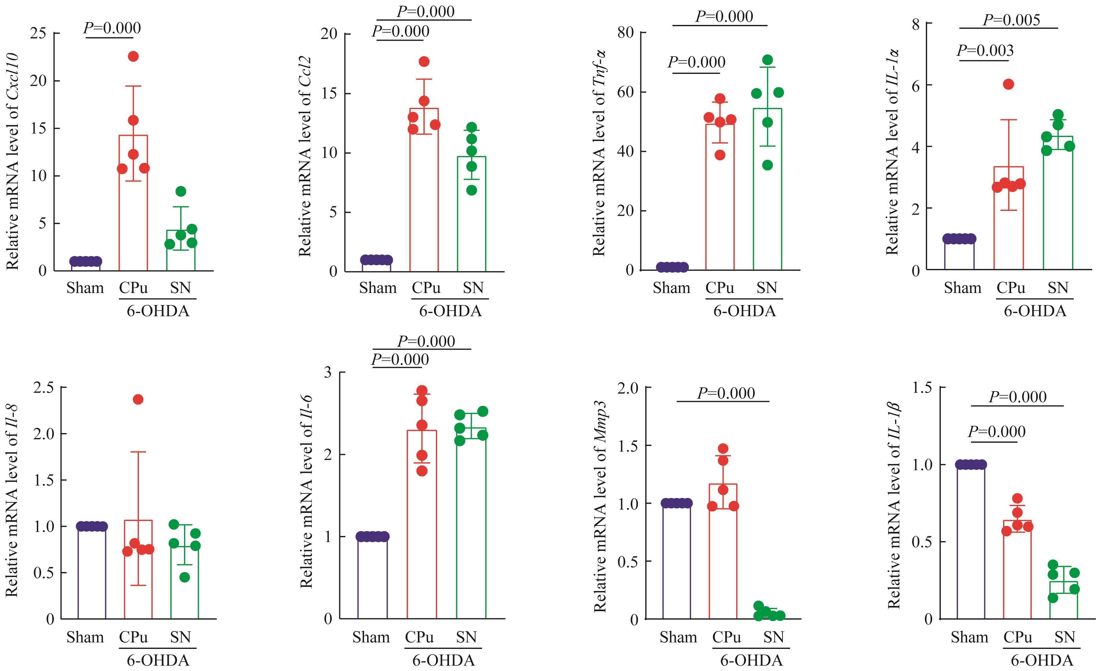
目的·探索6-羟基多巴胺(6-hydroxydopamine,6-OHDA)诱导的帕金森病小鼠模型是否具有衰老表现及胶质细胞的衰老变化。方法·将30只雄性9~10月龄C57BL/6J小鼠随机分为假手术组(Sham组)和6-OHDA组,每组15只。通过6-OHDA纹状体立体定位注射建立帕金森病模型。采用阿扑吗啡诱导的旋转实验、转棒实验评估小鼠造模后第21日的神经功能损伤情况;末次行为学实验后,取小鼠脑组织,通过蛋白质印迹法检测脑组织纹状体及黑质区域酪氨酸羟化酶(tyrosine hydroxylase,TH)蛋白含量;免疫荧光染色观察纹状体及黑质区域衰老标志物p16Ink4a、p21的表达变化以及胶质细胞、衰老标志物双阳性的细胞数量变化;采用实时荧光定量PCR(RT-qPCR)检测纹状体及黑质区域的细胞周期蛋白依赖性激酶抑制剂p16Ink4ap15Ink4bp19Ink4dp21p27Kip1,以及衰老相关分泌表型(senescence-associated secretory phenotype, SASP)包括Cxcl10Ccl2、肿瘤坏死因子α(tumor necrosis factor-α,Tnf-α)、白介素1α(interleukin-1 α,Il-1α)、Il-1βIl-6、基质金属蛋白酶3(matrix metalloproteinase 3,Mmp3)的表达及变化。结果·转棒实验结果显示,与Sham组相比, 6-OHDA组小鼠在转棒上的停留时间较短(P=0.000);阿朴吗啡诱导的旋转实验结果显示,与Sham组相比,6-OHDA组小鼠总旋转次数较多(P=0.000);蛋白质印迹法检测结果显示,6-OHDA组纹状体及黑质区域TH蛋白表达量均显著低于Sham组(均P=0.000)。以上结果证实6-OHDA单侧纹状体小鼠帕金森病模型成功建立。免疫荧光染色结果显示:6-OHDA组纹状体和黑质区域p16Ink4a阳性细胞数量增多;p21在2组均无明显表达;6-OHDA组同时伴有衰老的星形胶质细胞、小胶质细胞、少突胶质细胞产生,且星形胶质细胞数量大于少突胶质细胞和小胶质细胞(均P<0.05)。RT-qPCR检测结果显示,与Sham组相比:6-OHDA组造模侧纹状体和黑质区域p16Ink4ap15Ink4bp19Ink4d的表达上调,差异具有统计学意义(均P<0.05);p21轻微上调,但差异均无统计学意义(均P?0.05);p27Kip1轻微上调,但仅在黑质区域的差异有统计学意义(P=0.016)。与Sham组相比,6-OHDA组造模侧纹状体区域的Cxcl10、Ccl2、Tnf-α、Il-1αIl-6显著上调,Il-1β显著下调(均P<0.05),黑质区域的Ccl2、Tnf-α、Il-1αIl-6显著上调(均P<0.05),Mmp3Il-1β均显著下调(均P<0.05)。结论·6-OHDA诱导的帕金森病小鼠模型表现出以p16上调和星形胶质细胞衰老为特征的衰老表型。

关键词: 帕金森病, 衰老, 6-羟基多巴胺, 动物模型, 星形胶质细胞

Abstract:

Objective·To explore whether 6-hydroxydopamine(6-OHDA)-induced Parkinson disease (PD) mouse models have senescent phenotypes and explore the changes of senescence-related glial cells.

Methods·Thirty 9?10 months old C57BL/6J male mice were divided into Sham-operated group (n=15) and 6-OHDA group (n=15). PD mouse models were established by stereotactic injection of 6-OHDA into striatum. The neurological function deficit was evaluated by rotarod test and apomorphine (APO)-induced rotational behavior on the 21st day after modeling. The brain was taken after the last behavioral experiment. Western blotting was used to detect the protein content of tyrosine hydroxylase (TH) in both caudate putamen (CPu) and substantia nigra (SN) regions. Immunofluorescence was used to observe the expression of glial cells and aging marker p16Ink4a / p21 in both CPu and SN regions. RT-qPCR was also used to detect the expression and changes of cyclin-dependent kinase inhibitor including p16Ink4a, p15Ink4b, p19Ink4d, p21 and p27Kip1, and senescence-associated secretory phenotype including Cxcl10, Ccl2, tumor necrosis factor-α (Tnf-α), interleukin-1α (Il-1α), Il-1β, Il-6 and matrix metalloproteinase 3 (MMP3).

Results·The decreased falling latency in rotarod test (P=0.000), the increased number of rotations induced by APO (P=0.000) and the loss of TH protein measured by Western blotting (P=0.000), suggested that 6-OHDA unilateral striatum mouse model of PD was successfully established. Immunofluorescence staining showed that the upregulation of an aging marker p16Ink4a in both CPu and SN regions of the 6-OHDA group compared with the Sham group, but p21 was not significantly expressed in both groups; senescent astrocytes, microglia and oligodendrocytes were accumulated in CPu and SN regions as well, while the number of astrocytes was larger than the other two types of glia cells (P<0.05). RT-qPCR results showed that, compared with those of the Sham group, p15Ink4b, p16Ink4a and p19Ink4d in CPu and SN regions of the 6-OHDA group were up-regulated (P<0.05); p21 was slightly up-regulated, but the difference was not statistically significant (P?0.05); p27kip1 was slightly up-regulated, but statistically significant difference only presented in SN region (P=0.016). RT-qPCR also showed that, compared with those of the Sham group, the levels of Cxcl10, Ccl2, Il-1α and Il-6 were significantly up-regulated while Il-1β was significantly decreased in CPu region of the 6-OHDA group (P<0.05); the levels of Ccl2, Tnf-α, Il-1α and Il-6 were significantly increased while Mmp3 and Il-1β were significantly decreased in SN region of the 6-OHDA group (P<0.05).

Conclusion·6-OHDA-induced PD mice exhibit senescent phenotypes characterized by upregulation of p16Ink4a and astrocyte senescence.

Key words: Parkinson disease (PD), senescence, 6-hydroxydopamine (6-OHDA), animal model, astrocyte

中图分类号: