Journal of Shanghai Jiao Tong University (Medical Science) ›› 2023, Vol. 43 ›› Issue (10): 1274-1281.doi: 10.3969/j.issn.1674-8115.2023.10.008
• Clinical research • Previous Articles
YU Xi1( ), SUN Junnan1, ZHANG Jiaojiao2, GAO Yue1, WANG Hu3, YU Yang1, WANG Hairong1(), HONG Wen4()
Received:2023-03-05
Accepted:2023-09-15
Online:2023-10-28
Published:2023-10-28
Contact:
WANG Hairong,HONG Wen
E-mail:yuxi199263@163.com;wanghairong@xinhuamed.com.cn;18930172044@189.com
CLC Number:
YU Xi, SUN Junnan, ZHANG Jiaojiao, GAO Yue, WANG Hu, YU Yang, WANG Hairong, HONG Wen. Efficacy of sternal cortical thickness ratio in adult chest CT in the diagnosis of osteopenia and osteoporosis[J]. Journal of Shanghai Jiao Tong University (Medical Science), 2023, 43(10): 1274-1281.
Add to citation manager EndNote|Ris|BibTeX
URL: https://xuebao.shsmu.edu.cn/EN/10.3969/j.issn.1674-8115.2023.10.008
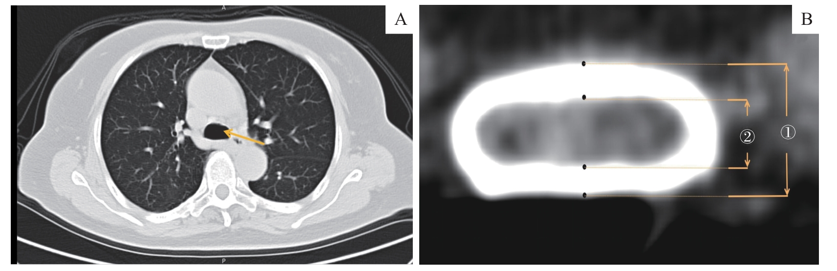
Fig 1 Measurement of cortical thickness in the sternum measurement plane
| Gender | >65 years | ≤65 years | ||||||||
|---|---|---|---|---|---|---|---|---|---|---|
| Normal bone mass group (n=33) | Osteopenia group (n=41) | Osteoporosis group (n=41) | χ2 value | P value | Normal bone mass group (n=45) | Osteopenia group (n=25) | Osteoporosis group (n=13) | χ2 value | P value | |
| Male | 21 (63.6) | 20 (48.8) | 14 (34.1) | 6.396 | 0.041 | 24 (53.3) | 9 (36.0) | 7 (53.8) | 2.131 | 0.345 |
| Female | 12 (36.4) | 21 (51.2) | 27 (65.9)① | 21 (46.7) | 16 (64.0) | 6 (46.2) | ||||
Tab 1 Gender distribution of patients with normal bone mass, osteopenia and osteoporosis [n(%)]
| Gender | >65 years | ≤65 years | ||||||||
|---|---|---|---|---|---|---|---|---|---|---|
| Normal bone mass group (n=33) | Osteopenia group (n=41) | Osteoporosis group (n=41) | χ2 value | P value | Normal bone mass group (n=45) | Osteopenia group (n=25) | Osteoporosis group (n=13) | χ2 value | P value | |
| Male | 21 (63.6) | 20 (48.8) | 14 (34.1) | 6.396 | 0.041 | 24 (53.3) | 9 (36.0) | 7 (53.8) | 2.131 | 0.345 |
| Female | 12 (36.4) | 21 (51.2) | 27 (65.9)① | 21 (46.7) | 16 (64.0) | 6 (46.2) | ||||
| Gender | >65 years | ≤65 years | ||||||||
|---|---|---|---|---|---|---|---|---|---|---|
| Normal bone mass group (n=33) | Osteopenia group (n=41) | Osteoporosis group (n=41) | F value | P value | Normal bone mass group (n=45) | Osteopenia group (n=25) | Osteoporosis group (n=13) | F value | P value | |
| Male | 75.14±7.50 | 75.40±9.38 | 82.57±8.06①② | 3.980 | 0.025 | 55.38±9.74 | 54.11±10.46 | 62.29±2.14 | 1.886 | 0.166 |
| Female | 71.58±4.56 | 73.38±5.19 | 80.00±6.90③④ | 11.522 | 0.000 | 56.29±8.34 | 56.69±8.24 | 58.33±9.87 | 0.080 | 0.924 |
Tab 2 Comparison of ages of patients with normal bone mass, osteopenia and osteoporosis (x±s)
| Gender | >65 years | ≤65 years | ||||||||
|---|---|---|---|---|---|---|---|---|---|---|
| Normal bone mass group (n=33) | Osteopenia group (n=41) | Osteoporosis group (n=41) | F value | P value | Normal bone mass group (n=45) | Osteopenia group (n=25) | Osteoporosis group (n=13) | F value | P value | |
| Male | 75.14±7.50 | 75.40±9.38 | 82.57±8.06①② | 3.980 | 0.025 | 55.38±9.74 | 54.11±10.46 | 62.29±2.14 | 1.886 | 0.166 |
| Female | 71.58±4.56 | 73.38±5.19 | 80.00±6.90③④ | 11.522 | 0.000 | 56.29±8.34 | 56.69±8.24 | 58.33±9.87 | 0.080 | 0.924 |
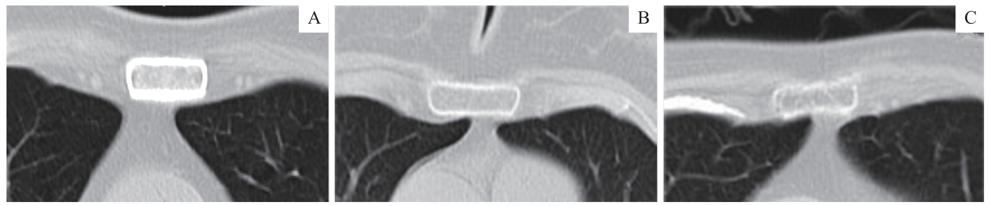
Fig 2 Typical chest CT imaging findings of sternum in patients with normal bone mass (A), osteopenia (B) and osteoporosis (C)
| Item | >65 years | ≤65 years | ||||||||
|---|---|---|---|---|---|---|---|---|---|---|
| Normal bone mass group (n=33) | Osteopenia group (n=41) | Osteoporosis group (n=41) | F value | P value | Normal bone mass group (n=45) | Osteopenia group (n=25) | Osteoporosis group (n=13) | F value | P value | |
| Male | 0.549±0.106 | 0.398±0.023① | 0.307±0.074①③ | 45.035 | 0.000 | 0.420±0.107 | 0.391±0.017② | 0.298±0.023①③ | 21.613 | 0.000 |
| Female | 0.464±0.119 | 0.319±0.046① | 0.214±0.058①④ | 52.734 | 0.000 | 0.396±0.063 | 0.348±0.038① | 0.280±0.016①⑤ | 15.915 | 0.000 |
| F value | 4.518 | 47.694 | 19.729 | 21.987 | 10.421 | 51.454 | ||||
| P value | 0.042 | 0.000 | 0.000 | 0.000 | 0.000 | 0.000 | ||||
Tab 3 Comparison of the sternal cortical thickness ratio in patients with normal bone mass, osteopenia and osteoporosis (x±s)
| Item | >65 years | ≤65 years | ||||||||
|---|---|---|---|---|---|---|---|---|---|---|
| Normal bone mass group (n=33) | Osteopenia group (n=41) | Osteoporosis group (n=41) | F value | P value | Normal bone mass group (n=45) | Osteopenia group (n=25) | Osteoporosis group (n=13) | F value | P value | |
| Male | 0.549±0.106 | 0.398±0.023① | 0.307±0.074①③ | 45.035 | 0.000 | 0.420±0.107 | 0.391±0.017② | 0.298±0.023①③ | 21.613 | 0.000 |
| Female | 0.464±0.119 | 0.319±0.046① | 0.214±0.058①④ | 52.734 | 0.000 | 0.396±0.063 | 0.348±0.038① | 0.280±0.016①⑤ | 15.915 | 0.000 |
| F value | 4.518 | 47.694 | 19.729 | 21.987 | 10.421 | 51.454 | ||||
| P value | 0.042 | 0.000 | 0.000 | 0.000 | 0.000 | 0.000 | ||||
| Gender | >65 years | ≤65 years | ||||||||
|---|---|---|---|---|---|---|---|---|---|---|
| r value | Spearman correlation coefficient | F value | T value | P value | r value | Spearman correlation coefficient | F value | T value | P value | |
| Male | 0.704 | 0.732 | 52.006 | 2.966 | 0.000 | 0.735 | 0.745 | 44.550 | 2.944 | 0.000 |
| Female | 0.785 | 0.819 | 93.213 | 9.310 | 0.000 | 0.479 | 0.732 | 11.337 | 3.857 | 0.000 |
Tab 4 Correlation analysis of sternal cortical thickness ratio and bone mineral density in male and female patients of different age groups
| Gender | >65 years | ≤65 years | ||||||||
|---|---|---|---|---|---|---|---|---|---|---|
| r value | Spearman correlation coefficient | F value | T value | P value | r value | Spearman correlation coefficient | F value | T value | P value | |
| Male | 0.704 | 0.732 | 52.006 | 2.966 | 0.000 | 0.735 | 0.745 | 44.550 | 2.944 | 0.000 |
| Female | 0.785 | 0.819 | 93.213 | 9.310 | 0.000 | 0.479 | 0.732 | 11.337 | 3.857 | 0.000 |
| Group | >65 years | ≤65 years | ||||||||
|---|---|---|---|---|---|---|---|---|---|---|
| AUC (95%CI) | Sensitivity/% | Specificity/% | Youden index | Cut-off value | AUC (95%CI) | Sensitivity/% | Specificity/% | Youden index | Cut-off value | |
| Male | ||||||||||
| Osteopenia | 0.924 (0.824‒0.977) | 90.5 | 95.0 | 0.855 | 0.44 | 0.741 (0.721‒0.962) | 75.0 | 72.4 | 0.474 | 0.42 |
| Osteoporosis | 0.813 (0.641‒0.984) | 96.6 | 76.5 | 0.714 | 0.34 | 0.846 (0.756‒0.927) | 82.4 | 83.3 | 0.765 | 0.32 |
| Female | ||||||||||
| Osteopenia | 0.897 (0.753‒0.962) | 83.3 | 90.2 | 0.833 | 0.39 | 0.768 (0.610‒0.926) | 61.9 | 93.7 | 0.556 | 0.39 |
| Osteoporosis | 0.930 (0.852‒0.987) | 90.5 | 85.2 | 0.757 | 0.28 | 0.937 (0.848‒0.998) | 80.0 | 83.3 | 0.677 | 0.28 |
Tab 5 Analysis of ROC curve characteristics of sternal cortical thickness ratio for the diagnosis of osteopenia and osteoporosis
| Group | >65 years | ≤65 years | ||||||||
|---|---|---|---|---|---|---|---|---|---|---|
| AUC (95%CI) | Sensitivity/% | Specificity/% | Youden index | Cut-off value | AUC (95%CI) | Sensitivity/% | Specificity/% | Youden index | Cut-off value | |
| Male | ||||||||||
| Osteopenia | 0.924 (0.824‒0.977) | 90.5 | 95.0 | 0.855 | 0.44 | 0.741 (0.721‒0.962) | 75.0 | 72.4 | 0.474 | 0.42 |
| Osteoporosis | 0.813 (0.641‒0.984) | 96.6 | 76.5 | 0.714 | 0.34 | 0.846 (0.756‒0.927) | 82.4 | 83.3 | 0.765 | 0.32 |
| Female | ||||||||||
| Osteopenia | 0.897 (0.753‒0.962) | 83.3 | 90.2 | 0.833 | 0.39 | 0.768 (0.610‒0.926) | 61.9 | 93.7 | 0.556 | 0.39 |
| Osteoporosis | 0.930 (0.852‒0.987) | 90.5 | 85.2 | 0.757 | 0.28 | 0.937 (0.848‒0.998) | 80.0 | 83.3 | 0.677 | 0.28 |
| Item | >65 years | ≤65 years | ||||
|---|---|---|---|---|---|---|
| Normal bone mass group (n=33) | Osteopenia group (n=41) | Osteoporosis group (n=41) | Normal bone mass group (n=45) | Osteopenia group (n=25) | Osteoporosis group (n=13) | |
| Male | ||||||
| Accuracy/% | 90.5 | 90.0 | 71.4 | 75.0 | 77.8 | 85.7 |
| False negative rate/% | 9.5 | 10.0 | 28.6 | 25.0 | 22.2 | 14.3 |
| False positive rate/% | 9.5 | 17.7 | 9.1 | 5.3 | 21.7 | 13.3 |
| Female | ||||||
| Accuracy/% | 83.3 | 90.8 | 85.2 | 76.2 | 87.5 | 83.3 |
| False negative rate/% | 16.7 | 9.5 | 14.8 | 23.8 | 12.5 | 16.7 |
| False positive rate/% | 15.4 | 17.4 | 4.2 | 5.9 | 15.8 | 14.3 |
Tab 6 Comparison of sternal cortical thickness ratio and DXA diagnostic results
| Item | >65 years | ≤65 years | ||||
|---|---|---|---|---|---|---|
| Normal bone mass group (n=33) | Osteopenia group (n=41) | Osteoporosis group (n=41) | Normal bone mass group (n=45) | Osteopenia group (n=25) | Osteoporosis group (n=13) | |
| Male | ||||||
| Accuracy/% | 90.5 | 90.0 | 71.4 | 75.0 | 77.8 | 85.7 |
| False negative rate/% | 9.5 | 10.0 | 28.6 | 25.0 | 22.2 | 14.3 |
| False positive rate/% | 9.5 | 17.7 | 9.1 | 5.3 | 21.7 | 13.3 |
| Female | ||||||
| Accuracy/% | 83.3 | 90.8 | 85.2 | 76.2 | 87.5 | 83.3 |
| False negative rate/% | 16.7 | 9.5 | 14.8 | 23.8 | 12.5 | 16.7 |
| False positive rate/% | 15.4 | 17.4 | 4.2 | 5.9 | 15.8 | 14.3 |
| 1 | BIJLSMA A Y, MESKERS C M, WESTENDORP R J, et al. Chronology of age-related disease definitions: osteoporosis and sarcopenia[J]. Ageing Res Rev, 2012, 11(2): 320-324. |
| 2 | NIH Consensus Development Panel on Osteoporosis Prevention, Diagnosis, and Therapy. Osteoporosis prevention, diagnosis, and therapy[J]. JAMA, 2001, 285(6): 785-795. |
| 3 | 中华医学会骨质疏松和骨矿盐疾病分会. 原发性骨质疏松症诊治指南(2011年)[J]. 中华骨质疏松和骨矿盐疾病杂志, 2011, 4(1): 2-17. |
| Osteoporosis and Bone Mineral Disease Branch of Chinese Medical Association. Guidelines for the diagnosis and treatment of primary osteoporosis (2011)[J]. Chinese Journal of Osteoporosis and Bone Mineral Research, 2011, 4(1): 2-17. | |
| 4 | 中华医学会骨质疏松和骨矿盐疾病分会. 原发性骨质疏松症诊疗指南(2017)[J]. 中国骨质疏松杂志, 2019, 25(3): 281-309. |
| Osteoporosis and Bone Mineral Disease Branch of Chinese Medical Association. Guidelines for the diagnosis and treatment of primary osteoporosis (2017)[J]. Chinese Journal of Osteoporosis, 2019, 25(3): 281-309. | |
| 5 | 马远征, 王以朋, 刘强, 等. 中国老年骨质疏松症诊疗指南(2018)[J]. 中国骨质疏松杂志, 2018, 24(12): 1541-1565. |
| MA Y Z, WANG Y P, LIU Q, et al. Guidelines for the diagnosis and treatment of osteoporosis in the elderly in China (2018)[J]. Chinese Journal of Osteoporosis, 2018, 24(12): 1541-1565. | |
| 6 | LI F Z, ECKSTROM E, HARMER P, et al. Exercise and fall prevention: narrowing the research-to-practice gap and enhancing integration of clinical and community practice[J]. J Am Geriatr Soc, 2016, 64(2): 425-431. |
| 7 | 刘琳. DXA影像评价与骨密度测定联合诊断原发性骨质疏松症的临床探讨[J]. 影像研究与医学应用, 2019, 3(17): 50-51. |
| LIU L. Clinical discussion of DXA imaging evaluation and bone densitometry in the diagnosis of primary osteoporosis[J]. Journal of Imaging Research and Medical Applications, 2019, 3(17): 50-51. | |
| 8 | RAMAN-WILMS L. Book review: guidelines for preclinical evaluation and clinical trials in osteoporosis[J]. Ann Pharmacother, 1999, 33: 1377-1378. |
| 9 | JANG S, GRAFFY P M, ZIEMLEWICZ T J, et al. Opportunistic osteoporosis screening at routine abdominal and thoracic CT: normative L1 trabecular attenuation values in more than 20 000 adults[J]. Radiology, 2019, 291(2): 360-367. |
| 10 | LESLIE W D, GIANGREGORIO L M, YOGENDRAN M, et al. A population-based analysis of the post-fracture care gap 1996‒2008: the situation is not improving[J]. Osteoporos Int, 2012, 23(5): 1623-1629. |
| 11 | KANIS J A. Diagnosis of osteoporosis and assessment of fracture risk[J]. Lancet, 2002, 359(9321): 1929-1936. |
| 12 | TINGART M J, APRELEVA M, VON STECHOW D, et al. The cortical thickness of the proximal humeral diaphysis predicts bone mineral density of the proximal humerus[J]. J Bone Joint Surg Br, 2003, 85(4): 611-617. |
| 13 | BLOOM R A. A comparative estimation of the combined cortical thickness of various bone sites[J]. Skeletal Radiol, 1980, 5(3): 167-170. |
| 14 | BARNETT E, NORDIN B E. The radiological diagnosis of osteoporosis: a new approach[J]. Clin Radiol, 1960, 11: 166-174. |
| 15 | YOSHII I, AKITA K. Cortical thickness relative to the transverse diameter of third metacarpal bone reflects bone mineral density in patients with rheumatoid arthritis[J]. Bone, 2020, 137: 115405. |
| 16 | MATHER J, MACDERMID J C, FABER K J, et al. Proximal humerus cortical bone thickness correlates with bone mineral density and can clinically rule out osteoporosis[J]. J Shoulder Elbow Surg, 2013, 22(6): 732-738. |
| 17 | PISTOIA W, VAN RIETBERGEN B, RÜEGSEGGER P. Mechanical consequences of different scenarios for simulated bone atrophy and recovery in the distal radius[J]. Bone, 2003, 33(6): 937-945. |
| 18 | RAUSCH S, KLOS K, GRAS F, et al. Utility of the cortical thickness of the distal radius as a predictor of distal-radius bone density[J]. Arch Trauma Res, 2013, 2(1): 11-15. |
| 19 | YE C X, GUO Y B, ZHENG Y H, et al. Distal radial cortical bone thickness correlates with bone mineral density and can predict osteoporosis: a cohort study[J]. Injury, 2020, 51(11): 2617-2621. |
| 20 | PICKHARDT P J, POOLER B D, LAUDER T, et al. Opportunistic screening for osteoporosis using abdominal computed tomography scans obtained for other indications[J]. Ann Intern Med, 2013, 158(8): 588-595. |
| 21 | LEE S J, BINKLEY N, LUBNER M G, et al. Opportunistic screening for osteoporosis using the sagittal reconstruction from routine abdominal CT for combined assessment of vertebral fractures and density[J]. Osteoporos Int, 2016, 27(3): 1131-1136. |
| 22 | NAZIA FATHIMA S M, TAMILSELVI R, PARISA BEHAM M, et al. Diagnosis of osteoporosis using modified U-net architecture with attention unit in DEXA and X-ray images[J]. J Xray Sci Technol, 2020, 28(5): 953-973. |
| 23 | YAO Q, LIU J, YUAN K, et al. Comparison of L1 CT-attenuation and cortical thickness in predicting osteoporosis by opportunistic CT[J]. J Xray Sci Technol, 2022, 30(3): 631-640. |
| 24 | MECZEKALSKI B, PODFIGURNA-STOPA A, GENAZZANI A R. Hypoestrogenism in young women and its influence on bone mass density[J]. Gynecol Endocrinol, 2010, 26(9): 652-657. |
| 25 | POSTNOV A A, VINOGRADOV A V, VAN DYCK D, et al. Quantitative analysis of bone mineral content by X-ray microtomography[J]. Physiol Meas, 2003, 24(1): 165-178. |
| 26 | NELSON M E, FISHER E C, CATSOS P D, et al. Diet and bone status in amenorrheic runners[J]. Am J Clin Nutr, 1986, 43(6): 910-916. |
| 27 | SHUFELT C L, TORBATI T, DUTRA E. Hypothalamic amenorrhea and the long-term health consequences[J]. Semin Reprod Med, 2017, 35(3): 256-262. |
| 28 | ITOH S, TOMIOKA H, TANAKA J, et al. Relationship between bone mineral density of the distal radius and ulna and fracture characteristics[J]. J Hand Surg Am, 2004, 29(1): 123-130. |
| 29 | KANIS J A, BORGSTROM F, DE LAET C, et al. Assessment of fracture risk[J]. Osteoporos Int, 2005, 16(6): 581-589. |
| 30 | LI Y L, WONG K H, LAW M W, et al. Opportunistic screening for osteoporosis in abdominal computed tomography for Chinese population[J]. Arch Osteoporos, 2018, 13(1): 76. |
| 31 | 马得廷, 王君霞, 李瑞光, 等. 基于多模态多层螺旋CT胸骨变异的观察[J]. 中华解剖与临床杂志, 2022, 27(2): 87-91. |
| MA D T, WANG J X, LI R G, et al. Observation of sternum variation based on multimodal multi-slice spiral CT[J]. Chinese Journal of Anatomy and Clinics, 2022, 27(2): 87-91. | |
| 32 | 徐俪筝, 陈鸣声, 司磊. 骨质疏松症卫生经济学评价研究的现状、挑战及建议[J]. 中华内分泌代谢杂志, 2021, 37(9): 859-862. |
| XU L Z, CHEN M S, SI L. Current status, challenges and suggestions of health economics evaluation research on osteoporosis[J].Chinese Journal of Endocrinology and Metabolism, 2021, 37(9): 859-862. |
| [1] | JIN Fangquan, FAN Chenghu, TANG Xiaodong, CHEN Yantong, QI Bingxian. Research progress in the relationship between mitochondrial dysfunction and osteoporosis [J]. Journal of Shanghai Jiao Tong University (Medical Science), 2023, 43(6): 761-767. |
| [2] | LI Xuran, TAO Shicong, GUO Shangchun. Ameliorative effects on osteoporosis of small extracellular vesicles derived from bone marrow mesenchymal stem cells [J]. Journal of Shanghai Jiao Tong University (Medical Science), 2023, 43(4): 406-416. |
| [3] | LIU Chenjun, YIN Bohao, SUN Hui, ZHANG Wei. Application of non-invasive methods of radiology to the osteoporosis [J]. Journal of Shanghai Jiao Tong University (Medical Science), 2023, 43(3): 385-390. |
| [4] | Hong-yan XUAN, Li-hua WANG, Hua-fang LI. Review of the factors influencing bone metabolism in schizophrenia [J]. JOURNAL OF SHANGHAI JIAOTONG UNIVERSITY (MEDICAL SCIENCE), 2021, 41(7): 972-976. |
| [5] | Miao-miao CAI, Yan-hong GAO. Research progress of osteosarcopenia [J]. JOURNAL OF SHANGHAI JIAOTONG UNIVERSITY (MEDICAL SCIENCE), 2021, 41(5): 678-683. |
| [6] | WANG Feng-wei, SHEN Qiu-ming, SHI Yue, ZHANG Shu-xian, WANG Hu-wen, CHANG Rui-jie, YANG Ying-hua, WAN He-ping, SHEN Tian, CAI Yong. Analysis on the prevention of osteoporosis in middle-aged and elderly residents of Shanghai community [J]. , 2020, 40(4): 525-. |
| [7] | CUI Ya-qi, BAI Yu-bing, XU Yi-chen, TAN Xin-chen, LI Meng-ying, JIA Hao. Research progress of adipose-derived mesenchymal stem cell and its extracellular vesicles in osteogenesis [J]. JOURNAL OF SHANGHAI JIAOTONG UNIVERSITY (MEDICAL SCIENCE), 2020, 40(12): 1672-1676. |
| [8] | YANG Yi-qi, TANG Ting-ting. SIRT1 signaling pathway in bone metabolism [J]. , 2019, 39(11): 1335-. |
| [9] | LI Zi-lin1, GU Wen-qing2, SHEN Tian1. Advances in research on relationship between osteoporosis and intestinal microbe [J]. , 2019, 39(10): 1214-. |
| [10] | SHI Yue1, WANG Ze-zhou1, SHEN Qiu-ming1, Lhakpa Tsamlag1, WAN He-ping2, YANG Ying-hua3, SHEN Tian1, CAI Yong1. Validity and reliability of osteoporosis prevention and control behavior scale for health care workers in community [J]. , 2018, 38(4): 439-. |
| [11] | YANG Qian-hao, ZHU Dao-yu, CHEN Yi-xuan, GAO You-shui, ZHANG Chang-qing. Research progress of roles of mammalian target of rapamycin signaling in bone homeostasis and associated diseases [J]. , 2018, 38(11): 1391-. |
| [12] | GENG Yan-lai1, 2, NI Lei1, 3, SHI Guo-chao1, 3. Diagnostic value of D-dimer combined with echocardiography and vascular ultrasonography of lower extremity in pulmonary thromboembolism [J]. , 2018, 38(10): 1219-. |
| [13] | XU Zi-jun, CUI Ying-chao, Lü Cheng, WU Ying-yan, CAI Yong, SHEN Tian. Health behavior intervention models and theories for osteoporosis patients [J]. , 2017, 37(1): 118-. |
| [14] | HU Yan, GAO Yan-hong. Research progress of bone-targeting estrogen-like drugs in treatment of osteoporosis [J]. , 2016, 36(3): 437-. |
| [15] | CHANG An-jin, QIAN Ying, CHEN Xiao-nong, et al. Prevalence of low bone mineral density in patients undergoing maintenance hemodialysis and relevant factors [J]. , 2016, 36(1): 59-. |
| Viewed | ||||||
|
Full text |
|
|||||
|
Abstract |
|
|||||