| 1 |
LIN P Y, NIIMI H, OHSUGI Y, et al. Application of ligature-induced periodontitis in mice to explore the molecular mechanism of periodontal disease[J]. Int J Mol Sci, 2021, 22(16): 8900.
|
| 2 |
HAJISHENGALLIS G, CHAVAKIS T. Local and systemic mechanisms linking periodontal disease and inflammatory comorbidities[J]. Nat Rev Immunol, 2021, 21(7): 426-440.
|
| 3 |
World Health Organization. Global oral health status report: towards universal health coverage for oral health by 2030 [EB/OL]. [2024-08-31]. https://www.who.int/team/noncommunicable-diseases/global-status-report-on-oral-health-2022.
|
| 4 |
OZ H S, PULEO D A. Animal models for periodontal disease[J]. J Biomed Biotechnol, 2011, 2011: 754857.
|
| 5 |
KANTARCI A, HASTURK H, VAN DYKE T E. Animal models for periodontal regeneration and peri-implant responses[J]. Periodontol 2000, 2015, 68(1): 66-82.
|
| 6 |
HAJISHENGALLIS G. Illuminating the oral microbiome and its host interactions: animal models of disease[J]. FEMS Microbiol Rev, 2023, 47(3): fuad018.
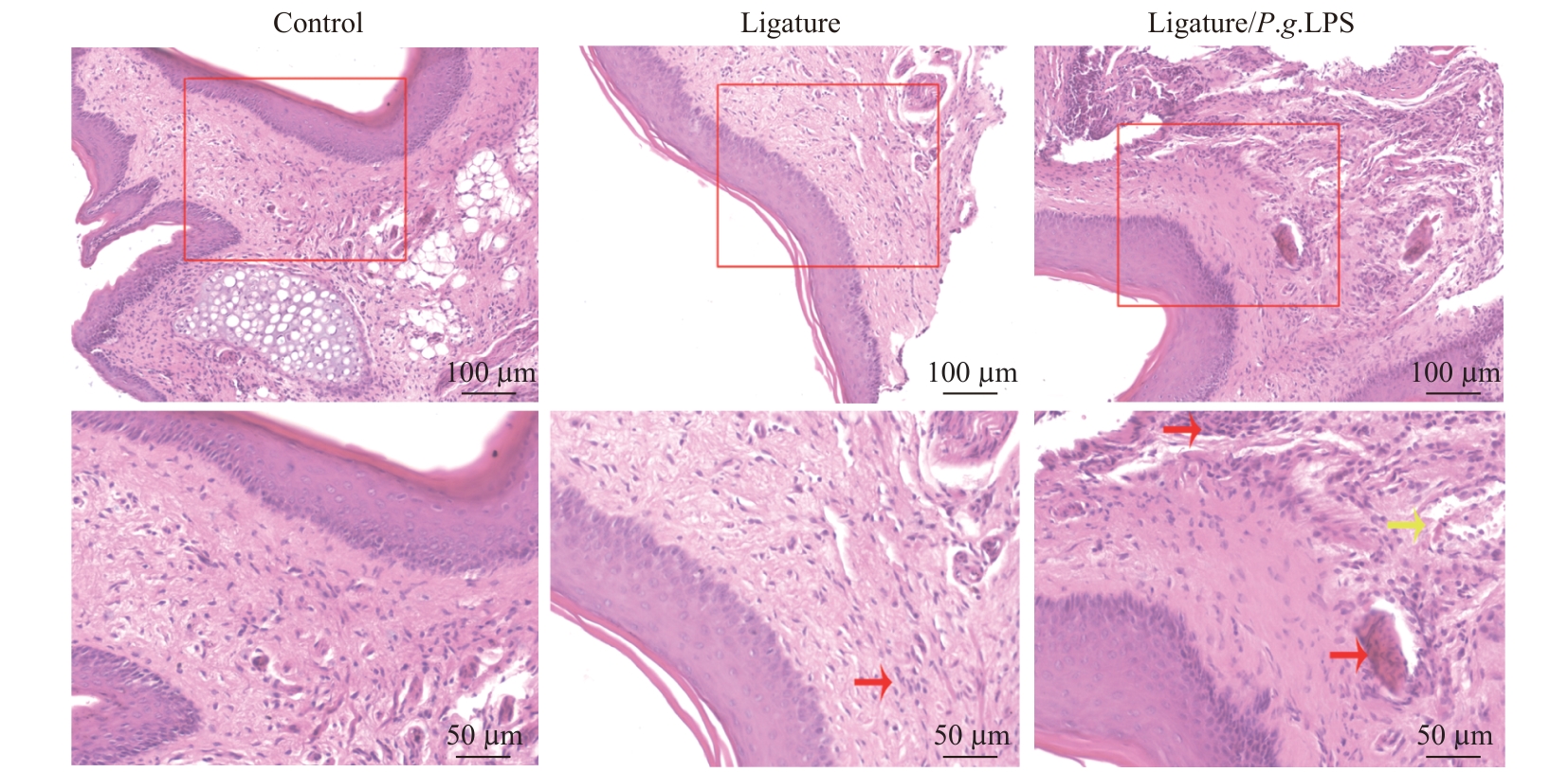
|
| 7 |
ROJAS C, GARCÍA M P, POLANCO A F, et al. Humanized mouse models for the study of periodontitis: an opportunity to elucidate unresolved aspects of its immunopathogenesis and analyze new immunotherapeutic strategies[J]. Front Immunol, 2021, 12: 663328.
|
| 8 |
高丽, 于晓潜, 蔡宇. 丝线结扎及局部涂抹牙龈卟啉单胞菌对小鼠牙槽骨骨吸收的影响[J]. 北京大学学报(医学版), 2017, 49(1): 31-35.
|
|
GAO L, YU X Q, CAI Y. Effect of molar ligation and local Porphyromonas gingivalis inoculation on alveolar bone loss in the mouse[J]. Journal of Peking University (Health Sciences), 2017, 49(1): 31-35.
|
| 9 |
国家质量监督检验检疫总局, 中国国家标准化管理委员会. 实验室生物安全通用要求: GB 19489—2008[S]. 北京: 中国标准出版社, 2009.
|
|
General Administration of Quality Supervision, Inspection and Quarantine of the People's Republic of China, National Standardization Administration. Laboratories general requirements for biosafety: GB19489—2008[S]. Beijing: Standards Press of China, 2009.
|
| 10 |
WANG X, TONG Y X, ZHANG J Y, et al. Neuroinflammation changes with periodontal inflammation status during periodontitis in wild-type mice[J]. Oral Dis, 2021, 27(4): 1001-1011.
|
| 11 |
SOUZA J A C, MAGALHÃES F A C, OLIVEIRA G J P L, et al. Pam2CSK4 (TLR2 agonist) induces periodontal destruction in mice[J]. Braz Oral Res, 2020, 34: e012.
|
| 12 |
束蓉, 倪靖. 2018牙周病和植体周病国际新分类: 牙周炎分期分级疾病定义系统临床应用体会[J]. 口腔医学, 2020, 40(1): 1-6.
|
|
SHU R, NI J. 2018 International classification of periodontal diseases and implant diseases: clinical application of staging and grading of periodontitis[J]. Stomatology, 2020, 40(1): 1-6.
|
| 13 |
MARCHESAN J, GIRNARY M S, JING L, et al. An experimental murine model to study periodontitis[J]. Nat Protoc, 2018, 13: 2247-2267.
|
| 14 |
SCANU A, GIRAUDO C, GALUPPINI F, et al. Periodontal injection of lipopolysaccharide promotes arthritis development in mice[J]. Inflammation, 2019, 42(3): 1117-1128.
|
| 15 |
BAI L, CHEN B Y, LIU Y, et al. A mouse periodontitis model with humanized oral bacterial community[J]. Front Cell Infect Microbiol, 2022, 12: 842845.
|
| 16 |
HARIYANI N, HALIMAH A N, AL-JUNAID M, et al. Mouse periodontitis models using whole Porphyromonas gingivalis bacteria induction[J]. Saudi Dent J, 2021, 33(8): 819-825.
|
| 17 |
de MOLON R S, PARK C H, JIN Q M, et al. Characterization of ligature-induced experimental periodontitis[J]. Microsc Res Tech, 2018, 81(12): 1412-1421.
|
| 18 |
LI S Y, ZENG W M, LIU G J, et al. Evaluation of morphological, histological, and immune-related cellular changes in ligature-induced experimental periodontitis in mice[J]. J Dent Sci, 2023, 18(4): 1716-1722.
|
| 19 |
SUH J S, KIM S, BOSTRÖM K I, et al. Periodontitis-induced systemic inflammation exacerbates atherosclerosis partly via endothelial-mesenchymal transition in mice[J]. Int J Oral Sci, 2019, 11(3): 21.
|
| 20 |
BLASCO-BAQUE V, GARIDOU L, POMIÉ C, et al. Periodontitis induced by Porphyromonas gingivalis drives periodontal microbiota dysbiosis and insulin resistance via an impaired adaptive immune response[J]. Gut, 2017, 66(5): 872-885.
|
| 21 |
CHEN X T, WAN Z, YANG L, et al. Exosomes derived from reparative M2-like macrophages prevent bone loss in murine periodontitis models via IL-10 mRNA[J]. J Nanobiotechnology, 2022, 20(1): 110.
|
| 22 |
TANG Y, QI Y D, CHEN Y, et al. Erythrocyte-mimicking nanovesicle targeting Porphyromonas gingivalis for periodontitis[J]. ACS Nano, 2024, 18(32): 21077-21090.
|
| 23 |
HUANG H Y, PAN W Y, WANG Y F, et al. Nanoparticulate cell-free DNA scavenger for treating inflammatory bone loss in periodontitis[J]. Nat Commun, 2022, 13(1): 5925.
|
 ), ZHANG Qianqian, SUI Baiyan, LIU Xin(